. 2008 May;82(10):4793-806.
doi: 10.1128/JVI.01587-07. Epub 2008 Mar 12.

Human herpesvirus 8 infects and replicates in primary cultures of activated B lymphocytes through DC-SIGN

Affiliations

Human herpesvirus 8 infects and replicates in primary cultures of activated B lymphocytes through DC-SIGN

Giovanna Rappocciolo et al. J Virol. 2008 May.

Abstract

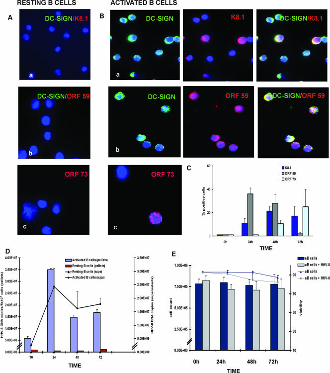
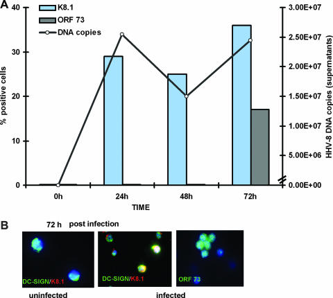
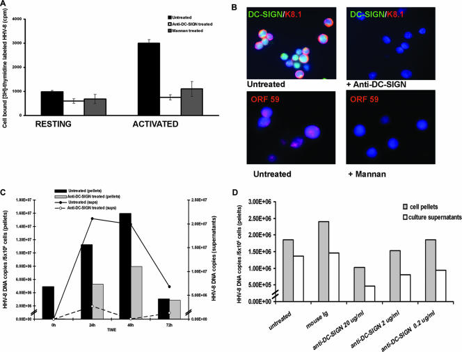
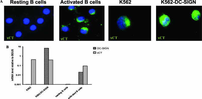
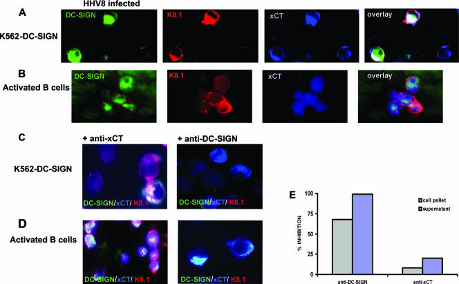
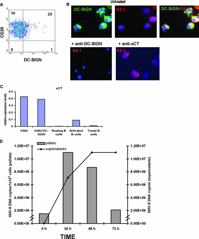
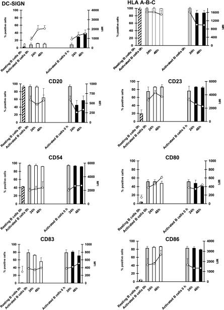
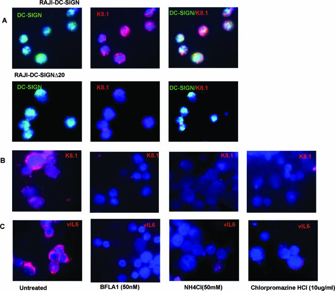
Human herpesvirus 8 (HHV-8) is the etiological agent of Kaposi's sarcoma, primary effusion lymphoma, and some forms of multicentric Castleman's disease. Although latent HHV-8 DNA can be detected in B cells from persons with these cancers, there is little information on the replication of HHV-8 in B cells. Indeed, B cells are relatively resistant to HHV-8 infection in vitro. We have recently shown that DC-SIGN, a C-type lectin first identified on dendritic cells (DC), is an entry receptor for HHV-8 on DC and macrophages. We have also demonstrated previously that B lymphocytes from peripheral blood and tonsils express DC-SIGN and that this expression increases after B-cell activation. Here we show that activated blood and tonsillar B cells can be productively infected with HHV-8, as measured by an increase in viral DNA, the expression of viral lytic and latency proteins, and the production of infectious virus. The infection of B cells with HHV-8 was blocked by the pretreatment of the cells with antibody specific for DC-SIGN or with mannan but not antibody specific for xCT, a cystine/glutamate exchange transporter that has been implicated in HHV-8 fusion to cells. The infection of B cells with HHV-8 resulted in increased expression of DC-SIGN and a decrease in the expression of CD20 and major histocompatibility complex class I. HHV-8 could also infect and replicate in B-cell lines transduced to express full-length DC-SIGN but not in B-cell lines transduced to express DC-SIGN lacking the transmembrane domain, demonstrating that the entry of HHV-8 into B cells is related to DC-SIGN-mediated endocytosis. The role of endocytosis in viral entry into activated B cells was confirmed by blocking HHV-8 infection with endocytic pathway inhibitors. Thus, the expression of DC-SIGN is essential for productive HHV-8 infection of and replication in B cells.


Figures

FIG. 1.
FIG. 1.
HHV-8 infects and replicates in B cells. (A and B) rB cells and aB cells were infected with HHV-8 and stained with MAb against DC-SIGN (green fluorescence) and anti-K8.1 (24 h) or anti-ORF59 protein (48 h) (red fluorescence) or with MAb against ORF73 protein (72 h; red fluorescence) (panels a, b, and c, respectively). Uninfected B cells were negative for each of the three viral proteins (data not shown). (C) Expression of viral lytic and latency proteins in HHV-8-infected aB cells over time (bars represent means ± standard errors [SE]; K8.1, n = 11; ORF59 protein, n = 4; ORF73 protein, n = 5). For each slide, at least four independent fields were counted and the results were averaged. (D) Quantitative HHV-8 DNA results from seven separate experiments with B cells from seven different donors. B cells were assayed by real-time PCR analyses of extracts of rB-cell or aB-cell pellets and the corresponding DNase-resistant, concentrated cell culture supernatants (sups). Both uninfected cell pellets and supernatants were negative for HHV-8 DNA (data not shown). (E) Cell counts (bars) and viability (lines) for uninfected (blue) and HHV-8-infected (gray) aB cells (means ± SE; n = 13).
FIG. 2.
FIG. 2.
HHV-8 particles derived from infected B-cell primary cultures are infectious. (A) Supernatants from aB cells infected with HHV-8 for 24 h were harvested, centrifuged, and used to infect freshly aB cells. At the indicated time points, cell pellets and supernatants were harvested and the amount of HHV-8 DNA was determined by real-time PCR (line). Bars indicate the percentages of cells expressing lytic (K8.1) or latency (ORF73) viral proteins. The results shown are representative of two independent experiments. (B) Cells were stained for the expression of DC-SIGN and of K8.1 and ORF73 viral proteins at 72 h postinfection.
FIG. 3.
FIG. 3.
HHV-8 binds and infects B cells through DC-SIGN. (A) Radioactively labeled HHV-8 was incubated with rB or aB cells that were left untreated or were pretreated with anti-DC-SIGN MAb or mannan. Each bar represents the mean of results from duplicate experiments (± SE). (B) aB cells were left untreated or treated with anti-DC-SIGN MAb or mannan prior to infection with HHV-8. Cells were stained for DC-SIGN (green) and K8.1 (red) or ORF59 protein (red) and counterstained with DAPI (4′,6′-diamidino-2-phenylindole; blue) at 24 h postinfection. (C) rB or aB cells were left untreated or incubated with anti-DC-SIGN MAb prior to infection with HHV-8. HHV-8 DNA in both cell pellets and cell culture supernatants (sups) was measured by real-time PCR as described in the legend to Fig. 1. No HHV-8 DNA was detected in uninfected B-cell cultures used as controls (data not shown). (D) aB cells were left untreated or incubated with anti-DC-SIGN MAb at different concentrations prior to infection with HHV-8. Mouse IgGs were used as controls at the highest concentration used for the anti-DC-SIGN MAb treatment (20 μg/ml). HHV-8 DNA in the cell pellets and supernatants at 24 h postinfection was measured. The results shown are representative of two independent experiments.
FIG. 4.
FIG. 4.
HHV-8 infects and replicates in DC-SIGN-transfected Raji and K562 cells. (A) Raji cells or Raji-DC-SIGN cells were infected with HHV-8 for 24 h as described in Materials and Methods and stained for DC-SIGN (green) and lytic protein K8.1 (red) and counterstained with DAPI (blue). (B) Quantitative viral DNA results from samples that were extracted from Raji-DC-SIGN cells and the corresponding DNase-resistant, concentrated cell culture supernatants (sups). Both uninfected and wild-type cell pellets and supernatants were negative for HHV-8 DNA (data not shown). (C) K562 cells or K562-DC-SIGN cells were infected with HHV-8 for 24 h as described in Materials and Methods and stained for DC-SIGN (green) and lytic protein K8.1 (red) and counterstained with DAPI (blue). (D) Quantitative viral DNA results from samples that were extracted from K562-DC-SIGN cells and the corresponding DNase-resistant, concentrated cell culture supernatants. Both uninfected and wild-type cell pellets and supernatants were negative for HHV-8 DNA (data not shown).The results shown are representative of two independent experiments.
FIG. 5.
FIG. 5.
Expression of xCT on B cells and K562 cells. (A) Freshly isolated rB or aB cells or K562 or K562-DC-SIGN cells were incubated with rabbit antisera against xCT peptide and goat anti-rabbit IgG-FITC and counterstained with DAPI. (B) DC-SIGN- and xCT-specific mRNA levels in K562, K562-DC-SIGN, rB, and aB cells. The levels are expressed relative to mRNA levels for the β-GUS gene (an internal housekeeping gene) in these cells.
FIG. 6.
FIG. 6.
Effect of anti-xCT and anti-DC-SIGN Ab treatment on HHV-8 infection. (A and B) K562-DC-SIGN cells (A) or aB cells (B) were infected with HHV-8 and stained for DC-SIGN (green), xCT (blue), or K8.1 (red). Each of the first three panels in each row shows the individual dual combinations, while the fourth panel shows the three-color overlay. (C and D) K562-DC-SIGN cells (C) or aB cells (D) were treated with anti-xCT rabbit antiserum (left panels) or anti-DC-SIGN MAb (right panels) or left untreated (A and B) and were infected with HHV-8 for 24 h and then stained for the expression of DC-SIGN, xCT, and K8.1. Each panel represents a three-color overlay as in panels A and B. (E) Relative levels of HHV-8 DNA in the cell pellets and supernatants of cultures pretreated with either anti-DC-SIGN MAb or anti-xCT rabbit antiserum. The levels of inhibition were measured against those of cultures pretreated with mouse IgG or preimmune rabbit antiserum, respectively.
FIG. 7.
FIG. 7.
HHV-8 infects and replicates in fresh tonsillar B cells. (A) B cells were isolated from tonsils (as described in Materials and Methods) by magnetic bead purification. Cells were then stained for the expression of CD20 and DC-SIGN and analyzed by fluorescence-activated cell sorting. (B) Fresh tonsillar B cells were left untreated or treated with anti-DC-SIGN MAb or anti-xCT antisera prior to infection with HHV-8, cultured for 24 h, and stained with anti-DC-SIGN (green) or anti-K8.1 (red) MAb. Nuclei are stained blue with DAPI. (C) xCT-specific mRNA levels in K562, K562-DC-SIGN, aB, and tonsillar B cells. The levels are expressed relative to mRNA levels for the β-GUS gene (an internal housekeeping gene) in these cells. The results shown are representative of two independent experiments. (D) Quantitative DNA results from real-time PCR assays. HHV-8 DNA was extracted from tonsillar B cells and the corresponding DNase-resistant, concentrated cell culture supernatants. Both uninfected cell pellets and supernatants were negative for HHV-8 DNA (data not shown).
FIG. 8.
FIG. 8.
Expression of cellular markers in activated, HHV-8-infected B cells. aB cells were left untreated (white bars) or infected with HHV-8 (black bars) and cultured for 48 h. At the indicated time points, cells were stained for the expression of B-cell markers and analyzed by flow cytometry. rB and aB cells were also analyzed at the onset of infection (0 h). Bars and lines represent the means (± SE) of results from six independent experiments and refer to percentages of positive cells and MFIs, respectively.
FIG. 9.
FIG. 9.
HHV-8 endocytosis in B-cell lines and aB cells. (A) Raji-DC-SIGN or Raji-DC-SIGN-Δ20 cells were infected with HHV-8, cultured for 24 h, and then stained for the expression of HHV-8 protein K8.1 (red) or DC-SIGN (green). Nuclei were stained with DAPI. (B and C) aB cells were left untreated or pretreated with BFLA, NH4Cl, or chlorpromazine HCl (as described in Materials and Methods) and infected with HHV-8. Cells were washed, cultured for 24 h, and stained for the expression of lytic proteins K8.1 and vIL-6.

Similar articles

Cited by

References

    1. Akula, S. M., P. P. Naranatt, N. S. Walia, F. Z. Wang, B. Fegley, and B. Chandran. 2003. Kaposi's sarcoma-associated herpesvirus (human herpesvirus 8) infection of human fibroblast cells occurs through endocytosis. J. Virol. 777978-7990. - PMC - PubMed
    1. Akula, S. M., N. P. Pramod, F. Z. Wang, and B. Chandran. 2001. Human herpesvirus 8 envelope-associated glycoprotein B interacts with heparan sulfate-like moieties. Virology 284235-249. - PubMed
    1. Akula, S. M., N. P. Pramod, F. Z. Wang, and B. Chandran. 2002. Integrin α3β1 (CD 49c/29) is a cellular receptor for Kaposi's sarcoma-associated herpesvirus (KSHV/HHV-8) entry into the target cells. Cell 108407-419. - PubMed
    1. Akula, S. M., F. Z. Wang, J. Vieira, and B. Chandran. 2001. Human herpesvirus 8 interaction with target cells involves heparan sulfate. Virology 282245-255. - PubMed
    1. Ambroziak, J. A., D. J. Blackbourn, B. G. Herndier, R. G. Glogau, J. H. Gullett, A. R. McDonald, E. T. Lennette, and J. A. Levy. 1995. Herpes-like sequences in HIV-infected and uninfected Kaposi's sarcoma patients. Science 268582-583. - PubMed

Publication types

MeSH terms

LinkOut - more resources