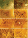
Prevalence and morphology of druse types in the macula and periphery of eyes with age-related maculopathy
- PMID: 18326750
- PMCID: PMC2279189
- DOI: 10.1167/iovs.07-1466
Prevalence and morphology of druse types in the macula and periphery of eyes with age-related maculopathy
Abstract
Purpose: Macular drusen are hallmarks of age-related maculopathy (ARM), but these focal extracellular lesions also appear with age in the peripheral retina. The present study was conducted to determine regional differences in morphology that contribute to the higher vulnerability of the macula to advanced disease.
Methods: Drusen from the macula (n = 133) and periphery (n = 282) were isolated and concentrated from nine ARM-affected eyes. A semiquantitative light microscopic evaluation of 1-mum-thick sections included 12 parameters.
Results: Significant differences were found between the macula and periphery in ease of isolation, distribution of druse type, composition qualities, and substructures. On harvesting, macular drusen were friable, with liquefied or crystallized contents. Peripheral drusen were resilient and never crystallized. On examination, soft drusen appeared in the macula only, had homogeneous content without significant substructures, and had abundant basal laminar deposits (BlamD). Several substructures, previously postulated as signatures of druse biogenesis, were found primarily in hard drusen. Specific to hard drusen, which appeared everywhere, were central subregions and reduced RPE coverage. Macular hard drusen with a rich substructure profile differed from primarily homogeneous peripheral hard drusen. Compound drusen, found in the periphery only, exhibited a composition profile that was not intermediate between hard and soft.
Conclusions: The data confirm regional differences in druse morphology, composition, and physical properties, most likely based on different formative mechanisms that may contribute to macular susceptibility for ARM progression. Two other reasons that only the macula is at high risk despite having relatively few drusen are the exclusive presence of soft drusen and the abundant BlamD in this region.
Figures


Similar articles
-
The Age-Related Macular Degeneration Complex: Linking Epidemiology and Histopathology Using the Minnesota Grading System (The Inaugural Frederick C. Blodi Lecture).Trans Am Ophthalmol Soc. 2015 Sep;113:Blodi. Trans Am Ophthalmol Soc. 2015. PMID: 27895380 Free PMC article.
-
Age-related changes in the macula. A histopathological study of fifty Indian donor eyes.Indian J Ophthalmol. 2002 Sep;50(3):201-4. Indian J Ophthalmol. 2002. PMID: 12355694
-
Early deterioration in ellipsoid zone in eyes with non-neovascular age-related macular degeneration.Int Ophthalmol. 2017 Aug;37(4):801-806. doi: 10.1007/s10792-016-0331-3. Epub 2016 Sep 3. Int Ophthalmol. 2017. PMID: 27591785 Clinical Trial.
-
Drusen in age-related macular degeneration: pathogenesis, natural course, and laser photocoagulation-induced regression.Surv Ophthalmol. 1999 Jul-Aug;44(1):1-29. doi: 10.1016/s0039-6257(99)00072-7. Surv Ophthalmol. 1999. PMID: 10466585 Review.
-
Antecedents of Soft Drusen, the Specific Deposits of Age-Related Macular Degeneration, in the Biology of Human Macula.Invest Ophthalmol Vis Sci. 2018 Mar 20;59(4):AMD182-AMD194. doi: 10.1167/iovs.18-24883. Invest Ophthalmol Vis Sci. 2018. PMID: 30357337 Free PMC article. Review.
Cited by
-
Developing an image-based grading scale for peripheral drusen to investigate associations of peripheral drusen type with age-related macular degeneration.Sci Rep. 2024 Aug 29;14(1):20041. doi: 10.1038/s41598-024-70352-3. Sci Rep. 2024. PMID: 39198593 Free PMC article.
-
Choriocapillaris Impairment, Visual Function, and Distance to Fovea in Aging and Age-Related Macular Degeneration: ALSTAR2 Baseline.Invest Ophthalmol Vis Sci. 2024 Jul 1;65(8):40. doi: 10.1167/iovs.65.8.40. Invest Ophthalmol Vis Sci. 2024. PMID: 39042400 Free PMC article.
-
Antioxidants and Mechanistic Insights for Managing Dry Age-Related Macular Degeneration.Antioxidants (Basel). 2024 May 4;13(5):568. doi: 10.3390/antiox13050568. Antioxidants (Basel). 2024. PMID: 38790673 Free PMC article. Review.
-
Complement regulation in the eye: implications for age-related macular degeneration.J Clin Invest. 2024 May 1;134(9):e178296. doi: 10.1172/JCI178296. J Clin Invest. 2024. PMID: 38690727 Free PMC article. Review.
-
Age-Related Macular Degeneration, a Mathematically Tractable Disease.Invest Ophthalmol Vis Sci. 2024 Mar 5;65(3):4. doi: 10.1167/iovs.65.3.4. Invest Ophthalmol Vis Sci. 2024. PMID: 38466281 Free PMC article. Review.
References
-
- Green WR. Histopathology of age-related macular degeneration. Mol Vis. 1999;5:27. - PubMed
-
- Abdelsalam A, Del Priore L, Zarbin MA. Drusen in age-related macular degeneration: pathogenesis, natural course, and laser photocoagulation-induced regression. Surv Ophthalmol. 1999;44:1–29. - PubMed
-
- Klein R, Klein BE, Linton KL. Prevalence of age-related maculopathy. The Beaver Dam Eye Study. Ophthalmology. 1992;99:933–943. - PubMed
-
- Bressler NM, Bressler SB, West SK, Fine SL, Taylor HR. The grading and prevalence of macular degeneration in Chesapeake Bay watermen. Arch Ophthalmol. 1989;107:847–852. - PubMed
-
- Lengyel I, Tufail A, Hosaini HA, Luthert P, Bird AC, Jeffery G. Association of drusen deposition with choroidal intercapillary pillars in the aging human eye. Invest Ophthalmol Vis Sci. 2004;45:2886–2892. - PubMed
Publication types
MeSH terms
Grants and funding
LinkOut - more resources
Full Text Sources
Medical

