. 2008 May 2;283(18):12324-32.
doi: 10.1074/jbc.M707898200. Epub 2008 Mar 4.

Stabilization of RelB requires multidomain interactions with p100/p52

Affiliations

Stabilization of RelB requires multidomain interactions with p100/p52

Amanda J Fusco et al. J Biol Chem. .

Abstract

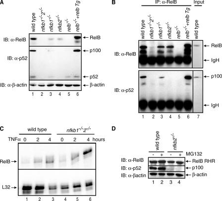
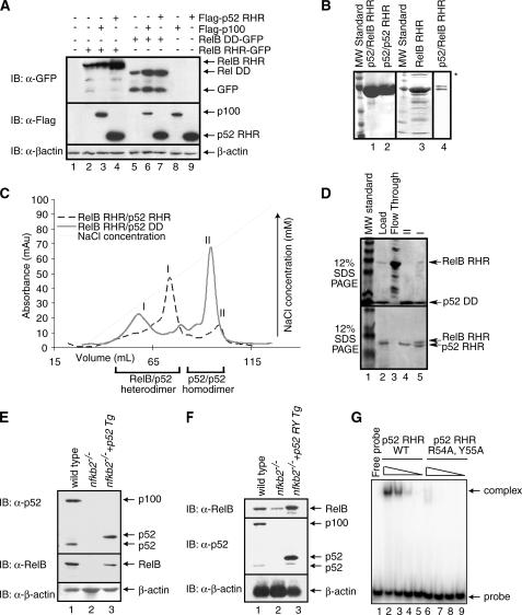
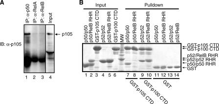
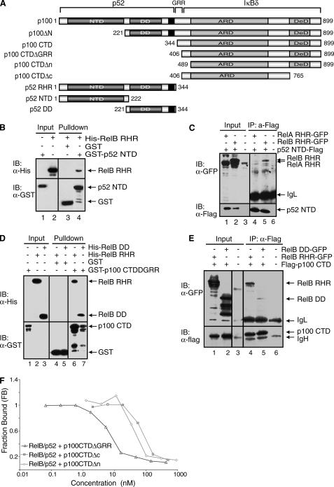
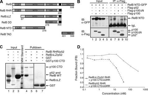
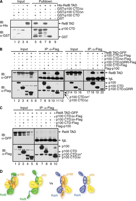
The NF-kappaB family member RelB has many properties not shared by other family members such as restricted subunit association and lack of regulation by the classical IkappaB proteins. We show that the protein level of RelB is significantly reduced in the absence of p100 and reduced even more when both p100 and p105 are absent. RelB stabilizes itself by directly interacting with p100, p105, and their processed products. However, RelB forms complexes with its partners using different interaction modes. Although the C-terminal ankyrin repeat domain of p105 is not involved in the RelB-p105 complex formation, all domains and flexible regions of each protein are engaged in the RelB-p100 complex. In several respects the RelB-p52 and RelB-p100 complexes are unique in the NF-kappaB family. The N-terminal domain of p100/p52 interacts with RelB but not RelA. The transcriptional activation domain of RelB, but not RelA, directly interacts with the processing region of p100. These unique protein-protein contacts explain why RelB prefers p52 as its dimeric partner for transcriptional activity and is retained in the cytoplasm as an inhibited complex by p100. This association-mediated stabilization of RelB implies a possible role for RelB in the processing of p100 into p52.


Figures

FIGURE 1.
FIGURE 1.
Stability of RelB depends on the presence of p100 protein. A, extracts of wild type, nfkb1-/-, nfkb2-/-, nfkb1-/-/nfkb2-/-, relb-/-, and RelB transgene (Tg) in relb-/- MEF cells were separated by SDS-PAGE followed by immunoblot with RelB antibody (top panel), p52 antibody (middle panel), and β-actin antibody (bottom panel). B, cell extracts from the same cells as in A were used to immunoprecipitate RelB-bound proteins using RelB antibody, and the complexes were separated by SDS-PAGE followed by immunoblot with RelB antibody (top panel) and p52 antibody (bottom panel). C, RNase protection assay of RelB mRNA in wt and nfkb1-/-2-/- MEF cells in resting and tumor necrosis factor α-induced cells in the top panel and a control mRNA (ribosomal protein L32) in the bottom panel. D, extracts of wild type and nfkb2-/- MEF cells were separated by SDS-PAGE followed by immunoblot with RelB antibody (top panel), p52 antibody (middle panel), and β-actin antibody (bottom panel).
FIGURE 2.
FIGURE 2.
RelB RHR stabilization by p52 in vitro. A, Western blot analysis of the steady state levels of RelB RHR and RelB DD proteins in HEK 293 cells transfected with FLAG-p100, FLAG-p52, and RelB-GFP in different combinations. In the presence of p100 and p52, RelB protein levels are enhanced. B, Coomassie-stained SDS-PAGE showing purity of p52-RelB heterodimer, p52 homodimer, and RelB RHR homodimer. A lower concentration of p52-RelB heterodimer is shown in lane 4 to visualize the separation between the two proteins. RelB continuously degrades during purification, and the degradation products are seen in the gel (lane 4). Aggregated species of RelB in lane 3 is marked by an asterisk. C, co-refolded mixtures of RelB RHR and p52 RHR (black), and RelB RHR and p52 DD (gray) were separated by cation exchange (S-Sepharose column) chromatography. D, Coomassie-stained SDS-PAGE of samples from the S column chromatography of the RelB RHR-p52 DD (top panel) and RelB RHR-p52 RHR (bottom panel). The load appears to contain excess p52, which masks the RelB RHR. E, Western blot analysis of the steady state levels of p100/p52 (top panel) and RelB (middle panel) in wt, nfkb2-/- MEF and p52-reconstituted nfkb2-/- cells. Reconstituted p52 migrates higher due to the exact site of processing of p100 is unknown. F, DNA binding-defective p52 mutant stabilizes RelB. Western blot analysis of the steady state levels of p100/p52 (top panel) and RelB (middle panel) in wt, nfkb2-/- MEF, and p52-reconstituted nfkb2-/- cells. Reconstituted p52 migrates higher due to the exact site of processing of p100 is unknown. G, electrophoretic mobility shift assay analysis of wild type p52 and p52 R54A,Y55A double mutant. Lanes 2 and 6, 3 and 7, 4 and 8, and 5 and 9 contain 1100, 250, 25, and 2.5 nm wild type and double mutant p52, respectively.
FIGURE 3.
FIGURE 3.
p105 interacts with RelB in a distinct mode. A, cell extract from HeLa cells were used to immunoprecipitate RelB, p50, and RelA-bound p105 using RelB, p50, and RelA antibodies. The complexes were separated by SDS-PAGE followed by immunoblot with p105 antibody. B, in vitro GST pulldown experiments demonstrating the difference in the binding interaction between p100 CTD (IκBδ) and p105 CTD (IκBγ) with p52-RelB. Lane 8 demonstrates the lack of interaction of p105 CTD with p50/RelB versus the stable interaction between p100 CTD and p52-RelB (lane 10). Lanes 7 and 9 are positive controls of p105 CTD interacting with p50 and p100 CTD with p52, respectively. Lanes 1–6 show the inputs, and lanes 11–14 show the controls.
FIGURE 4.
FIGURE 4.
All domains and flexible regions of p100 contact RelB. A, schematic representation of p100 and its deletion mutants. The black regions represent the nuclear localization signals. B, Western blot analysis of in vitro GST pulldown experiments demonstrating the binding interaction between the NTD of p52 and RelB RHR, followed by immunoblot with α-GST or α-His antibodies. C, co-IP experiments showing the interaction between p52 NTD-FLAG and RelB RHR-GFP from the extracts of cotransfected HEK 293 cells. p52 NTD was isolated by IP and analyzed by IB (bottom panel), and the co-precipitated RelB was analyzed by IB (top panel). An asterisk indicates a nonspecific band. D, Western blot analysis of in vitro GST-pulldown experiments showing binding interaction between the GST-tagged CTD of p100 and His-RelB RHR and His-RelB DD. E, co-IP experiments showing the binding interaction between the FLAG tagged CTD of p100 and RelB RHR-GFP and RelB DD-GFP. These experiments were done similarly as described in C. F, fluorescence polarization assay showing the effect of the regions flanking the ARD of p100 in inhibition of DNA binding by the RelB-p52 heterodimer. Constant amounts of RelB-p52 RHR bound to a fluorescently labeled DNA was titrated with increasing amounts of GST-p100 CTD proteins.
FIGURE 5.
FIGURE 5.
The LZ domain of RelB is involved in p100 binding. A, schematic representations of RelB domains and the deletion mutants of RelB used in the binding experiments. The black regions represent the nuclear localization signals. B, co-IP experiments showing binding interaction between RelB NTD-GFP and wt or deletion mutants of p100 with the FLAG peptide. The cells were cotransfected with RelB NTD-GFP and p100 mutants, extracts were immunoprecipitated by α-FLAG antibody and immunoblotted with FLAG (bottom panel) and GFP (top panel). An asterisk indicates a nonspecific band. C, in vitro GST pulldown experiments were done using equal amounts of pure recombinant proteins showing that the N-terminal LZ domain of RelB is involved in the binding interaction with the CTD of p100. Input and pulldown samples were separated by SDS-PAGE followed by Coomassie staining. Please note that His-RelB RHR and p52 RHR co-migrate in the SDS-PAGE (lanes 3 and 4). At position 64 of RelB, a cryptic thrombin cleavage site is located; therefore the first 64 residues are removed (lanes 1 and 5). D, fluorescence polarization experiments showing the functional role of the LZ domain of RelB in binding the p100 CTDΔGRR. Fluorescence polarization assay was done the same as in Fig. 3F.
FIGURE 6.
FIGURE 6.
The TAD of RelB contacts the p100 processing site. A, Western blot analysis of in vitro GST pulldown experiments showing the interaction between the TAD of RelB with the GST-tagged CTD of p100. Input and pulldown samples were separated by SDS-PAGE followed by IB. B, co-IP showing the same interaction as in A but in cotransfected HEK 293 cells. FLAG-p100 or mutants were co-expressed with RelB TAD. IPed extracts were separated by SDS-PAGE, and the presence of co-precipitated RelB was analyzed by IB (top panel). The level of p100 and p100 CTD were analyzed by IB (bottom panel). An asterisk indicates that p100 CTDΔn migrates as the same size as the IgH band. C, the TAD of RelA does not bind p100. D, model of the interactions between RelB and p52/p100 as compared with the interactions between p52/p100 and p65.

Similar articles

Cited by

References

    1. Baldwin, A. S., Jr. (1996) Annu. Rev. Immunol. 14 649-683 - PubMed
    1. Ghosh, S., May, M. J., and Kopp, E. B. (1998) Annu. Rev. Immunol. 16 225-260 - PubMed
    1. Ghosh, S., and Karin, M. (2002) Cell 109 (Suppl.) S81-S96 - PubMed
    1. Karin, M., and Ben-Neriah, Y. (2000) Annu. Rev. Immunol. 18 621-663 - PubMed
    1. Solan, N. J., Miyoshi, H., Carmona, E. M., Bren, G. D., and Paya, C. V. (2002) J. Biol. Chem. 277 1405-1418 - PubMed

Publication types

Substances