. 2008 Feb;7(2):148-58.
doi: 10.1016/j.cmet.2007.11.015.

Regulation of ceramide biosynthesis by TOR complex 2

Affiliations

Regulation of ceramide biosynthesis by TOR complex 2

Sofia Aronova et al. Cell Metab. 2008 Feb.

Abstract

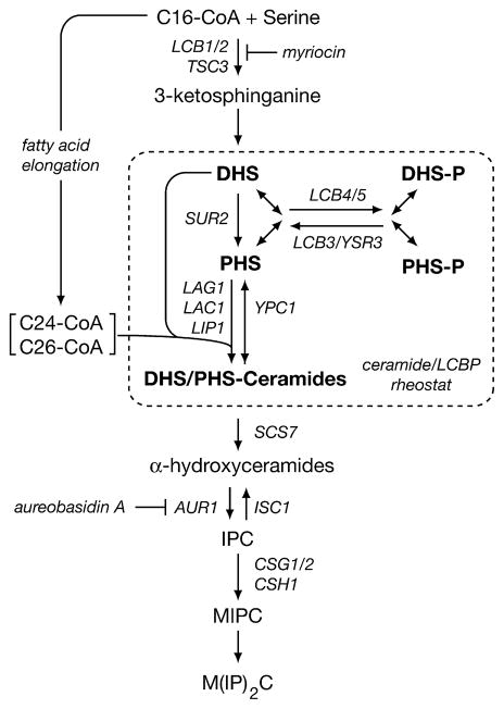
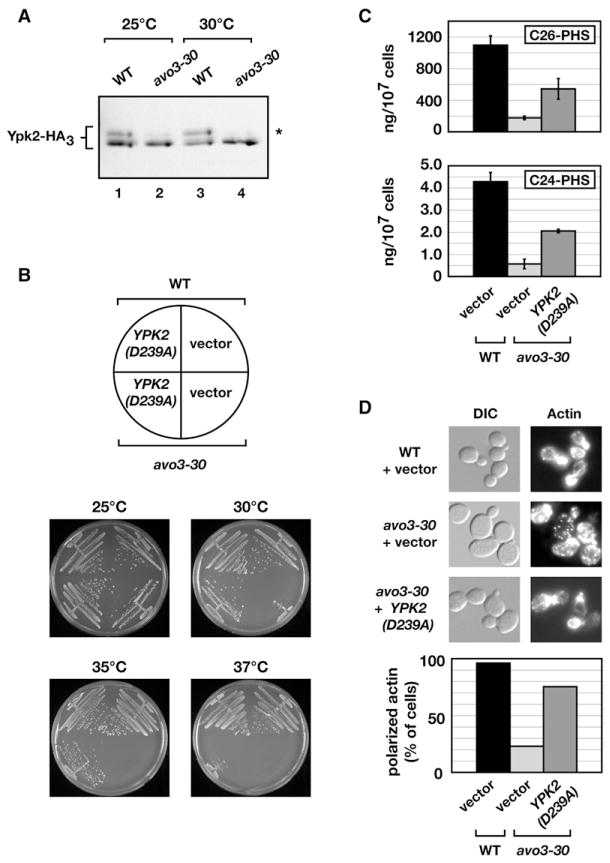
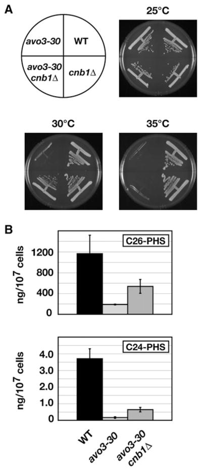
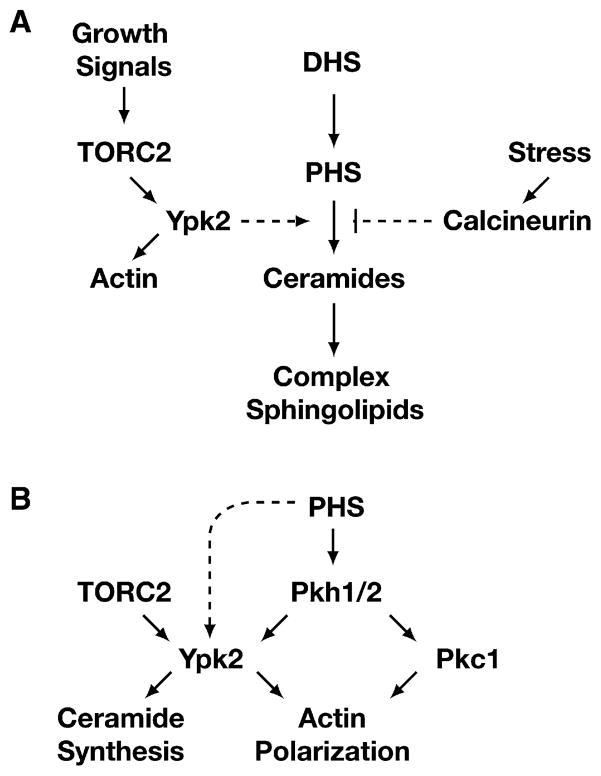
Ceramides and sphingoid long-chain bases (LCBs) are precursors to more complex sphingolipids and play distinct signaling roles crucial for cell growth and survival. Conserved reactions within the sphingolipid biosynthetic pathway are responsible for the formation of these intermediates. Components of target of rapamycin complex 2 (TORC2) have been implicated in the biosynthesis of sphingolipids in S. cerevisiae; however, the precise step regulated by this complex remains unknown. Here we demonstrate that yeast cells deficient in TORC2 activity are impaired for de novo ceramide biosynthesis both in vivo and in vitro. We find that TORC2 regulates this step in part by activating the AGC kinase Ypk2 and that this step is antagonized by the Ca2+/calmodulin-dependent phosphatase calcineurin. Because Ypk2 is activated independently by LCBs, the direct precursors to ceramides, our data suggest a model wherein TORC2 signaling is coupled with LCB levels to control Ypk2 activity and, ultimately, regulate ceramide formation.


Figures

Figure 1
Figure 1. Simplified Schematic Diagram of the Sphingolipid Biosynthetic Pathway in S. cerevisiae
Shown are some of the major intermediates of the pathway as well as a selected set of genes encoding proteins required for specific steps of the pathway considered here. Not shown is Ydc1, an alkaline ceramidase that catabolizes DHS-ceramides.
Figure 2
Figure 2. Characterization of avo3-30, a Temperature-Sensitive Allele of AVO3
(A) Phenotype of avo3-30 cells on solid medium. Wild-type (WT; LHY291) and avo3-30 cells were streaked onto YPD agar plates and incubated at the indicated temperatures for 2–3 days. (B) The temperature-sensitive (ts) phenotype of avo3-30 is rescued by plasmid-expressed WT AVO3. WT and avo3-30 cells were transformed with either the control vector pRS313 (Sikorski and Heiter, 1989) or pAVO3 and grown on SCD agar plates lacking histidine at the indicated temperatures for 2–3 days. (C) Cell integrity-related phenotypes of avo3-30 cells. Cells were streaked onto YPD agar plates or plates containing 0.8 M sorbitol (+Sorb), 1 mM caffeine (+Caff), or 0.2 μg/ml rapamycin (+Rap) and incubated at 30°C or 37°C, as indicated. (D) Hypersensitivity of avo3-30 cells to inhibitors of the sphingolipid biosynthetic pathway. Cells were streaked onto YPD agar plates or plates containing 0.5 μM myriocin (+Myr) or 9 nM aureobasidin A (+Aur) and incubated at 25°C. (E) Protein levels of Avo3 in WT and avo3-30 cells. Cells were grown at 25°C and were either left at 25°C or shifted to 30°C and 37°C for a further 5 hr. Aliquots were collected each hour and processed for western blot analysis. Tor1 protein levels were analyzed as a loading control. (F) Growth curves of WT and avo3-30 cells in liquid culture. Cells were grown in YPD medium at 25°C, diluted to OD600 = 0.08, and shifted to 30°C or 37°C for 8 hr. Growth was monitored by A600, and growth curves were plotted on a semilogarithmic scale.
Figure 3
Figure 3. Monitoring Intermediates in the Sphingolipid Biosynthetic Pathway in Wild-Type and avo3-30 Cells
(A) Levels of phytoceramides were analyzed in extracts derived from WT and avo3-30 cells grown in YPD medium at 25°C following a shift to 30°C for 3 hr. (B) Levels of LCBs (DHS and PHS) and LCBPs (DHS-P and PHS-P) were analyzed in extracts derived from WT and avo3-30 cells in YPD medium following a shift to 30°C for 3 hr. (C) Levels of DHS and PHS were analyzed in extracts derived from WT, avo3-30, and avo3-30 lcb4Δ cells in YPD medium following a shift to 30°C for 3 hr. In (A–C), error bars indicate the SD of separately extracted samples (n = 3). (D) Monitoring ceramide synthesis in vitro. Microsomal membranes were isolated and ceramide synthase activity was measured as described in Experimental Procedures. Left panel: schematic diagram of the in vitro reaction, showing the structure of the C18-SPHC17 ceramide product and the substrates SPHC17 and C18-CoA used for this reaction. Right panel: C18-CoA concentration dependence of ceramide synthase activity in microsomal membranes isolated from WT versus avo3-30 cells.
Figure 4
Figure 4. Beneficial Effects of Deleting LCB4 and/or Adding Exogenous PHS with Respect to avo3-30 Phenotypes
(A and B) Growth in liquid YPD medium (A) and on YPD agar plates (B) of indicated strains in the absence or presence of different concentrations of PHS and at different temperatures, as indicated. (C) Effect of PHS addition on actin polarization in the indicated strains grown at 30°C (upper panels). Bar graph represents the quantification of this experiment and includes data for WT and avo3-30 cells grown at 25°C for comparison. (D) Influence of LCB4 deletion and/or PHS addition on viability of avo3-30 cells. The indicated strains were grown to early log phase in liquid media that either lacked or contained the indicated concentration of PHS at 25°C and were then transferred to 30°C for 4 hr. Cultures were diluted and plated onto solid YPD medium at 25°C to determine colony-forming units (expressed as % of WT). (E) Addition of PHS partially rescues phytoceramide levels in avo3-30 and avo3-30 lcb4Δ cells. Cells were grown at 25°C and shifted to 30°C for 3 hr. PHS was then added to a final concentration of 4 μM, and cells were incubated for an additional 3 hr at 30°C. Sphingolipids were extracted, and ceramide levels were analyzed by LC-MS/MS. Error bars indicate the SD of separately extracted samples (n = 3).
Figure 5
Figure 5. Expression of a Constitutively Active Allele of YPK2 Rescues Multiple Phenotypes of the avo3-30 Mutant
(A) WT and avo3-30 cells expressing a HA3-tagged version of Ypk2 were grown in YPD medium at 25°C and either shifted to 30°C for 3 hr or left at 25°C and were then processed for western blot analysis, probing for Ypk2 using anti-HA antibody. The asterisk indicates the phosphorylated form of Ypk2 (Kamada et al., 2005). (B) WT and avo3-30 cells harboring either pYE352[YPK2D239A] or vector plasmid were streaked onto selective dropout plates and incubated at the indicated temperatures for 3–4 days. (C) WT and avo3-30 cells harboring either pYE352[YPK2D239A] or vector plasmid were grown in selective dropout medium at 25°C overnight, followed by a shift to 30°C for 3 hr. Sphingolipids were extracted and ceramide levels were analyzed by LC-MS/MS as described in Experimental Procedures. Error bars indicate the SD of separately extracted samples (n = 3). (D) WT and avo3-30 cells harboring either pYE352[YPK2D239A] or vector plasmid were grown as described in (C), fixed, and stained for actin with rhodamine-coupled phalloidin. The graph below shows the percentage of cells with completely polarized actin patches.
Figure 6
Figure 6. Deletion of the Calcineurin Regulatory Subunit CNB1 Restores a Subset of the Phenotypes of the avo3-30 Mutant
(A) WT, avo3-30, avo3-30 cnb1Δ, and cnb1Δ cells were streaked onto YPD plates and incubated at the indicated temperatures. (B) avo3-30 and avo3-30 cnb1Δ cells were grown in liquid YPD medium to mid-log phase, sphingolipids were extracted, and ceramide levels were analyzed by LC-MS/MS. Error bars indicate the SD of separately extracted samples (n = 3).
Figure 7
Figure 7. Models Illustrating Connections between TORC2 Signaling and Regulation of the Sphingolipid Biosynthetic Pathway
(A) TORC2 signals to Ypk2 to positively regulate de novo synthesis of ceramides at a step that is antagonized by calcineurin. (B) Interplay between TORC2 and PHS for activation of Ypk2 for ceramide synthesis. See text for details.

Comment in

Similar articles

Cited by

References

    1. Audhya A, Loewith R, Parsons AB, Gao L, Tabuchi M, Zhou H, Boone C, Hall MN, Emr SD. Genome-wide lethality screen identifies new PI4,5P2 effectors that regulate the actin cytoskeleton. EMBO J. 2004;23:3747–3757. - PMC - PubMed
    1. Beeler T, Bacikova D, Gable K, Hopkins L, Johnson C, Slife H, Dunn T. The Saccharomyces cerevisiae TSC10/YBR265w gene encoding 3-ketosphinganine reductase is identified in a screen for temperature-sensitive suppressors of the Ca2+-sensitive csg2Delta mutant. J Biol Chem. 1998;273:30688–30694. - PubMed
    1. Bultynck G, Heath VL, Majeed AP, Galan JM, Haguenauer-Tsapis R, Cyert MS. Slm1 and slm2 are novel substrates of the calcineurin phosphatase required for heat stress-induced endocytosis of the yeast uracil permease. Mol Cell Biol. 2006;26:4729–4745. - PMC - PubMed
    1. Canton DA, Litchfield DW. The shape of things to come: an emerging role for protein kinase CK2 in the regulation of cell morphology and the cytoskeleton. Cell Signal. 2006;18:267–275. - PubMed
    1. Clarke CJ, Hannun YA. Neutral sphingomyelinases and nSMase2: bridging the gaps. Biochim Biophys Acta. 2006;1758:1893–1901. - PubMed

Publication types

MeSH terms