Down-regulation of NKG2D and NKp80 ligands by Kaposi's sarcoma-associated herpesvirus K5 protects against NK cell cytotoxicity
- PMID: 18230726
- PMCID: PMC2234200
- DOI: 10.1073/pnas.0707883105
Down-regulation of NKG2D and NKp80 ligands by Kaposi's sarcoma-associated herpesvirus K5 protects against NK cell cytotoxicity
Abstract
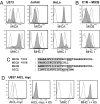
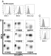
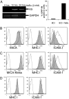
Natural killer (NK) cells are important early mediators of host immunity to viral infections. The NK activatory receptors NKG2D and NKp80, both C-type lectin-like homodimeric receptors, stimulate NK cell cytotoxicity toward target cells. Like other herpesviruses, Kaposi's sarcoma-associated herpesvirus (KSHV) down-regulates MHC class I molecules to avoid detection by cytotoxic T lymphocytes but renders cells susceptible to NK cell cytotoxicity. We now show that the KSHV immune evasion gene, K5, reduces cell surface expression of the NKG2D ligands MHC class I-related chain A (MICA), MICB, and the newly defined ligand for NKp80, activation-induced C-type lectin (AICL). Down-regulation of both MICA and AICL requires the ubiquitin E3 ligase activity of K5 to target substrate cytoplasmic tail lysine residues. The common MICA *008 allele has a frameshift mutation leading to a premature stop codon and is resistant to down-regulation because of the loss of lysine residues. K5-mediated ubiquitylation signals internalization but not degradation of MICA and causes a potent reduction in NK cell-mediated cytotoxicity. The down-regulation of ligands for both the NKG2D and NKp80 activation pathways provides KSHV with a powerful mechanism for evasion of NK cell antiviral functions.
Conflict of interest statement
The authors declare no conflict of interest.
Figures


Similar articles
-
Natural killer cell evasion by an E3 ubiquitin ligase from Kaposi's sarcoma-associated herpesvirus.Biochem Soc Trans. 2008 Jun;36(Pt 3):459-63. doi: 10.1042/BST0360459. Biochem Soc Trans. 2008. PMID: 18481981 Review.
-
Kaposi's sarcoma-associated herpesvirus ubiquitin ligases downregulate cell surface expression of l-selectin.J Gen Virol. 2021 Nov;102(11). doi: 10.1099/jgv.0.001678. J Gen Virol. 2021. PMID: 34726593
-
Inhibition of natural killer cell-mediated cytotoxicity by Kaposi's sarcoma-associated herpesvirus K5 protein.Immunity. 2000 Sep;13(3):365-74. doi: 10.1016/s1074-7613(00)00036-4. Immunity. 2000. PMID: 11021534
-
Human cytomegalovirus glycoprotein UL16 causes intracellular sequestration of NKG2D ligands, protecting against natural killer cell cytotoxicity.J Exp Med. 2003 Jun 2;197(11):1427-39. doi: 10.1084/jem.20022059. J Exp Med. 2003. PMID: 12782710 Free PMC article.
-
The trafficking and regulation of membrane receptors by the RING-CH ubiquitin E3 ligases.Exp Cell Res. 2009 May 15;315(9):1593-600. doi: 10.1016/j.yexcr.2008.10.026. Epub 2008 Nov 5. Exp Cell Res. 2009. PMID: 19013150 Review.
Cited by
-
KSHV RTA antagonizes SMC5/6 complex-induced viral chromatin compaction by hijacking the ubiquitin-proteasome system.PLoS Pathog. 2022 Aug 1;18(8):e1010744. doi: 10.1371/journal.ppat.1010744. eCollection 2022 Aug. PLoS Pathog. 2022. PMID: 35914008 Free PMC article.
-
Infection and immune control of human oncogenic γ-herpesviruses in humanized mice.Philos Trans R Soc Lond B Biol Sci. 2019 May 27;374(1773):20180296. doi: 10.1098/rstb.2018.0296. Philos Trans R Soc Lond B Biol Sci. 2019. PMID: 30955487 Free PMC article. Review.
-
Dynamic Natural Killer Cell and T Cell Responses to Influenza Infection.Front Cell Infect Microbiol. 2020 Aug 18;10:425. doi: 10.3389/fcimb.2020.00425. eCollection 2020. Front Cell Infect Microbiol. 2020. PMID: 32974217 Free PMC article. Review.
-
The RING-CH ligase K5 antagonizes restriction of KSHV and HIV-1 particle release by mediating ubiquitin-dependent endosomal degradation of tetherin.PLoS Pathog. 2010 Apr 15;6(4):e1000843. doi: 10.1371/journal.ppat.1000843. PLoS Pathog. 2010. PMID: 20419159 Free PMC article.
-
Intracellular assembly and trafficking of MHC class I molecules.Traffic. 2009 Dec;10(12):1745-52. doi: 10.1111/j.1600-0854.2009.00979.x. Epub 2009 Sep 2. Traffic. 2009. PMID: 19761542 Free PMC article. Review.
References
Publication types
MeSH terms
Substances
Grants and funding
LinkOut - more resources
Full Text Sources
Other Literature Sources
Molecular Biology Databases
Research Materials

