. 2007 Dec;81(23):12836-45.
doi: 10.1128/JVI.01804-07. Epub 2007 Sep 19.

Kaposi's sarcoma-associated herpesvirus encodes an ortholog of miR-155

Affiliations

Kaposi's sarcoma-associated herpesvirus encodes an ortholog of miR-155

Rebecca L Skalsky et al. J Virol. 2007 Dec.

Abstract

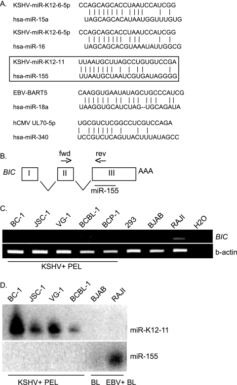
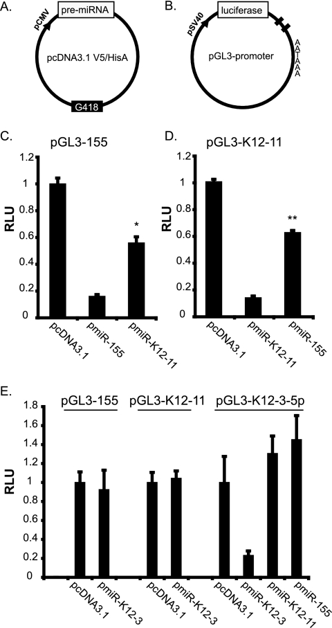
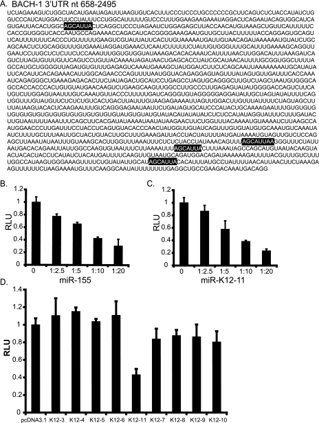
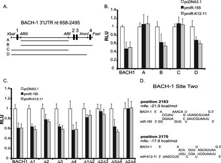
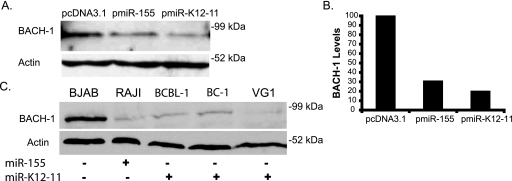
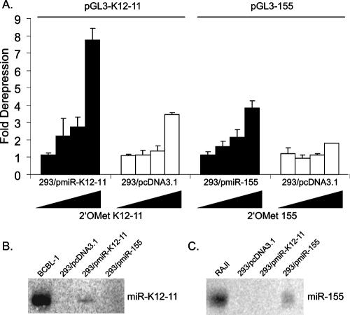
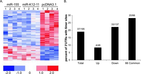
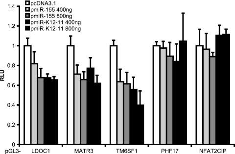
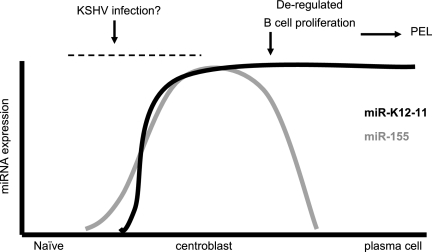
MicroRNAs (miRNAs) are small noncoding RNAs that posttranscriptionally regulate gene expression by binding to 3'-untranslated regions (3'UTRs) of target mRNAs. Kaposi's sarcoma-associated herpesvirus (KSHV), a virus linked to malignancies including primary effusion lymphoma (PEL), encodes 12 miRNA genes, but only a few regulatory targets are known. We found that KSHV-miR-K12-11 shares 100% seed sequence homology with hsa-miR-155, an miRNA frequently found to be up-regulated in lymphomas and critically important for B-cell development. Based on this seed sequence homology, we hypothesized that both miRNAs regulate a common set of target genes and, as a result, could have similar biological activities. Examination of five PEL lines showed that PELs do not express miR-155 but do express high levels of miR-K12-11. Bioinformatic tools predicted the transcriptional repressor BACH-1 to be targeted by both miRNAs, and ectopic expression of either miR-155 or miR-K12-11 inhibited a BACH-1 3'UTR-containing reporter. Furthermore, BACH-1 protein levels are low in cells expressing either miRNA. Gene expression profiling of miRNA-expressing stable cell lines revealed 66 genes that were commonly down-regulated. For select genes, miRNA targeting was confirmed by reporter assays. Thus, based on our in silico predictions, reporter assays, and expression profiling data, miR-K12-11 and miR-155 regulate a common set of cellular targets. Given the role of miR-155 during B-cell maturation, we speculate that miR-K12-11 may contribute to the distinct developmental phenotype of PEL cells, which are blocked in a late stage of B-cell development. Together, these findings indicate that KSHV miR-K12-11 is an ortholog of miR-155.


Figures

FIG. 1.
FIG. 1.
Herpesvirus miRNAs share seed sequence homology with human miRNAs. (A) miRNA sequences from KSHV, EBV, and human cytomegalovirus were aligned with sequences in the human miRNA database. Shown are those viral miRNAs exhibiting the greatest sequence homology to human miRNAs. (B) miR-155 is expressed from exon 3 of the non-protein-coding BIC. Primers (forward [fwd] and reverse [rev]) for RT-PCR analysis of BIC expression are indicated. (C) RT-PCR analysis of BIC expression in KSHV-infected PEL cell lines. The RAJI cell line, an EBV-infected BL cell line, was used as a positive control. H2O indicates the no-template control. (D) Northern blot analysis of KSHV-miR-K12-11 and hsa-miR-155 expression in PEL and BL cells. Twenty-five micrograms of total RNA was loaded per lane and hybridized to probes for either miR-K12-11 or miR-155.
FIG. 2.
FIG. 2.
Importance of the seed sequence for miRNA targeting. (A) Schematic of miRNA expression vector. A region encompassing the pre-miRNA for either miR-155 or miR-K12-11 was PCR amplified and cloned downstream of the cytomegalovirus promoter in pcDNA3.1. (B) Schematic of miRNA sensor vector. Two antisense complementary binding sites for each miRNA were inserted into the 3′UTR of pGL3-Promoter (Promega) downstream of the luciferase gene. (C and D) Luciferase expression from miRNA sensors (pGL3-155 or pGL3-K12-11) is down-regulated in response to ectopic miRNA expression. 293 cells were transfected with 40 ng of the indicated sensor and 800 ng miRNA expression vector using Lipofectamine 2000 (Invitrogen). Lysates were analyzed at 72 h posttransfection (Promega). Relative light units (RLU) are normalized to total protein determined by BCA (Pierce). * indicates a P value of <0.01, and ** indicates a P value of <0.01 by Student's t test. (E) Luciferase assays to control for ectopic miRNA effects. miRNA expression and sensor vectors for KSHV-miR-K12-3-5p were cotransfected with plasmids as indicated in C and D.
FIG. 3.
FIG. 3.
BACH-1 is targeted by miR-K12-11 and miR-155. (A) The BACH-1 3′UTR contains four seed match sites (highlighted) based on three target prediction programs (miRanda, PicTar, and TargetScan). (B and C) A region of the BACH-1 3′UTR (nt 658 to 2495) encompassing the predicted miRNA binding sites was cloned downstream of luciferase (pGL3-BACH1). 293 cells were cotransfected with 40 ng pGL3-BACH-1 and increasing amounts (100, 200, 400, and 800 ng) of pmiR-155 or pmiR-K12-11. Ratios on the x axis indicate the amount of miRNA vector to reporter. pcDNA3.1 was used as filler. Relative light units (RLU) are normalized to total protein determined by BCA (Pierce). (D) Additional KSHV miRNAs do not target the BACH-1 3′UTR. 293 cells were cotransfected with 40 ng pGL3-BACH-1, 10 ng pEF-RL (Renilla), and 800 ng of either pcDNA3.1 or KSHV miRNA expression vector. Light units are normalized to Renilla luciferase. For B to D, average data from three independent transfections are shown.
FIG. 4.
FIG. 4.
Seed match site 2 is essential for combinatorial regulation of BACH-1 by miR-155 and miR-K12-11. (A) Schematic of the BACH-1 3′UTR with potential miRNA seed match binding sites (sites 1 to 4) and restriction enzyme sites. “A” to “D” are truncation mutants generated by restriction enzyme digestion. (B) Mapping of miRNA binding sites. BACH-1 fragments A to D were inserted into pGL3-Promoter, and 40 ng of each reporter was cotransfected with 10 ng pEF-RL and 800 ng either pcDNA3.1, pmiR-155, or pmiR-K12-11 into 293 cells. (C) Site-directed mutagenesis to validate mapping data. Individual or double seed match sites within pGL3-BACH-1 were mutated to an XhoI site. Forty nanograms of each reporter was cotransfected with 10 ng pEF-RL and 800 ng miRNA expression vectors as described above (B). For B and C, lysates were harvested at 72 h, and relative light units (RLU) were normalized to Renilla luciferase. Shown are average data for five to eight independent transfections. (D) Potential miR-155 and miR-K12-11 binding to site 2 within the BACH-1 3′UTR (RNA-hybrid) (53). The binding position within the 3′UTR and the minimum free hybridization energy (mfe) for each miRNA are shown.
FIG. 5.
FIG. 5.
BACH-1 is down-regulated in miR-155- and miR-K12-11-expressing cells. (A) Transient expression of miR-155 or miR-K12-11 affects BACH-1 protein levels. BJAB cells were transfected with 2 μg miRNA expression vector or pcDNA3.1 control (nucleofection) (Amaxa, Inc.). Lysates were harvested at 72 h and probed for endogenous BACH-1 or actin. (B) Graph of BACH-1 levels normalized to actin (analyzed by NIH ImageJ) for transfected BJAB cells shown in A. (C) PEL cells expressing miR-K12-11 and BL cells expressing miR-155 express low amounts of BACH-1 protein. Lysates from 1 × 10⁵ BJAB, RAJI, BCBL-1, BC-1, or VG-1 cells were probed for endogenous BACH-1 and actin.
FIG. 6.
FIG. 6.
miRNA expression in stable cell lines. (A) Luciferase derepression assays were performed using 293/pmiR-K12-11, 293/pmiR-155, or 293/pcDNA3.1 cells using 40 ng of the indicated sensor vector and increasing amounts (20 to 80 pmol) of a 2′OMe antagomir specific to the miRNA of interest. 2′OMe K12-10 and 100 ng of pcDNA3.1 were used as filler. Light units are normalized to total protein by BCA (Pierce). (B and C) Northern blot analysis of miRNA expression in stable cell lines. Thirty micrograms of total RNA was loaded per lane and hybridized to a probe for either miR-K12-11 (B) or miR-155 (C). RNA from BCBL-1 and RAJI cells was used as a positive control.
FIG. 7.
FIG. 7.
Gene expression profiling reveals a common set of target genes. (A) Genes are down-regulated in response to miRNA expression. Colors represent changes in variance-normalized gene expression differences for individual genes represented by the probe sets as indicated on the color scale. Four samples were used for each cell line. One hundred ninety-five genes were changed in response to miRNA expression: 137 genes were down-regulated, and 48 were up-regulated. Sixty-four genes showed >75% cross-validation. (B) The 3′UTRs of down-regulated genes are enriched for seed match sites. 3′UTRs of altered genes were scanned for seed match sites. Shown is the percentage of 3′UTRs with seed match sites for either the total number of altered genes (total), up-regulated (up) or down-regulated (down) genes, or the 66 commonly down-regulated genes (66 common). Thirty-three out of 137 down-regulated genes contained 6-mer seed match sites within their 3′UTRs, while 20 out of the 66 commonly down-regulated genes contained seed match sites.
FIG. 8.
FIG. 8.
Validation of candidate target genes. The 3′UTRs of LDOC1, MATR3, TM6SF1, PHF17, and NFAT2CIP contained at least one seed match site and were cloned into pGL3-Promoter downstream of luciferase. 3′UTR reporters were cotransfected into 293 cells with 10 ng pEF-RL and either 800 ng pcDNA3.1 or the indicated amounts of miRNA expression vector to determine miRNA targeting. Four hundred nanograms of pcDNA3.1 was used as filler for the transfection of 400 ng miRNA expression vector. Lysates were harvested at 72 h, and relative light units (RLU) were normalized to Renilla luciferase. For pGL3-LDOC1, pGL3-MATR3, and pGL3-TM6SF1, a 1.5- to 2.5-fold decrease in luciferase was observed with miR-155 and miR-K12-11 expression. Shown are average data from three to six independent transfections.
FIG. 9.
FIG. 9.
Model of a potential role for miR-K12-11 in lymphomagenesis. Germinal-center-dependent B-cell maturation is dependent on a precise miR-155 expression burst whereby both induction and shutoff are equally important (54, 63). In contrast, after naïve B cells are infected with KSHV, sustained miR-K12-11 expression could contribute to proliferation in the absence of terminal differentiation into a mature antibody-producing B cell, a phenotype which is congruent with that characterized for PEL cells (29).

Similar articles

Cited by

References

    1. Abbott, A. L., E. Alvarez-Saavedra, E. A. Miska, N. C. Lau, D. P. Bartel, H. R. Horvitz, and V. Ambros. 2005. The let-7 MicroRNA family members mir-48, mir-84, and mir-241 function together to regulate developmental timing in Caenorhabditis elegans. Dev. Cell 9:403-414. - PMC - PubMed
    1. Adams, J. M., and S. Cory. 2007. The Bcl-2 apoptotic switch in cancer development and therapy. Oncogene 26:1324-1337. - PMC - PubMed
    1. Ambros, V. 2004. The functions of animal microRNAs. Nature 431:350-355. - PubMed
    1. An, F. Q., H. M. Folarin, N. Compitello, J. Roth, S. L. Gerson, K. R. McCrae, F. D. Fakhari, D. P. Dittmer, and R. Renne. 2006. Long-term-infected telomerase-immortalized endothelial cells: a model for Kaposi's sarcoma-associated herpesvirus latency in vitro and in vivo. J. Virol. 80:4833-4846. - PMC - PubMed
    1. Bartel, D. P. 2004. MicroRNAs: genomics, biogenesis, mechanism, and function. Cell 116:281-297. - PubMed

Publication types

MeSH terms