Comparative Study
. 2007 Aug 1;27(31):8250-60.
doi: 10.1523/JNEUROSCI.1800-07.2007.

Reducing agents sensitize C-type nociceptors by relieving high-affinity zinc inhibition of T-type calcium channels

Affiliations
Comparative Study

Reducing agents sensitize C-type nociceptors by relieving high-affinity zinc inhibition of T-type calcium channels

Michael T Nelson et al. J Neurosci. .

Abstract

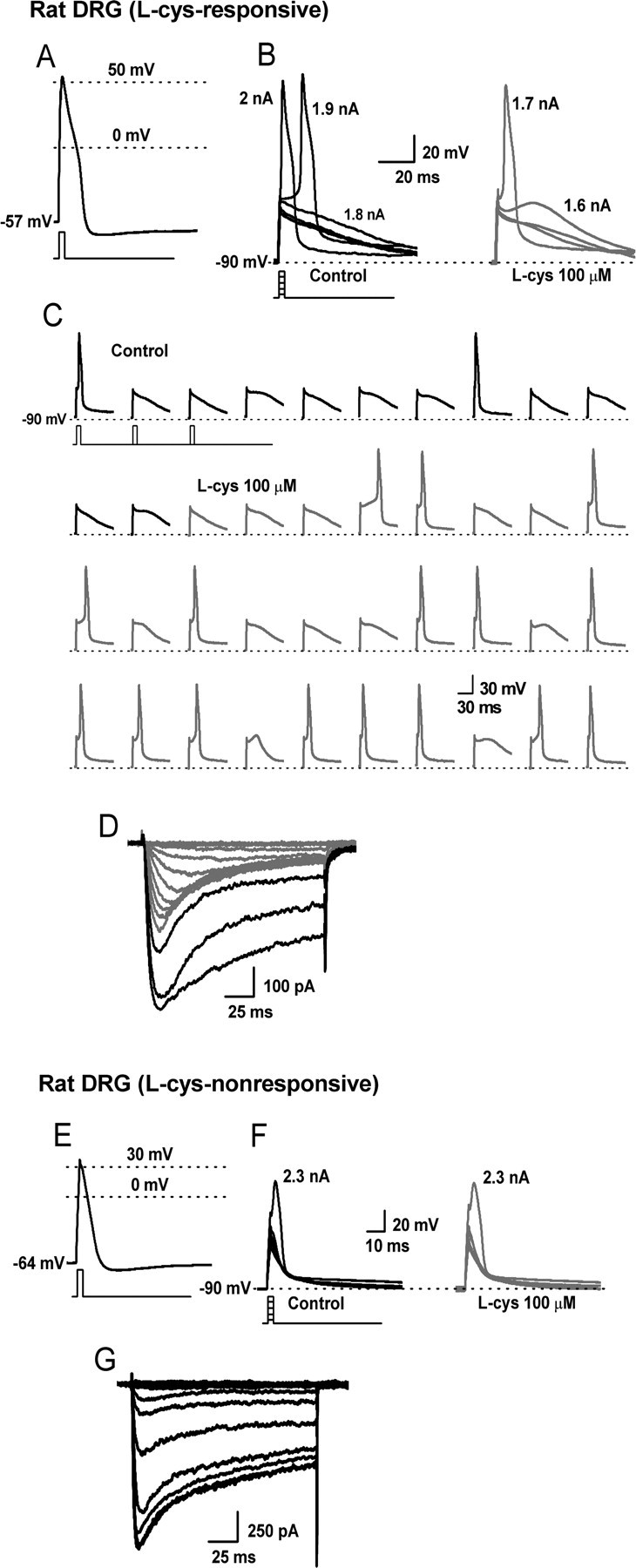
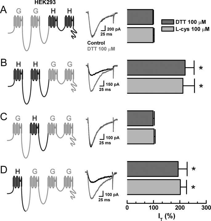
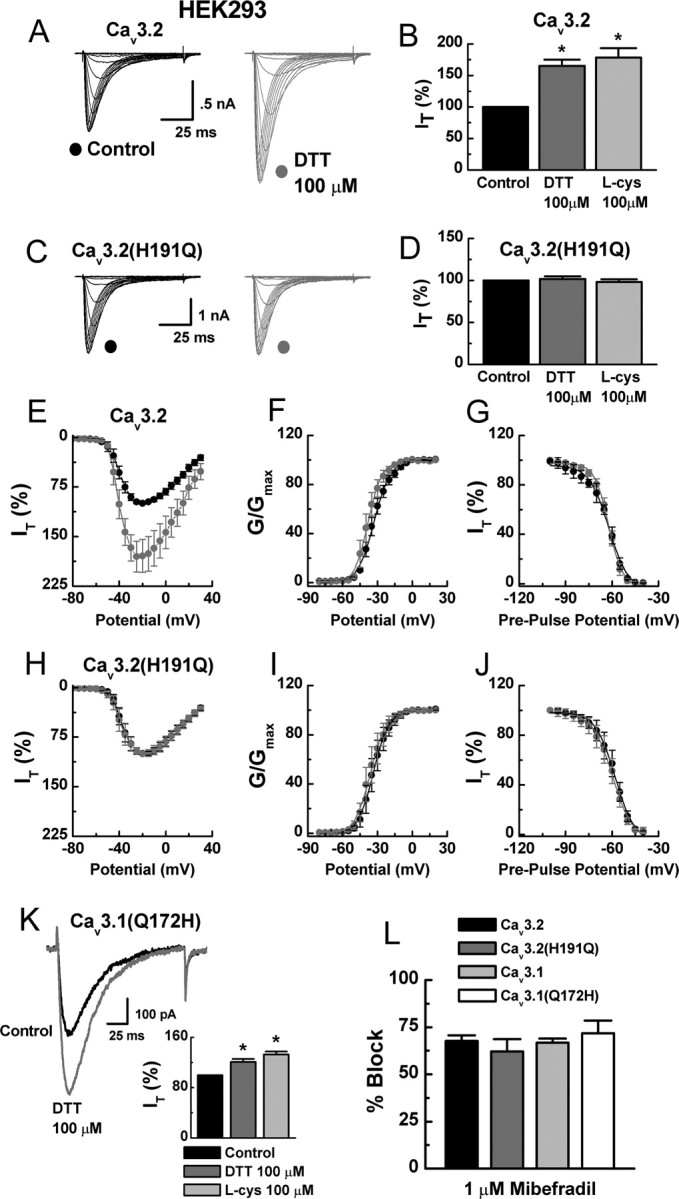
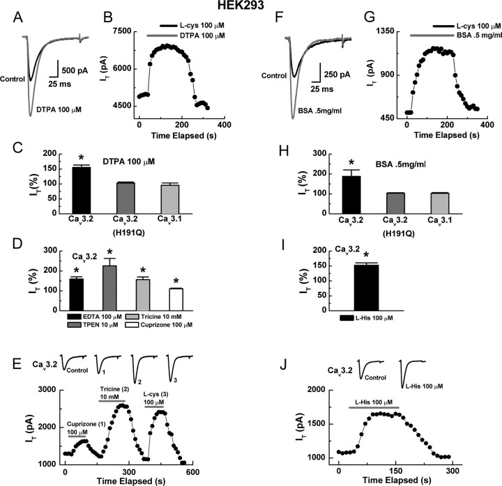
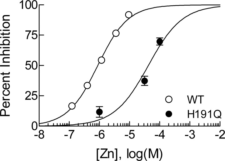
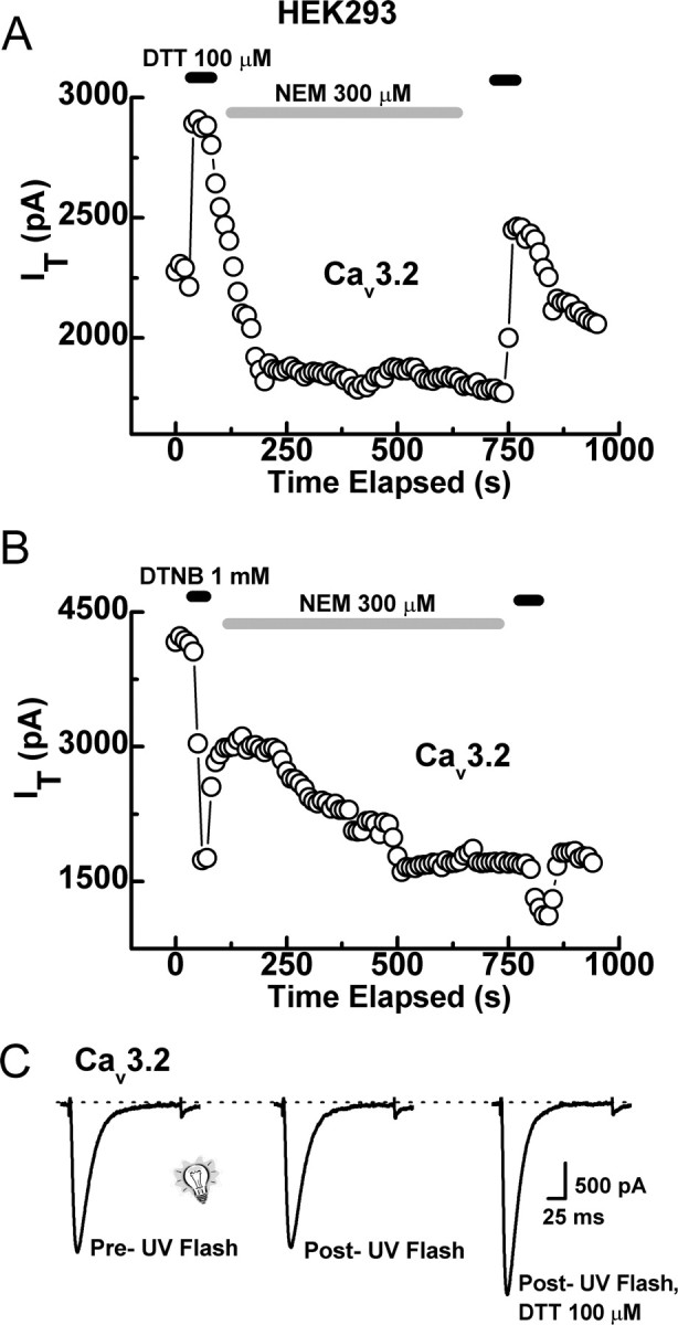
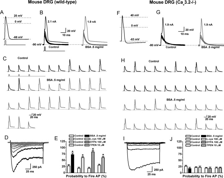
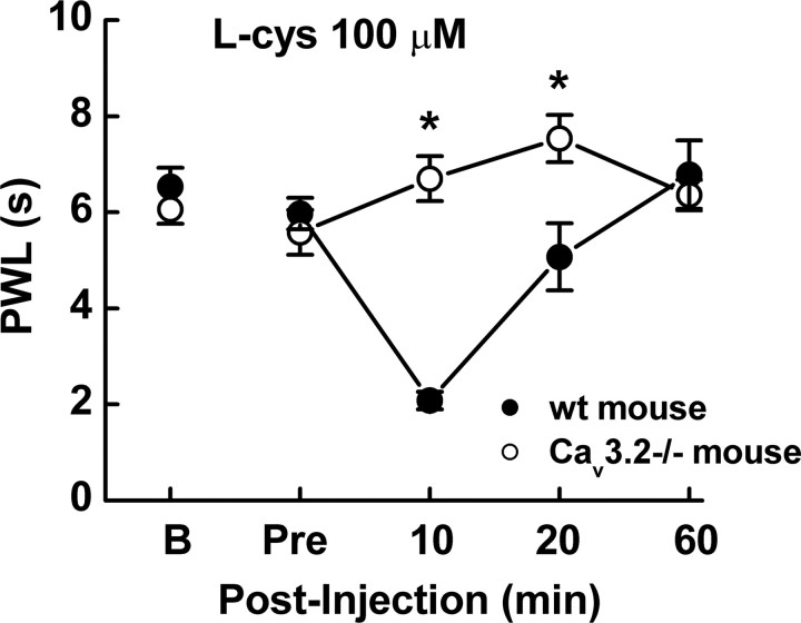
Recent studies have demonstrated an important role for T-type Ca2+ channels (T-channels) in controlling the excitability of peripheral pain-sensing neurons (nociceptors). However, the molecular mechanisms underlying the functions of T-channels in nociceptors are poorly understood. Here, we demonstrate that reducing agents as well as endogenous metal chelators sensitize C-type dorsal root ganglion nociceptors by chelating Zn2+ ions off specific extracellular histidine residues on Ca(v)3.2 T-channels, thus relieving tonic channel inhibition, enhancing Ca(v)3.2 currents, and lowering the threshold for nociceptor excitability in vitro and in vivo. Collectively, these findings describe a novel mechanism of nociceptor sensitization and firmly establish reducing agents, as well as Zn2+, Zn2+-chelating amino acids, and Zn2+-chelating proteins as endogenous modulators of Ca(v)3.2 and nociceptor excitability.


Figures

Figure 1.
Figure 1.
The effects of l-cys on the excitability of T-current-containing and T-current-deficient rat C-type nociceptors. A–D, Traces are from a single, acutely dissociated, l-cys-sensitive rat DRG neuron. E–G, Traces from another, acutely dissociated, l-cys-insensitive rat DRG neuron. A, AP elicited by a 1 ms, 2 nA current injection at the RMP of the cell. B, The cell was manually hyperpolarized to −90 mV and progressively greater current injections (Δ100 pA) delivered every 10 s to determine the threshold for AP firing. Threshold was 1.9 nA in control (left) and 1.7 nA in the presence of l-cys (right). C, Continuous segment of an experiment showing that l-cys increases the probability (percentage) to fire APs in response to trains of subthreshold stimuli (stimuli were 1 ms, 1.7 nA delivered every 10 s). D, I–V traces recorded in Ca2+ current-isolating external solution after current-clamp recording. Note the presence of both T-type (rapidly inactivating; gray lines) and HVA (slowly inactivating; black lines) currents. E, AP elicited by a 1 ms, 2.5 nA current injection at the RMP of the cell. F, Threshold was unaffected by the application of l-cys. G, I–V traces; note the presence of only HVA currents.
Figure 2.
Figure 2.
Reducing agent sensitivity of serial chimeras constructed from Cav3.1 (α1G) and Cav3.2 (α1H) T-channels. Currents were evoked from HEK293 cells expressing the indicated constructs by steps from −90 to −30 mV, before and during exposure to DTT. Histograms display averaged effects of DTT and l-cys in each construct expressed as percentage of control (n = 5–9). A, GGHH: DTT, 98 ± 2%; l-cys, 99 ± 4%. B, HHGG: DTT, 220 ± 34%, *p < 0.01; l-cys, 211 ± 44%, p < 0.01. C, GHGG: DTT, 99 ± 5%; l-cys, 102 ± 4%. D, HGGG: DTT, 193 ± 34%, *p < 0.01; l-cys, 202 ± 24%, p < 0.01.
Figure 3.
Figure 3.
H191 is required for the reducing agent sensitivity of Cav3.2. A, Currents evoked from HEK293 cells expressing Cav3.2 by steps from −90 to 0 mV (Δ5 mV), before and during exposure to DTT. B, Averaged effects of DTT and l-cys on Cav3.2 currents expressed as percentage of control: DTT, 165 ± 10%, n = 32, *p < 0.01; l-cys, 178 ± 15%, n = 17, *p < 0.01. C, Currents evoked from HEK293 cells expressing Cav3.2(H191Q), before and during exposure to DTT. D, Averaged effects of DTT and l-cys on Cav3.2(H191Q) currents expressed as percentage of control: DTT, 102 ± 3%, n = 15; l-cys, 98 ± 3%, n = 11. E, Averaged effects of DTT on Cav3.2 currents evoked by steps from −90 to −80 through 30 mV (n = 7). F, Effects of DTT on voltage-dependent activation of Cav3.2 currents calculated from I–V data and fit with Equation 1: control, V50 of −33.0 ± 0.3 mV, k = 7.0 ± 0.3; DTT, V50 of −38.0 ± 0.4 mV, k = 6.0 ± 0.3. G, Effects of DTT on steady-state inactivation of Cav3.2 currents. Currents were recorded at −30 mV after 3.5-s-long prepulses to potentials ranging from −100 to −40 mV. Averaged data were fit with Equation 2: control, V50 of −62.0 ± 0.6 mV, k = 6.0 ± 0.6; DTT, V50 of −62.0 ± 0.4 mV, k = 5.0 ± 0.4; n = 6. H, Averaged effects of DTT on Cav3.2(H191Q) currents evoked by steps from −90 to −80 through 30 mV (n = 6). I, Effects of DTT on voltage-dependent activation of Cav3.2(H191Q) currents: control, V50 of −34.0 ± 0.4 mV, k = 6.0 ± 0.2; DTT, V50 of −35.0 ± 0.4 mV, k = 7.0 ± 0.3. J, Effects of DTT on steady-state inactivation of Cav3.2(H191Q) currents: control, V50 of −58.0 ± 0.6 mV, k = 6.0 ± 0.4; DTT, V50 of −60.0 ± 0.6 mV, k = 6.0 ± 0.4; n = 6. K, Raw traces and averaged effects of DTT and l-cys on Cav3.1(Q172H) currents expressed as percentage of control: DTT, 121 ± 5%, n = 11, *p < 0.01; l-cys, 132 ± 5%, n = 5, *p < 0.01. L, Mibefradil inhibition of currents evoked from the indicated constructs expressed as percentage of control: Cav3.2, 68 ± 4%; Cav3.2(H191Q), 62 ± 7%; Cav3.1, 67 ± 2%; Cav3.1(Q172H), 71 ± 7%; n = 5–6.
Figure 4.
Figure 4.
Synthetic and endogenous metal chelators mimic and occlude the effects of reducing agents. A, Currents evoked from HEK293 cells expressing Cav3.2 by steps from −90 to −30 mV, before and during exposure to DTPA. B, Time course showing that application of DTPA occludes the effect of subsequently applied l-cys; similar results were obtained in four additional cells. C, Averaged effects of DTPA on currents from the indicated constructs expressed as percentage of control: Cav3.2, 155 ± 8%, n = 5, *p < 0.01; Cav3.2(H191Q), 107 ± 4%, n = 6; Cav3.1 95 ± 8%, n = 5. D, Averaged effects of various chelators on Cav3.2 currents expressed as percentage of control: EDTA, 159 ± 11%; TPEN, 225 ± 37%; tricine, 156 ± 14%; cuprizone, 111 ± 3%; n = 6–8, *p < 0.01. E, Time course and raw traces comparing the effects of cuprizone, tricine, and l-cys on Cav3.2 currents. F, Currents evoked from Cav3.2 before and during exposure to BSA. G, Time course showing that application of BSA occludes the effect of subsequently applied l-cys. H, Averaged effects of BSA on currents from the indicated constructs expressed as percentage of control: Cav3.2, 188 ± 32%, n = 4, *p < 0.01; Cav3.2(H191Q), 103 ± 3%, n = 5; Cav3.1, 103 ± 4%, n = 5. I, Averaged effects of l-his on Cav3.2 currents expressed as percentage of control: 152 ± 8%, n = 6, *p < 0.01. J, Time course and raw traces showing the effects of l-his on Cav3.2 currents.
Figure 5.
Figure 5.
Cav3.2(H191Q) has reduced sensitivity to Zn2+. Concentration–response curve for inhibition of Cav3.2 and Cav3.2(H191Q) by Zn2+ in HEK293 cells. Fitted values for IC50 and Hill–Langmuir coefficient (h) are as follows: Cav3.2, IC50 of 0.89 ± 0.05 μm, h = 0.87 ± 0.04, n = 4–8; Cav3.2(H191Q), IC50 of 42 ± 7 μm, h = 0.77 ± 0.15, n = 4–8. Cav3.2 data were obtained in the presence of 10 mm tricine to establish a nominally Zn2+-free baseline, as well as to buffer free Zn2+ concentrations (see Materials and Methods). Cav3.2(H191Q) data were obtained without tricine because this channel is insensitive to chelators (Fig. 4), rendering Zn2+ buffering irrelevant. WT, Wild type.
Figure 6.
Figure 6.
Cysteine-modifying agents do not disrupt the effects of reducing agents. A, Time course showing that application of NEM does not disrupt the enhancement of Cav3.2 currents by DTT. For these experiments, DTT was applied first, followed by a 1–2 min wash, NEM was then applied for 8–15 min, followed by another wash, and DTT was then applied for a second time. We then compared the magnitude of the first DTT effect with that of the second (for calculation of the second effect, we considered the steady-state inhibition by NEM as a new baseline). In similar experiments from five cells, the current enhancement from the second application of DTT was 53 ± 8% compared with 45 ± 4% for the first application, indicating no inhibition of DTT by NEM. B, In contrast, NEM significantly attenuated the inhibition by DTNB. The first application of DTNB produced an average inhibition of 48 ± 10% and the second only 22 ± 2% (n = 5; p < 0.01). C, Representative traces showing that UV light (1 s pulse) does not modify Cav3.2 currents independently or prevent subsequent modulation by DTT. In similar experiments from four cells, DTT increased currents by 48 ± 6% after exposure to UV light.
Figure 7.
Figure 7.
The effects of chelators and reducing agents on the excitability of C-type nociceptors isolated from wild-type or Cav3.2−/− mice. A–D are from a single wild-type cell; F–I are from a single Cav3.2−/− cell. A, AP elicited by a 1 ms, 3 nA current injection at the RMP of the cell. B, The cell was manually hyperpolarized to −90 mV and progressively greater current injections (Δ100 pA) delivered every 10 s to determine the threshold for AP firing. Threshold was 2.1 nA under control conditions (left) and 1.9 nA in the presence of BSA (right). C, Continuous segment of an experiment showing that BSA increased the probability to fire APs in response to trains of subthreshold stimuli (stimuli were 1 ms, 1.9 nA delivered every 10 s). D, I–V traces recorded in Ca2+ current-isolating external solution after current-clamp recording. Note the presence of both T-type (rapidly inactivating; gray lines) and HVA (slowly inactivating; black lines) currents. E, Average effect of BSA, l-cys, DTPA, and TPEN on the probability to fire APs (percentage) in response to trains of subthreshold stimuli in wild-type cells (BSA: control, 27.7 ± 5.0; BSA, 72.6 ± 6.0; p < 0.01; L-cys: control, 18.3 ± 4.0; l-cys, 60.7 ± 4.0; p < 0.01; DTPA: control, 18.1 ± 4.0; DTPA, 83.5 ± 6.0; p < 0.01; TPEN: control, 30.6 ± 3.8; TPEN, 60.8 ± 4.6; p < 0.01; n = 5–9). F, AP elicited from a Cav3.2−/− cell by a 1 ms, 3 nA current injection at the RMP cell. G, Threshold was unaffected by application of BSA. H, Continuous segment of an experiment showing that BSA has little effect on the probability to fire APs in response to trains of subthreshold stimuli (stimuli were 1 ms, 1.8 nA delivered every 10 s). I, I–V traces; note the presence of only HVA currents. J, Average effect of BSA, l-cys, DTPA, and TPEN on the probability to fire APs (percentage) in response to trains of subthreshold stimuli in Cav3.2−/− cells (BSA: control, 26.5 ± 4.0; BSA, 17.6 ± 5.0; l-cys: control, 21.6 ± 3.0, l-cys, 22.2 ± 5.0; DTPA: control, 19.1 ± 3.0; DTPA, 18.2 ± 5.0; TPEN: control, 19.9 ± 3.7; TPEN, 20.2 ± 3.0; n = 7–8).
Figure 8.
Figure 8.
l-cys induces thermal hyperalgesia in wild-type but not Cav3.2−/− mice in vivo. l-cys induced a decrease in PWL in response to low-intensity infrared radiation (IR30) in wild-type (wt; littermate) mice but not in Cav3.2−/− mice. l-cys significantly decreased PWL at both 10 and 20 min after injection (n = 10; *p < 0.05). Wild-type PWLs reverted to baseline by 60 min after injection. “B” shows baseline PWL obtained 1 d before test; “Pre” shows the value obtained immediately before injection.

Similar articles

Cited by

References

    1. Bhave G, Gereau RW., IV Posttranslational mechanisms of peripheral sensitization. J Neurobiol. 2004;61:88–106. - PubMed
    1. Bourinet E, Alloui A, Monteil A, Barrere C, Couette B, Poirot O, Pages A, McRory J, Snutch TP, Eschalier A, Nargeot J. Silencing of the Cav3.2 T-type calcium channel gene in sensory neurons demonstrates its major role in nociception. EMBO J. 2005;24:315–324. - PMC - PubMed
    1. Campbell JN, Meyer RA. Mechanisms of neuropathic pain. Neuron. 2006;52:77–92. - PMC - PubMed
    1. Cardenas CG, Del Mar LP, Scroggs RS. Variation in serotonergic inhibition of calcium channel currents in four types of rat sensory neurons differentiated by membrane properties. J Neurophysiol. 1995;74:1870–1879. - PubMed
    1. Chen CC, Lamping KG, Nuno DW, Barresi R, Prouty SJ, Lavoie JL, Cribbs LL, England SK, Sigmund CD, Weiss RM, Williamson RA, Hill JA, Campbell KP. Abnormal coronary function in mice deficient in alpha1H T-type Ca2+ channels. Science. 2003;302:1416–1418. - PubMed

Publication types