. 2007 Sep;27(17):6068-83.
doi: 10.1128/MCB.00664-07. Epub 2007 Jun 25.

Arkadia activates Smad3/Smad4-dependent transcription by triggering signal-induced SnoN degradation

Affiliations

Arkadia activates Smad3/Smad4-dependent transcription by triggering signal-induced SnoN degradation

Laurence Levy et al. Mol Cell Biol. 2007 Sep.

Abstract

E3 ubiquitin ligases play important roles in regulating transforming growth factor beta (TGF-beta)/Smad signaling. Screening of an E3 ubiquitin ligase small interfering RNA library, using TGF-beta induction of a Smad3/Smad4-dependent luciferase reporter as a readout, revealed that Arkadia is an E3 ubiquitin ligase that is absolutely required for this TGF-beta response. Knockdown of Arkadia or overexpression of a dominant-negative mutant completely abolishes transcription from Smad3/Smad4-dependent reporters, but not from Smad1/Smad4-dependent reporters or from reporters driven by Smad2/Smad4/FoxH1 complexes. We show that Arkadia specifically activates transcription via Smad3/Smad4 binding sites by inducing degradation of the transcriptional repressor SnoN. Arkadia is essential for TGF-beta-induced SnoN degradation, but it has little effect on SnoN levels in the absence of signal. Arkadia interacts with SnoN and induces its ubiquitination irrespective of TGF-beta/Activin signaling, but SnoN is efficiently degraded only when it forms a complex with both Arkadia and phosphorylated Smad2 or Smad3. Finally, we describe an esophageal cancer cell line (SEG-1) that we show has lost Arkadia expression and is deficient for SnoN degradation. Reintroduction of wild-type Arkadia restores TGF-beta-induced Smad3/Smad4-dependent transcription and SnoN degradation in these cells, raising the possibility that loss of Arkadia function may be relevant in cancer.


Figures
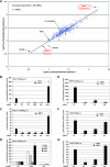
FIG. 1.
FIG. 1.
Loss of Arkadia completely and specifically inhibits the Smad3-dependent TGF-β pathway. (A) 289 siRNA SMARTpools were screened in duplicate using the HaCaT CAGA12-Luc/TK-Renilla cell line. Luciferase levels were analyzed and normalized to Renilla levels (Luciferase/Renilla). The two duplicate experiments are represented on a dot plot using a logarithmic (log10) scale which provides an easier representation of the range of values on the same graph. The negative control was a nontargeting siRNA, and positive controls were siRNAs against Smad3, Smad4, and TβRII as indicated. The dots corresponding to the Luciferase/Renilla values for siRNAs that target RNF111 (Arkadia) and TRIM33 (Ectodermin) are also indicated. (B and C) Plots of Luciferase/Renilla values for HaCaT CAGA12-Luc/TK-Renilla cells (B) or luciferase only values for c-JunSBR6-Luc cells (C) transfected with the indicated siRNA SMARTpools and then treated with TGF-β (+ TGF-β) or not treated with TGF-β (− TGF-β). (D) Plots of Luciferase/Renilla values for HaCaT CAGA12-Luc/TK-Renilla cells transfected with the four single siRNAs corresponding to the deconvolution of the nontargeting SMARTpool or those that target Ectodermin or Arkadia. (E to G) NIH 3T3 cells were transfected with the indicated mouse siRNA SMARTpools followed by transfection with plasmids encoding the TK-Renilla reporter and either CAGA12-Luc (E) or ARE-Luc together with the plasmid encoding xFoxH1a (F) or BRE-Luc (G). Cells were treated with TGF-β or BMP4 or not treated with TGF-β or BMP4 as indicated. Luciferase activity was normalized to Renilla activity. Abbreviations in panels B to G: Ctl, nontargeting control siRNA; S3, Smad3; S4, Smad4; Ark, Arkadia; Ecto, Ectodermin; oligo, oligonucleotide.
FIG. 2.
FIG. 2.
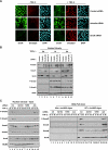
Arkadia acts downstream of TGF-β-induced Smad phosphorylation and nuclear accumulation. (A) HaCaT cells were transfected with the indicated siRNA SMARTpools, then treated with TGF-β (+ TGF-β) for 1 h or not treated with TGF-β (− TGF-β), and processed for immunofluorescence using an anti-Smad2/3 antibody and an anti-phosphorylated-Smad3 (anti-P-Smad3) antibody. Nuclei were visualized with 4′,6′-diamidino-2-phenylindole (DAPI). (B) HaCaT cells were transfected with the indicated siRNA SMARTpools and then treated with TGF-β or not treated with TGF-β for the indicated times. Whole-cell extracts were analyzed by Western blotting using antibodies against Smad3, P-Smad3, and against the Smad3 target genes PAI-1 and p21. Grb2 was used as a loading control. (C) Plots of Luciferase/Renilla values for HaCaT CAGA12-Luc/TK-Renilla cells transfected with the indicated siRNA SMARTpools and then treated with TGF-β or not treated with TGF-β.
FIG. 3.
FIG. 3.
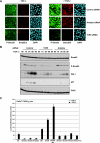
TGF-β-induced SnoN degradation requires Arkadia. (A) HaCaT cells were transfected with the indicated siRNA SMARTpools, then treated with TGF-β for 1 h (+ TGF-β) or not treated with TGF-β (− TGF-β), and processed for immunofluorescence using an anti-Smad2/3 antibody and an anti-SnoN antibody. Nuclei were visualized with 4′,6′-diamidino-2-phenylindole (DAPI). (B) HaCaT cells were transfected with the indicated siRNA SMARTpools and then treated with TGF-β for the times indicated or not treated with TGF-β. Nuclear extracts were analyzed directly by Western blotting using antibodies against Arkadia, Smurf1, SnoN, phosphorylated Smad2 (P-Smad2), P-Smad3, and poly(ADP-ribose) polymerase (PARP) as a control. (C) HaCaT cells were transfected with the indicated siRNA SMARTpools and then treated with TGF-β or not treated with TGF-β for the times indicated. Nuclear extracts were either analyzed directly by Western blotting using antibodies against SnoN, Smad3, P-Smad2, P-Smad3, and MCM6 as a control (left blots) or by DNA pull-down assay using the wild-type c-JunSBR oligonucleotide or a version mutated in the Smad3/Smad4 binding sites (right blots). Abbreviations: Ctl, nontargeting control siRNA; S2+S3, Smad2 and Smad3; Ark, Arkadia; Mut and WT c-JunSBR oligos, mutant and wild-type c-JunSBR oligonucleotides, respectively.
FIG. 4.
FIG. 4.
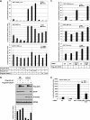
Arkadia mutated in its RING domain inhibits Smad3-dependent transcription by preventing TGF-β-induced SnoN degradation. (A) 293T or NIH 3T3 cells were cotransfected with the TK-Renilla reporter and either CAGA12-Luc (top graph) or ARE-Luc together with the plasmid encoding xFoxH1a (middle graph) or with BRE-Luc (bottom graph), with increasing amounts of plasmids encoding Flag-SnoN-wt (Flag-tagged wild-type SnoN), Flag-Ark-wt (Flag-tagged wild-type Arkadia), or Flag-Ark-C937A as indicated (−, none). 293T cells were treated with SB-431542 (SBI) overnight to abolish autocrine signaling before induction or not with Activin. NIH 3T3 cells were treated with BMP4 (+BMP4) or not treated with BMP4 (−BMP4). (B) 293T cells were transfected with plasmids encoding Flag-Ark-wt or Flag-Ark-C937A. Cells were treated with SBI and Activin as described above for panel A. Nuclear extracts were analyzed by Western blotting using antibodies against the Flag tag, SnoN, phosphorylated Smad2 (P-Smad2), and MCM6, as a loading control. Quantification of SnoN levels relative to MCM6 levels is shown below the blots. (C) 293T cells were transfected with the indicated siRNA SMARTpools, followed by transfection with the TK-Renilla reporter and either CAGA12-Luc, ARE-Luc, or Pitx2-Luc together with a plasmid encoding xFoxH1a and in the presence (+) or absence (−) of Flag-Ark-C937A. Cells were treated with SBI and Activin as described above for panel A. (D) HaCaT CAGA12-Luc/TK-Renilla cells were transfected with the indicated siRNA SMARTpools and treated with TGF-β (+TGF-β) or not treated with TGF-β (−TGF-β). Ctl, nontargeting control siRNA; Ark, Arkadia.
FIG. 5.
FIG. 5.
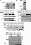
Arkadia binds and ubiquitinates SnoN independently of phosphorylated Smad2/3, but Arkadia-mediated SnoN degradation requires phosphorylated Smad2/3. (A) 293T cells were transfected with an empty vector (−) or with Flag-Ark-C937A (+) as indicated. Cells were treated with SB-431542 (SBI) overnight, before induction with Activin (Act) for 1 h. Nuclear extracts were analyzed by Western blotting using anti-Flag, anti-SnoN, or anti-phosphorylated Smad2 (anti-P-Smad2) antibodies, either directly (input) or after immunoprecipitation (IP) with anti-Flag beads (Flag-beads) or empty beads (Beads). (B to D) 293T cells were transfected with Flag-Ark-wt (Flag-tagged wild-type Arkadia) (B) or Flag-Ark-C937, HA-Smad2, or HA-Smad3 as indicated (C) or with HA-SnoN-wt, HA-SnoN-mS23, or Flag-Ark-C937A (D) as indicated. Cells were treated as described above for panel A, but in addition, in panel B, cells were pretreated with MG132 for 4 h prior to Activin induction. Whole-cell extracts were analyzed by Western blotting using the indicated antibodies either directly (Input blots) or after IP with anti-Flag beads (Flag-IP). The arrowheads indicate the bands that correspond to the analyzed proteins. (E) 293T cells were transfected with the indicated plasmids, and cells were treated with SBI overnight and then either treated with MG132 (25 μM) for 3 h and induced with Activin for another hour (MG132 4 h + Activin 1 h) or with Activin only for 1 h (Activin 1 h) or kept in the presence of SBI. Whole-cell extracts were analyzed by Western blotting using anti-Flag and anti-HA antibodies. Wild-type Arkadia migrates as two bands. The top band corresponds to self-ubiquitinated Arkadia. (F) 293T cells were transfected and treated with SBI as described above and then treated with MG132 (50 μM) for 4 h prior to induction with Activin for another hour. Total lysates were immunoprecipitated with an anti-HA antibody. Ubiquitination of HA-SnoN was then assayed by Western blotting the IP with an anti-His antibody and also by overexposure of the top region of the HA blot. Levels of transfected Arkadia and endogenous P-Smad2 were analyzed by Western blotting of the total lysate. Ub, ubiquitinated.
FIG. 6.
FIG. 6.
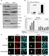
Arkadia expression restores the TGF-β-induced Smad3/Smad4 transcriptional response and SnoN degradation in the Barrett's-associated esophageal adenocarcinoma cell line SEG-1. (A) HaCaT and SEG-1 cells were treated with TGF-β for the indicated times (from 0 h to 30 min [30′] to 6 h), and nuclear extracts were analyzed by Western blotting using the indicated antibodies. MCM6 levels acted as a loading control. P-Smad2, phosphorylated Smad2. (B) SEG-1 cells were transfected with the control nontargeting siRNA SMARTpool (Ctl) or with siRNA SMARTpools targeting Ski and SnoN, followed by transfection with CAGA12-Luc and TK-Renilla and treatment with TGF-β (+TGF-β) or not (−TGF-β). (C) SEG-1 cells were transfected with TK-Renilla and either the Smad3-dependent reporter CAGA12-Luc or the Smad2-dependent reporter ARE-Luc together with the plasmid encoding xFoxH1a and in the presence (+) or absence (−) of Flag-Arkwt (Flag-tagged wild-type Arkadia) or Flag-Ark-C937A. Cells were treated with TGF-β (+TGF-β) or not treated with TGF-β (−TGF-β). (D) SEG-1 cells were transfected with plasmids expressing green fluorescent protein (GFP), GFP-Arkwt (GFP-tagged wild-type Arkadia), or GFP-Ark-ΔRING (GFP-tagged Arkadia deleted in the RING domain) for 24 h and then treated with TGF-β for 1 h or not treated with TGF-β. The cells were fixed, the GFP was visualized directly, and SnoN was detected by immunofluorescence. Nuclei were visualized with 4′,6′-diamidino-2-phenylindole (DAPI). White arrows indicate transfected cells.
FIG. 7.
FIG. 7.
Model of mechanism of Arkadia action. (A) In the nuclei of unstimulated cells, SnoN is complexed to Arkadia (Ark), and SnoN is also bound to repeated AGAC elements (or the reverse complement [GTCT]) with Smad4, forming a transcriptionally repressive complex. In the absence of signal, SnoN is ubiquitinated by Arkadia, but it is not efficiently degraded. Ub, ubiquitin. (B) Upon TGF-β/Activin/Nodal stimulation, phosphorylated Smad2/3 (P-Smad2/3) interacts with Arkadia (Ark) and SnoN, leading to degradation of SnoN via the proteasome. This allows phosphorylated Smad3 (or Smad2Δexon3) complexed with Smad4 to bind the AGAC sites and activate transcription of target genes. (C) One such target gene is SnoN (shown in red), which will act with Smad4 to repress transcription again. For further details, see Discussion.

Similar articles

Cited by

References

    1. Bonni, S., H. R. Wang, C. G. Causing, P. Kavsak, S. L. Stroschein, K. Luo, and J. L. Wrana. 2001. TGF-β induces assembly of a Smad2-Smurf2 ubiquitin ligase complex that targets SnoN for degradation. Nat. Cell Biol. 3:587-595. - PubMed
    1. Boyer, P. L., C. Colmenares, E. Stavnezer, and S. H. Hughes. 1993. Sequence and biological activity of chicken snoN cDNA clones. Oncogene 8:457-466. - PubMed
    1. Chen, X., M. J. Rubock, and M. Whitman. 1996. A transcriptional partner for MAD proteins in TGF-β signalling. Nature 383:691-696. - PubMed
    1. Cohen, S. B., R. Nicol, and E. Stavnezer. 1998. A domain necessary for the transforming activity of SnoN is required for specific DNA binding, transcriptional repression and interaction with TAF(II)110. Oncogene 17:2505-2513. - PubMed
    1. Dennler, S., S. Huet, and J. M. Gauthier. 1999. A short amino-acid sequence in MH1 domain is responsible for functional differences between Smad2 and Smad3. Oncogene 18:1643-1648. - PubMed

Publication types

MeSH terms

LinkOut - more resources