. 2007 Mar;81(5):2117-27.
doi: 10.1128/JVI.01961-06. Epub 2006 Dec 13.

Downregulation of gamma interferon receptor 1 by Kaposi's sarcoma-associated herpesvirus K3 and K5

Affiliations

Downregulation of gamma interferon receptor 1 by Kaposi's sarcoma-associated herpesvirus K3 and K5

Qinglin Li et al. J Virol. 2007 Mar.

Abstract

Upon viral infection, the major defense mounted by the host immune system is activation of the interferon (IFN)-mediated antiviral pathway. In order to complete their life cycles, viruses must modulate the host IFN-mediated immune response. The K3 and K5 proteins of a human tumor-inducing herpesvirus, Kaposi's sarcoma-associated herpesvirus (KSHV), have been shown to downregulate the surface expression of host immune modulatory receptors by increasing their endocytosis rates, which leads to suppression of cell-mediated immunity. In this report, we demonstrate that K3 and K5 both specifically target gamma interferon receptor 1 (IFN-gammaR1) and induce its ubiquitination, endocytosis, and degradation, resulting in downregulation of IFN-gammaR1 surface expression and, thereby, inhibition of IFN-gamma action. Mutational analysis indicated that K5 appeared to downregulate IFN-gammaR1 more strongly than K3 and that the amino-terminal ring finger motif and the carboxyl-terminal region of K5 were necessary for IFN-gammaR1 downregulation. These results suggest that KSHV K3 and K5 suppress both cytokine-mediated and cell-mediated immunity, which ensures efficient viral avoidance of host immune controls.


Figures

FIG. 1.
FIG. 1.
Downregulation of IFN-γR1 surface expression by transient transfection of K3 and K5. BJAB cells were electroporated with GFP, GFP-K3, or GFP-K5 vector. The cell surface levels of MHC-I, IFN-γR1, ICAM-1, and MHC class II in the GFP-positive populations were assessed at 48 h posttransfection by flow cytometry. The dotted line in each histogram represents the isotype control. Data were reproduced in three independent experiments.
FIG. 2.
FIG. 2.
Downregulation of endogenous IFN-γR1 on cells stably expressing K3 or K5. (A) Downregulation of IFN-γR1 by K3 or K5. Puromycin-resistant cells were stained with antibodies against MHC class I, IFN-γR1, ICAM-1, and MHC class II and analyzed by flow cytometry. The dotted line in each histogram represents the isotype control. (B) Expression of K3, K5, and lymphocyte surface proteins. The same amounts of BJAB-vector, BJAB-K3, and BJAB-K5 cell lysates were used for immunoblotting with various antibodies. Anti-tubulin antibody was included as a loading control.
FIG. 3.
FIG. 3.
Downregulation of exogenously expressed Flag-tagged IFN-γR1 by K3 or K5. (A) BJAB-EF, BJAB-K3, and BJAB-K5 cells were electroporated with pTracer-F-IFN-γR1 vector. The cell surface levels of F-IFN-γR1 in the GFP-positive cell population was assessed at 48 h posttransfection by staining them with IFN-γR1 or MHC class I antibody, followed by flow cytometry. The data were reproduced in three independent experiments. (B) BJAB-EF and BJAB-F-IFN-γR1 cells were electroporated with pTracer-GFP reporter, pTracer-K3, or pTracer-K5 vector. The cell surface levels of IFN-γR1 in the GFP-positive cell population were assessed at 48 h posttransfection by staining them with Flag or IFN-γR1antibody by flow cytometry. The data were reproduced in three independent experiments.
FIG. 4.
FIG. 4.
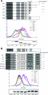
Mutational analysis of K5 for IFN-γR1 downregulation. (A) Point mutations of the N-terminal zinc finger motifs and deletion mutations in the C-terminal region of K5. A schematic of the locations of various mutations of the K5 protein is shown at the top. Deletion mutations in the C-terminal region of K5 were generated as follows: ΔC1, deletion of residues 233 to 256; ΔC2, deletion of residues 198 to 256; ΔC3, deletion of residues 194 to 256; and ΔC4, deletion of residues 178 to 256. The cysteine residues at 15, 18, 30, 32, 56, and 59 in the putative zinc finger motifs in the N-terminal region of K5 were replaced with serine residues to generate the mZn mutant. The abilities of wild-type K5 and each mutant to downregulate IFN-γR1, as assessed by flow cytometry, were given relative scores. ++++, ++, and − indicate very strong, strong, and no activity of the K5 mutant in IFN-γR1 downregulation, respectively. To examine the abilities of K5 and its mutants to downregulate IFN-γR1, BJAB cells were electroporated with pTracer-GFP-K5 (WT), GFP-K5 mZn (mZn), or one of the deletion mutants. Cell surface levels of IFN-γR1 in the GFP-positive populations were assessed 48 h posttransfection by staining them with an IFN-γR1-specific antibody. The data were reproduced in at least two independent experiments. (B) Mutations in sequence-specific motifs of K5 and IFN-γR1 downregulation. The schematic at the top shows the locations of various mutations of the K5 protein. Six mutations, Y156F (Y/F), Y156/A (Y/A), P/A, DE1/QQ, DE2/QQ, and DE12/QQ, were introduced into K5. The locations of these mutations are described in greater detail in the text. Along the right side of the schematic, the abilities of wild-type K5 and each mutant to downregulate IFN-γR1, as shown in the below the schematic, were given relative scores. +++ and − indicate very strong and no activity of the K5 mutant in IFN-γR1 downregulation, respectively. Below are shown the differential IFN-γR1 downregulation activities of various K5 mutants. The detailed procedure is described in the legend to panel A. The data were reproduced in at least two independent experiments. (C) The NTRV sequence in the cytoplasmic region of K5 is required for IFN-γR1 downregulation. A schematic of the locations of various mutations of the K5 protein is shown at the top. Four mutations, K5 N160A (N/A), K5 T161A (T/A), K5 R162A (R/A), and K5 V163A (V/A), were introduced into K5. Each mutation is described in greater detail in the text. Along the right side, the abilities of wild-type K5 and each mutant to downregulate IFN-γR1, as shown below the schematic, were given relative scores. +++, +, and − indicate very strong, weak, and no activity of the K5 mutant in IFN-γR1 downregulation, respectively. Below are shown differential IFN-γR1 downregulation activities of various K5 mutants. The detailed procedure is described in the legend to panel A. The data were reproduced in at least two independent experiments. Cell lysates were used for anti-K5 immunoblot analysis to demonstrate the equivalent expressions of wt K5 and its mutants.
FIG. 4.
FIG. 4.
Mutational analysis of K5 for IFN-γR1 downregulation. (A) Point mutations of the N-terminal zinc finger motifs and deletion mutations in the C-terminal region of K5. A schematic of the locations of various mutations of the K5 protein is shown at the top. Deletion mutations in the C-terminal region of K5 were generated as follows: ΔC1, deletion of residues 233 to 256; ΔC2, deletion of residues 198 to 256; ΔC3, deletion of residues 194 to 256; and ΔC4, deletion of residues 178 to 256. The cysteine residues at 15, 18, 30, 32, 56, and 59 in the putative zinc finger motifs in the N-terminal region of K5 were replaced with serine residues to generate the mZn mutant. The abilities of wild-type K5 and each mutant to downregulate IFN-γR1, as assessed by flow cytometry, were given relative scores. ++++, ++, and − indicate very strong, strong, and no activity of the K5 mutant in IFN-γR1 downregulation, respectively. To examine the abilities of K5 and its mutants to downregulate IFN-γR1, BJAB cells were electroporated with pTracer-GFP-K5 (WT), GFP-K5 mZn (mZn), or one of the deletion mutants. Cell surface levels of IFN-γR1 in the GFP-positive populations were assessed 48 h posttransfection by staining them with an IFN-γR1-specific antibody. The data were reproduced in at least two independent experiments. (B) Mutations in sequence-specific motifs of K5 and IFN-γR1 downregulation. The schematic at the top shows the locations of various mutations of the K5 protein. Six mutations, Y156F (Y/F), Y156/A (Y/A), P/A, DE1/QQ, DE2/QQ, and DE12/QQ, were introduced into K5. The locations of these mutations are described in greater detail in the text. Along the right side of the schematic, the abilities of wild-type K5 and each mutant to downregulate IFN-γR1, as shown in the below the schematic, were given relative scores. +++ and − indicate very strong and no activity of the K5 mutant in IFN-γR1 downregulation, respectively. Below are shown the differential IFN-γR1 downregulation activities of various K5 mutants. The detailed procedure is described in the legend to panel A. The data were reproduced in at least two independent experiments. (C) The NTRV sequence in the cytoplasmic region of K5 is required for IFN-γR1 downregulation. A schematic of the locations of various mutations of the K5 protein is shown at the top. Four mutations, K5 N160A (N/A), K5 T161A (T/A), K5 R162A (R/A), and K5 V163A (V/A), were introduced into K5. Each mutation is described in greater detail in the text. Along the right side, the abilities of wild-type K5 and each mutant to downregulate IFN-γR1, as shown below the schematic, were given relative scores. +++, +, and − indicate very strong, weak, and no activity of the K5 mutant in IFN-γR1 downregulation, respectively. Below are shown differential IFN-γR1 downregulation activities of various K5 mutants. The detailed procedure is described in the legend to panel A. The data were reproduced in at least two independent experiments. Cell lysates were used for anti-K5 immunoblot analysis to demonstrate the equivalent expressions of wt K5 and its mutants.
FIG. 5.
FIG. 5.
Increased IFN-γR1 endocytosis and ubiquitination by K3 and K5. (A) Increased IFN-γR1 endocytosis rates. BJAB-EF, BJAB-K3, and BJAB-K5 cells were used to measure the IFN-γR1 endocytosis rate. Internalized rates of IFN-γR1 were determined using a FACS-based endocytosis assay. The results are representative of two independent experiments. (B) Increases of IFN-γR1 ubiquitination. 293T cells were transfected with K3, K5, IFN-γR1 (γR1), and ubiquitin (Ub) in various combinations and incubated with or without treatment with MG132. At 48 h posttransfection, cell lysates were immunoprecipitated (IP) with anti-IFN-γR1 antibody, followed by immunoblotting (IB) with anti-Ub antibody. Cell lysates were also used for immunoblotting with anti-IFN-γR1 antibody (bottom). Vec, vector.
FIG. 6.
FIG. 6.
Suppression of IFN-γR signal transduction by K3 and K5. (A) Suppression of STAT1 phosphorylation by K3 and K5 expression. BJAB-EF, BJAB-K3, and BJAB-K5 cell extracts were prepared at various time points after IFN-γ stimulation, followed by immunoblotting with either the pY701 phosphospecific STAT1 antibody or an antibody recognizing all forms of STAT1 as a loading control. (B) JAK and STAT expression. Normalized BJAB-EF, BJAB-K3, and BJAB-K5 cell extracts were used for immunoblotting assays with antibodies to STAT1, STAT2, JAK1, JAK2, or TYK2. (C) Inhibition of IFN-γ-induced GAS promoter activation by K3 and K5. BJAB-EF, BJAB-K3, and BJAB-K5 cells were transfected with a luciferase reporter plasmid (GAS or ISRE) and a control β-galactosidase plasmid, pGK-β-Gal. At 24 h posttransfection, the cells were incubated for 8 h in the presence or absence of IFN-α or IFN-γ. Luciferase activity was then measured and normalized for transfection efficiency by β-galactosidase activity. The error bars indicate standard deviations.

Similar articles

Cited by

References

    1. Bach, E. A., M. Aguet, and R. D. Schreiber. 1997. The IFN gamma receptor: a paradigm for cytokine receptor signaling. Annu. Rev. Immunol. 15:563-591. - PubMed
    1. Bandres, J. C., A. S. Shaw, and L. Ratner. 1995. HIV-1 Nef protein downregulation of CD4 surface expression: relevance of the lck binding domain of CD4. Virology 207:338-341. - PubMed
    1. Barnes, B., B. Lubyova, and P. M. Pitha. 2002. On the role of IRF in host defense. J. Interferon Cytokine Res. 22:59-71. - PubMed
    1. Billiau, A. 1996. Interferon-gamma: biology and role in pathogenesis. Adv. Immunol. 62:61-130. - PubMed
    1. Boname, J. M., and P. G. Stevenson. 2001. MHC class I ubiquitination by a viral PHD/LAP finger protein. Immunity 15:627-636. - PubMed

Publication types

MeSH terms