Continuum of prion protein structures enciphers a multitude of prion isolate-specified phenotypes
- PMID: 17142317
- PMCID: PMC1748184
- DOI: 10.1073/pnas.0608970103
Continuum of prion protein structures enciphers a multitude of prion isolate-specified phenotypes
Abstract
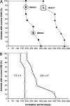
On passaging synthetic prions, two isolates emerged with incubation times differing by nearly 100 days. Using conformational-stability assays, we determined the guanidine hydrochloride (Gdn.HCl) concentration required to denature 50% of disease-causing prion protein (PrP(Sc)) molecules, denoted as the [Gdn.HCl](1/2) value. For the two prion isolates enciphering shorter and longer incubation times, [Gdn.HCl](1/2) values of 2.9 and 3.7 M, respectively, were found. Intrigued by this result, we measured the conformational stabilities of 30 prion isolates from synthetic and naturally occurring sources that had been passaged in mice. When the incubation times were plotted as a function of the [Gdn.HCl](1/2) values, a linear relationship was found with a correlation coefficient of 0.93. These findings demonstrate that (i) less stable prions replicate more rapidly than do stable prions, and (ii) a continuum of PrP(Sc) structural states enciphers a multitude of incubation-time phenotypes. Our data argue that cellular machinery must exist for propagating a large number of different PrP(Sc) conformers, each of which enciphers a distinct biological phenotype as reflected by a specific incubation time. The biophysical explanation for the unprecedented plasticity of PrP(Sc) remains to be determined.
Conflict of interest statement
Conflict of interest statement: G.L., D.P., S.J.D., and S.B.P. have a financial interest in InPro Biotechnology.
Figures


Similar articles
-
Synthetic Prion Selection and Adaptation.Mol Neurobiol. 2019 Apr;56(4):2978-2989. doi: 10.1007/s12035-018-1279-2. Epub 2018 Aug 3. Mol Neurobiol. 2019. PMID: 30074230
-
Protease-sensitive synthetic prions.PLoS Pathog. 2010 Jan 22;6(1):e1000736. doi: 10.1371/journal.ppat.1000736. PLoS Pathog. 2010. PMID: 20107515 Free PMC article.
-
Synthetic prions with novel strain-specified properties.PLoS Pathog. 2015 Dec 31;11(12):e1005354. doi: 10.1371/journal.ppat.1005354. eCollection 2015 Dec. PLoS Pathog. 2015. PMID: 26720726 Free PMC article.
-
Species-barrier phenomenon in prion transmissibility from a viewpoint of protein science.J Biochem. 2013 Feb;153(2):139-45. doi: 10.1093/jb/mvs148. Epub 2013 Jan 2. J Biochem. 2013. PMID: 23284000 Review.
-
Conformational conversion of prion protein in prion diseases.Acta Biochim Biophys Sin (Shanghai). 2013 Jun;45(6):465-76. doi: 10.1093/abbs/gmt027. Epub 2013 Apr 11. Acta Biochim Biophys Sin (Shanghai). 2013. PMID: 23580591 Review.
Cited by
-
Towards authentic transgenic mouse models of heritable PrP prion diseases.Acta Neuropathol. 2016 Oct;132(4):593-610. doi: 10.1007/s00401-016-1585-6. Epub 2016 Jun 28. Acta Neuropathol. 2016. PMID: 27350609 Free PMC article.
-
Prion disease: a tale of folds and strains.Brain Pathol. 2013 May;23(3):321-32. doi: 10.1111/bpa.12045. Brain Pathol. 2013. PMID: 23587138 Free PMC article. Review.
-
The effect of truncation on prion-like properties of α-synuclein.J Biol Chem. 2018 Sep 7;293(36):13910-13920. doi: 10.1074/jbc.RA118.001862. Epub 2018 Jul 20. J Biol Chem. 2018. PMID: 30030380 Free PMC article.
-
Coinfecting prion strains compete for a limiting cellular resource.J Virol. 2010 Jun;84(11):5706-14. doi: 10.1128/JVI.00243-10. Epub 2010 Mar 17. J Virol. 2010. PMID: 20237082 Free PMC article.
-
Mouse-adapted ovine scrapie prion strains are characterized by different conformers of PrPSc.J Virol. 2007 Nov;81(22):12119-27. doi: 10.1128/JVI.01434-07. Epub 2007 Aug 29. J Virol. 2007. PMID: 17728226 Free PMC article.
References
-
- Masters CL, Richardson EP., Jr Brain. 1978;101:333–344. - PubMed
-
- Prusiner SB. N Engl J Med. 2001;344:1516–1526. - PubMed
-
- Chesebro B. Science. 2004;305:1918–1921. - PubMed
-
- Nishida N, Katamine S, Manuelidis L. Science. 2005;310:493–496. - PubMed
-
- Jeffrey M, Gonzalez L, Espenes A, Press C, Martin S, Chaplin M, Davis L, Landsverk T, Macaldowie C, Eaton S, McGovern G. J Pathol. 2006;209:4–14. - PubMed
Publication types
MeSH terms
Substances
Grants and funding
LinkOut - more resources
Full Text Sources
Molecular Biology Databases
Research Materials

