. 2006 Nov;80(21):10772-86.
doi: 10.1128/JVI.00804-06. Epub 2006 Aug 23.

Kaposi's sarcoma-associated herpesvirus LANA-1 interacts with the short variant of BRD4 and releases cells from a BRD4- and BRD2/RING3-induced G1 cell cycle arrest

Affiliations

Kaposi's sarcoma-associated herpesvirus LANA-1 interacts with the short variant of BRD4 and releases cells from a BRD4- and BRD2/RING3-induced G1 cell cycle arrest

Matthias Ottinger et al. J Virol. 2006 Nov.

Abstract

The Kaposi's sarcoma-associated herpesvirus (KSHV) latency-associated nuclear antigen 1 (LANA-1) is required for the replication of episomal viral genomes. Regions in its N-terminal and C-terminal domains mediate the interaction with host cell chromatin. Several cellular nuclear proteins, e.g., BRD2/RING3, histones H2A and H2B, MeCP2, DEK, and HP1alpha, have been suggested to mediate this interaction. In this work, we identify the double-bromodomain proteins BRD4 and BRD3/ORFX as additional LANA-1 interaction partners. The carboxy-terminal region of the short variant of BRD4 (BRD4S) containing the highly conserved extraterminal domain directly interacts with an element in the LANA-1 carboxy-terminal domain. We show that ectopically expressed BRD4S and BRD2/RING3 delay progression into the S phase of the cell cycle in epithelial and B-cell lines and increase cyclin E promoter activity. LANA-1 partly releases epithelial and B cells from a BRD4S- and BRD2/RING3-induced G1 cell cycle arrest and also promotes S-phase entry in the presence of BRD4S and BRD2/RING3. This is accompanied by a reduction in BRD4S-mediated cyclin E promoter activity. Our data are in keeping with the notion that the direct interaction of KSHV LANA-1 with BRD4 and other BRD proteins could play a role in the G1/S phase-promoting functions of KSHV LANA-1. Further, our data support a model in which the LANA-1 C terminus contributes to a functional attachment to acetylated histones H3 and H4 via BRD4 and BRD2, in addition to the recently demonstrated direct interaction (A. J. Barbera, J. V. Chodaparambil, B. Kelley-Clarke, V. Joukov, J. C. Walter, K. Luger, and K. M. Kaye, Science 311:856-861, 2006) of the LANA-1 N terminus with histones H2A and H2B.


Figures
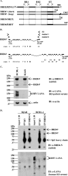
FIG. 1.
FIG. 1.
KSHV LANA-1 forms complexes with endogenous BRD4 proteins in the PEL cell line BCBL-1 as shown by coimmunoprecipitation. (A) Schematic diagram of human BET/fsh proteins. Amino acid positions of the highly conserved, ∼64 aa long, ET domains of RING3 (aa 640 to 703), BRD4 (aa 608 to 671), ORFX (aa 569 to 633), and BRDT (aa 509 to 571). BR1, bromodomain 1; BR2, bromodomain 2. (B) Schematic depiction of the BRD4S and BRD4L exon-intron structure. BRD4 is localized on human chromosome 19 (19p13.1). BRD4S is composed of 11 exons and gives rise to a 2,169-bp ORF, while BRD4L is composed of 19 exons with an alternative exon (11′) and gives rise to a 4,089-bp ORF. (C) Immunoblots (IB) showing the expression of endogenous BRD4 short and long variant proteins in the KSHV-negative Burkitt's lymphoma cell line BJAB and the PEL cell line BCBL-1. α, anti. (D) Detection of BRD4/LANA complexes in BCBL-1 cells by coimmunoprecipitation (co-IP) using a polyclonal anti-BRD4 antibody to precipitate endogenous BRD4S and BRD4L proteins from BJAB cells (negative control) or BCBL-1 lysates. Normal IgG served as a negative control. Four to 12% Bis-Tris PAGE and immunoblotting (IB) were performed. The upper panel shows a rabbit anti-BRD4 immunoblot. The lower panel shows a blot of KS patient serum to detect LANA in BCBL-1 cells. One-tenth of the lysates used for the co-IP was loaded as input control. With longer exposures, the long and the short forms of BRD4 become visible in the input lanes (compare with panel C, top panel). α, anti.
FIG. 2.
FIG. 2.
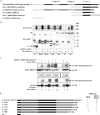
KSHV LANA-1 interacts with the C termini of BRD4S and BRD3/ORFX in vitro in GST pull-down assays. (A) Schematic representation of the GST-BET fusion proteins used in panels B and C. GST-RING3 B (aa 601 to 801), GST-BRD4S (aa 607 to 722), and GST-ORFX (aa 569 to 726) all contain the carboxy-terminal region of the respective BET protein. (B) Expression levels of GST fusion proteins used in panel C and as schematically depicted in panel A determined by anti-GST immunoblotting (IB). α, anti. (C) GST pull-down experiment showing the interaction of the KSHV LANA-1 carboxyterminal domain expressed in SF9 insect cells (arrow) with GST fusion proteins GST-RING3 B (middle panel, lane A), GST-BRD4S (middle panel, lane B), and GST-ORFX (middle panel, lane C) but not with GST alone (middle panel, lane D). The interaction of LANA-1 C with GST-RING3 B (37) served as positive control. One-tenth of the lysates used for the interaction assay were used as input control (left panel). +, presenceof.
FIG. 3.
FIG. 3.
KSHV LANA-1 coimmunoprecipitates with BRD4S and BRD3/ORFX in cotransfected cells. (A) Schematic diagram of the EGFP fusion proteins of full-length BRD2/RING3 and BRD4S and the C-terminal regions of BRD2/RING3, BRD4S, and BRD3/ORFX used in this experiment. (B) Coimmunoprecipitation experiments demonstrating the interaction of full-length LANA-1 with different EGFP-BRD fusion proteins. 293T cells were cotransfected with a full-length LANA-1 expression construct and one of the EGFP-tagged BRD expression constructs as depicted in panel A. Immunoprecipitation was performed using an anti (α)-GFP monoclonal antibody. After extensive washing, SDS-PAGE and immunoblotting (IB) were performed by probing the lower part of the membrane with an anti-GFP antibody (lower panel) to monitor the precipitation of EGFP-fusion proteins BRD2/RING3 (*, “R3”) and BRD4S (*, “4S”) or the carboxy-terminal domains of BRD2/RING3 (**, “R3-C”), BRD4S (***, “4S-C”), or BRD3/ORFX (**, “OX-C”) or EGFPC1 (****, “E”). The carboxy-terminal domains of the BRD proteins were consistently detected as two distinct bands (double and triple asterisks). The upper part of the membrane was probed with human KS patient serum to detect coimmunoprecipitated LANA-1 (upper panel). One-tenth of the lysate used for the IP is shown next to each IP lane as input (In). Bands corresponding to the immunoglobulin heavy chains are visible in all IP lanes around 50 kDa. +, presence of; −, absence of. (C) Coimmunoprecipitation experiments investigating the binding of LANA-1 full-length and carboxy-terminally truncated LANA-1 mutants L-1143, L-1139, and L-1133 to EGFP-tagged BRD4S (“4S”) full-length protein (upper panel) or EGFP alone (lower panel). +, presence of; −, absence of. IB, immunoblot; In, input; α, anti. (D) Schematic representation of C-terminally truncated LANA-1 mutants as used in panel C and a summary of their binding properties to BRD4S (this study) and BRD2/RING3 (^) (51). *, Binding data for L-1107 and L-1006 are not shown. The carboxy-terminal five amino acids of the respective LANA-1 construct are depicted.
FIG. 4.
FIG. 4.
Purified BRD4S aa444-722 and purified KSHV LANA-1 C-terminal domain directly interact in a GST pull-down assay and in an ELISA. (A, upper panel) Schematic depiction of the full-length BRD4S with its two bromodomains (BR1 and BR2) and the ET domain in comparison with the recombinant BRD4S fragment (amino acids 444 to 722) with an N-terminal Flag tag and a C-terminal six-histidine tag expressed in SF9 cells using a recombinant baculovirus. (Lower panel) Schematic representation of full-length KSHV LANA-1 and GST-LANAC protein representing the C-terminal domain of LANA-1 with amino acids 951 to 1162 N-terminally fused to GST. (B) Coomassie-stained SDS-PAGE gels depicting selected elution fractions of glutathione-Sepharose G affinity-purified recombinant GST-LANAC (left panel, right two lanes) expressed in E. coli or the Ni2+ affinity-purified recombinant BRD4S aa444-722 protein expressed in SF9 insect cells (right panel). The shown elution fractions of GST-LANAC and BRD4S aa444-722 were pooled, protein concentrations were determined, and these proteins and purified GST protein alone (not shown) were used for the GST pull-down (C) and ELISA (D) experiments. %, empty lanes. (C) Semiquantitative GST pull-down experiment with purified proteins from panel B and as depicted in panel A. Identical amounts of purified GST or GST-LANAC proteins were immobilized on glutathione 4B beads for 2 h at 4°C, washed, and incubated for 1 h at room temperature with increasing amounts of purified BRD4S aa444-722 protein as indicated in the figure. After extensive washing, SDS-PAGE and immunoblotting (IB) with an anti (α)-His antibody were performed. One-tenth of the input amounts were loaded as input control. Lanes were assembled from two gels (left and right). The figure shows one representative of three independent experiments. (D) ELISA with a fixed concentration of purified GST or GST-LANAC coated onto ELISA plates as described in Material and Methods. After washing steps, coated proteins were incubated with increasing concentrations of purified BRD4S aa444-722 protein. Unbound protein was washed off, and bound BRD4S aa444-722 was detected by using an anti-Flag monoclonal antibody and a secondary alkaline phosphatase-conjugated antibody. The figure shows one representative experiment out of four.
FIG. 5.
FIG. 5.
BRD4S and BRD2/RING3 induce G1/G0 cell cycle arrest and increase the activity of the cyclin E promoter in 293T cells. (A to D) Cell cycle analysis of EGFP (“E”), EGFP-BRD4S (“4S”), or EGFP-RING3 (“R3”) expressing cells using flow cytometry. Briefly, 293T cells in six-well plates were transfected with 1 μg of the respective expression plasmid and 1 μg of pcDNA3. Forty hours posttransfection, cells were BrdU pulsed for 1 h by adding BrdU to a final concentration of 10 μM. Subsequently, cells were paraformaldehyde fixed, permeabilized, and labeled with a primary BrdU-specific antibody and a secondary Cy5-conjugated antibody. Finally, cells were resuspended in PBS containing propidium iodide (PI) and flow cytometric analysis was performed. A total of 105 cells were measured in each sample by using a BD FACSCalibur. Data were analyzed using the software Windows Multiple Document Interface for Flow Cytometry 2.8 (WinMDI2.8). Four independently performed experiments showed similar results. pos, positive; neg, negative. (A) Example of gating procedure and cell cycle analysis for 293T cells transfected with EGFPC1. (A, left panel) Dot plot depicting 2,000 out of a total of 105 analyzed EGFP-transfected cells with the EGFP signal on the x axis (logarithmic scale) and the DNA content of the cells on the y axis (linear scale). With the gate R1, EGFP-positive cells with a DNA content ≥2n (diploid) and ≤4n (tetraploid) were included in subsequent analyses. (Middle panel) Dot plot. With the gate R2, exclusive analysis of single cells was assured. (Right panel) Cells within gates R1 and R2 were analyzed for BrdU positivity (y axis, log scale) in relation to their DNA content (x axis, linear scale) depicted as density plot. Quadrants were defined as shown and differentiate cells in the depicted cell cycle states. The percentage of cells in each individual cell cycle state is listed in each quadrant. (B) Table summarizing the analysis as outlined in panel A for EGFP-, EGFP-BRD4S-, or EGFP-RING3-expressing cells. (C) Histogram with the DNA content on the x axis and the number of cells on the y axis for EGFP-, EGFP-BRD4S-, or EGFP-RING3-expressing cells. (D) Same data as depicted in panel C, normalized to the total number of EGFP-positive cells. (E) Luciferase-based reporter assay. 293T cells in six-well plates were cotransfected with 50 ng of a human cyclin E promoter pGL2 basic reporter plasmid, together with 1 μg of the respective expression construct as depicted in Fig. 3A. Forty-eight hours posttransfection, cells were lysed and lysates were assayed for luciferase activity. Activities are depicted as relative activities relative to the empty vector EGFPC1 control. One representative of four independent experiments performed in duplicate is shown. Standard deviations (error bars) are depicted. No activation was observed with pGL2 basic as control reporter vector (data not shown).
FIG. 6.
FIG. 6.
KSHV LANA-1 decreases BRD4S-induced cyclin E promoter activity. Luciferase based reporter assay in HEK 293T cells. (A) HEK 293T cells in six-well plates were transfected with 50 ng of reporter plasmid (human cyclin E promoter) and 500 ng of expression plasmid for the myc-tagged BRD4S full-length protein (1) or different amounts of KSHV LANA-1 full-length (2), LANA Δ1108-1162 (L-1107) (3) or LANA Δ1007-1162 (L-1006) (4) expression constructs and lysed at confluence 48 h later. The total amount of DNA per transfection was adjusted to 1.05 μg using salmon sperm DNA. For details see Material and Methods. Absolute luciferase activities are shown with standard deviations (error bars). Additionally, mean relative activities are given as numbers on top of each column. (B) HEK 293T cells were cotransfected with 50 ng of reporter plasmid (human cyclin E promoter) and constant amounts of 500 ng of either empty vector pcDNA3 (mock) or BRD4S full-length expression plasmid in pcDNA3, together with increasing amounts (50 ng, 200 ng, and 500 ng) of pcDNA3 (1), full-length KSHV LANA-1 (2), LANA Δ1108-1162 (L-1107) (3), or LANA Δ1007-1162 (L-1006) (4) expression constructs, and lysed at confluence 48 h later. Mean luciferase activities in the absence of BRD4S expression were set at 1 for each amount (50 ng, 200 ng, or 500 ng) of the ORF 73 constructs or pcDNA3 (left three bars in panels 1, 2, 3, and 4). The relative activities in the presence of BRD4S are shown in the three right bars in panels 1, 2, 3, and 4. Mean relative luciferase activities are depicted and given as numbers on top of each column. Standard deviations (error bars) are shown. +, presence of; −, absence of.
FIG. 7.
FIG. 7.
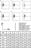
KSHV LANA-1 releases 293T cells from the BRD4S- or the BRD2/RING3-induced G1 cell cycle arrest. Flow cytometric cell cycle analysis was performed as described in the legend for Fig. 5 and Material and Methods. Cells were cotransfected with equal amounts (1 μg each) of EGFPC1 and pcDNA3 (A), EGFPC1 and a LANA-1 expression construct in pcDNA3 (LANA-pcDNA3) (B), EGFPC1-BRD4S and pcDNA3 (C), EGFPC1-BRD4S and LANA-pcDNA3 (D), EGFPC1-RING3 and pcDNA3 (E), or EGFPC1-RING3 and LANA-pcDNA3 (F). Briefly, 40 h posttransfection, 293T cells were pulsed with 10 μM BrdU for 1 h before fixing and labeling the cells. The upper part depicts density blots comparing EGFP-signal-positive cells from samples A, B, C, D, E, and F. DNA content measured as propidium iodide signal is depicted on the x axis. The y axis represents the BrdU signal. A total of 105 cells was analyzed per sample, and the cell cycle data are given in the table. The density blots are generated based on 2,000 analyzed cells of the initial population. The assay was repeated independently three times with similar results. pos, positive.
FIG. 8.
FIG. 8.
KSHV LANA-1 releases BJAB cells from the BRD4S- or the BRD2/RING3-induced G1 cell cycle arrest. BJAB cells were electroporated with 7.5 μg plus 7.5 μg of the plasmids as indicated in the legend to Fig. 7 (samples A, B, C, D, E, and F). Flow cytometric cell cycle analysis was performed as described in the legend for Fig. 5 and Material and Methods. Briefly, 40 h postelectroporation, BJAB cells were pulsed with 10 μM BrdU for 1.5 h before fixing and labeling. A total of 2 × 10⁵ cells was analyzed per sample, and the cell cycle data are given. The numbers represent means ± standard deviations of two independent experiments.

Similar articles

Cited by

References

    1. An, F. Q., N. Compitello, E. Horwitz, M. Sramkoski, E. S. Knudsen, and R. Renne. 2005. The latency-associated nuclear antigen of Kaposi's sarcoma-associated herpesvirus modulates cellular gene expression and protects lymphoid cells from p16 INK4A-induced cell cycle arrest. J. Biol. Chem. 280:3862-3874. - PubMed
    1. Barbera, A. J., M. E. Ballestas, and K. M. Kaye. 2004. The Kaposi's sarcoma-associated herpesvirus latency-associated nuclear antigen 1 N terminus is essential for chromosome association, DNA replication, and episome persistence. J. Virol. 78:294-301. - PMC - PubMed
    1. Barbera, A. J., J. V. Chodaparambil, B. Kelley-Clarke, V. Joukov, J. C. Walter, K. Luger, and K. M. Kaye. 2006. The nucleosomal surface as a docking station for Kaposi's sarcoma herpesvirus LANA. Science 311:856-861. - PubMed
    1. Bechtel, J. T., Y. Liang, J. Hvidding, and D. Ganem. 2003. Host range of Kaposi's sarcoma-associated herpesvirus in cultured cells. J. Virol. 77:6474-6481. - PMC - PubMed
    1. Cesarman, E., Y. Chang, P. S. Moore, J. W. Said, and D. M. Knowles. 1995. Kaposi's sarcoma-associated herpesvirus-like DNA sequences in AIDS-related body-cavity-based lymphomas. N. Engl. J. Med. 332:1186-1191. - PubMed

Publication types

MeSH terms

LinkOut - more resources