. 2006 Aug;80(16):8133-44.
doi: 10.1128/JVI.00278-06.

Epstein-Barr virus nuclear antigen 2 trans-activates the cellular antiapoptotic bfl-1 gene by a CBF1/RBPJ kappa-dependent pathway

Affiliations

Epstein-Barr virus nuclear antigen 2 trans-activates the cellular antiapoptotic bfl-1 gene by a CBF1/RBPJ kappa-dependent pathway

Pamela M Pegman et al. J Virol. 2006 Aug.

Abstract

The human herpesvirus Epstein-Barr virus (EBV) establishes latency and promotes the long-term survival of its host B cell by targeting the molecular machinery controlling cell fate decisions. The cellular antiapoptotic bfl-1 gene confers protection from apoptosis under conditions of growth factor deprivation when expressed ectopically in an EBV-negative Burkitt's lymphoma-derived cell line (B. D'Souza, M. Rowe, and D. Walls, J. Virol. 74:6652-6658, 2000), and the EBV latent membrane protein 1 (LMP1) and its cellular functional homologue CD40 can both drive bfl-1 via an NF-kappaB-dependent enhancer element in the bfl-1 promoter (B. N. D'Souza, L. C. Edelstein, P. M. Pegman, S. M. Smith, S. T. Loughran, A. Clarke, A. Mehl, M. Rowe, C. Gélinas, and D. Walls, J. Virol. 78:1800-1816, 2004). Here we show that the EBV nuclear antigen 2 (EBNA2) also upregulates bfl-1. EBNA2 trans-activation of bfl-1 requires CBF1 (or RBP-J kappa), a nuclear component of the Notch signaling pathway, and there is an essential role for a core consensus CBF1-binding site on the bfl-1 promoter. trans-activation is dependent on the EBNA2-CBF1 interaction, is modulated by other EBV gene products known to interact with the CBF1 corepressor complex, and does not involve activation of NF-kappaB. bfl-1 expression is induced and maintained at high levels by the EBV growth program in a lymphoblastoid cell line, and withdrawal of either EBNA2 or LMP1 does not lead to a reduction in bfl-1 mRNA levels in this context, whereas the simultaneous loss of both EBV proteins results in a major decrease in bfl-1 expression. These findings are relevant to our understanding of EBV persistence, its role in malignant disease, and the B-cell developmental process.


Figures
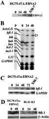
FIG. 1.
FIG. 1.
Expression of EBNA2 leads to increased bfl-1 mRNA and protein levels in BL-derived cell lines. (A) Western blot of DG75-tTA-EBNA2 cells induced to express EBNA2 by reculturing cells in the absence of tetracycline. Cells were harvested and analyzed for EBNA2 expression at various time points (indicated in hours above each lane); also included is the reference LCL X50-7. (B) RPA autoradiogram in which mRNA levels from the apoptosis-related genes bcl-x L/S, bfl-1, bik, bak, bax, bcl-2, and mcl-1 from the same experiment as that in panel A were analyzed. Unprotected 32P-labeled antisense riboprobes (5,000 cpm, lane P) were loaded alongside RPA-processed samples and are shown linked to their smaller RNase-protected fragments, which correspond to the steady-state levels of the corresponding mRNA in the samples. An increase in the steady-state level of bfl-1 mRNA (open arrow) is seen upon induction of EBNA2 expression. (C) Northern blot analysis of bfl-1 mRNA levels from a repeat EBNA2 induction experiment using DG75-EBNA2-tTA. Thirty-microgram samples of total RNA were loaded onto the gel, which was then blotted and probed with antisense bfl-1 riboprobe. The lower panel shows the same blot stripped and reprobed with a GAPDH antisense riboprobe. (D) Western blots showing upregulation of Bfl-1 expression in DG75-tTA-EBNA2 cells in response to EBNA2 induction following tetracycline withdrawal and in BL41-K3 cells in which ER-EBNA2 function was activated by addition of β-estradiol. The times postinduction/activation of EBNA2 (expressed in hours) are given above each lane. The lower panel shows the same blots probed with antibody to β-actin.
FIG. 2.
FIG. 2.
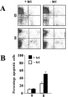
Comparative analysis of the apoptotic response of DG75-tTA-EBNA2 cells uninduced and induced to express EBNA2. (A) Representative fluorescence-activated cell sorter profiles of Syto 16 fluorescence (y axis) versus propidium iodide (PI) fluorescence (x axis) in DG75-tTA-EBNA2 uninduced (+ tet; left panels) and induced to express EBNA2 (− tet; right panels) for 2 days, followed by 0 and 8 days (as indicated on the left) of partial serum deprivation combined with ionomycin treatment. Viable cells (Syto 16-positive, PI-negative) are represented in each top left quadrant, apoptotic (Syto 16-negative, PI-negative) cells in the bottom left quadrants, and necrotic (Syto 16-negative, PI-positive) cells in the bottom right quadrants. (B) Summary of the average percentage of apoptotic cells in three repeat experiments, including that shown in panel A. The times at which samples were taken are indicated (in days) underneath the graph. Error bars indicate standard deviations.
FIG. 3.
FIG. 3.
EBNA2, but not Notch-IC, trans-activates the bfl-1 promoter in BL-derived cell lines. (A) DG75, BJAB, and BL41 cells were cotransfected with 7 μg of either pSG5 or pSGEBNA2 together with 1 μg of the bfl-1 promoter-reporter construct −1374/+81-luc. Cells were harvested at 24 h posttransfection and analyzed for luciferase activities, which were then normalized for transfection efficiency (based on β-galactosidase activity measured from acotransfected pCMVlacZ reporter which was included in all transfections). Luciferase values obtained from cotransfections with pSG5 were arbitrarily assigned a value of 1.0, and activation represents the relative normalized luciferase activities obtained upon cotransfection with pSGEBNA2. (B) Dose-dependent trans-activation of the bfl-1 promoter by EBNA2 but not EBNA2ww323sr, a mutant of EBNA2 that cannot bind CBF1. DG75 cells were cotransfected with increasing amounts of either pSGEBNA2 or pSGEBNA2ww323sr (effector plasmids; quantities are indicated underneath) and 1 μg of −1374/+81-luc. Cells were harvested at 24 h posttransfection and analyzed for luciferase activities, which were normalized and presented as described for panel A. (C) DG75 cells were transfected with 1 μg of either −1374/+81-luc or pGa981-6 and 5 μg of plasmid expressing the effector molecules indicated underneath each bar. The chimeric protein mNotch1IC:E2TANLS binds CBF1 via mNotch1IC and contains the additional trans-activation domain and nuclear localization sequence of EBNA2. Higher quantities of any of these effector plasmids (up to 12 μg) did not lead to increased bfl-1 promoter trans-activation (not shown). Cells were harvested at 24 h posttransfection and analyzed for luciferase activities, which were normalized and presented as described for panel A.
FIG. 4.
FIG. 4.
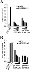
EBNA2-mediated trans-activation of the bfl-1 promoter is inhibited by EBNA3A, EBNA3B, EBNA3C, and the EBV latent gene product RPMS1. DG75 cells were cotransfected with 1 μg of −1374/+81-luc together with increasing amounts (in all cases 1, 3, 5, and 7 μg consecutively) of the expression vectors pCMV-EBNA3A, pCMV-EBNA3B, pCMV-EBNA3C, or pcDNA3-RPMS1/FLAG. Cells were harvested at 24 h posttransfection, and normalized luciferase values were expressed as activation levels (n-fold) relative to those of the corresponding control transfected cells in which the effector plasmids were pSGEBNA2 together with either p7CMV (5 μg) or pcDNA3.1 (5 μg).
FIG. 5.
FIG. 5.
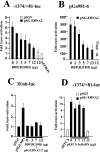
trans-activation of the bfl-1 promoter by EBNA2 is inhibited by coexpression of a dominant-negative mutant form of CBF1 and does not involve activation of NF-κB. The names of the reporter constructs are given above each figure. (A and B) Inhibition of EBNA2-mediated trans-activation of the bfl-1 promoter by a non-DNA-binding mutant of CBF1 [RBP(R218H)]. DG75 cells were cotransfected with 1 μg of −1374/+81-luc (A) or the reporter construct pGa981-6, in which transcription of the luciferase gene is regulated by CBF1 (B), and 7 μg of pSGEBNA2 together with increasing amounts of pEF-BOSneo-RBP(R218H) [indicated as RBP(R218H) underneath each bar]. (C) Overexpression of either EBNA2 or RBP(R218H) does not lead to increased NF-κB-dependent transcriptional activation in DG75 cells. Here, NF-κB activation was monitored by using the established NF-κB-dependent reporter construct, 3Enh-luc. (D) Coexpression of an IκBα mutant that inhibits activation of NF-κB did not significantly affect trans-activation of the bfl-1 promoter by EBNA2. In all cases (A to D), cells were harvested at 24 h posttransfection, and normalized luciferase values were expressed as activation levels (n-fold) relative to those of the corresponding control transfected cells with the relevant empty vectors as appropriate in each case.
FIG. 6.
FIG. 6.
Identification of a CBF1-like binding site on the bfl-1 promoter that mediates trans-activation by EBNA2. The figure shows a series of bfl-1-promoter-reporter constructs in which the promoter sequence has been progressively deleted from the 5′end (the coordinate of the 5′ end is given to the left of each construct). They all share a common 3′ terminus at 81 bp downstream from the transcription initiation site (indicated by a bent arrow), at which point they are joined to the luciferase gene (LUC). The relative location (black box) and sequence (open box) of a consensus CBF1-binding site is indicated, and the base changes made to this motif by site-directed mutagenesis to generate −1374/+81-luc(mCBF1) are underlined. DG75 cells were transfected with 7 μg of either pSG5 or pSGEBNA2 together with 2 μg of the individual reporter constructs (the names are given on the right). Cells were harvested at 24 h posttransfection and assayed for luciferase activity as previously described. Each normalized luciferase value was expressed as the activation level (n-fold) relative to the corresponding value obtained for each reporter construct when cotransfected with control pSG5.
FIG. 7.
FIG. 7.
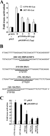
A role for Ets-family transcription factors in bfl-1 promoter trans-activation by EBNA2. (A) Sequence of the −301 to −111 region of the bfl-1 promoter showing the locations and sequences of candidate binding sites for relevant transcription factors. Three further reporter constructs were made by introducing base changes (underlined) to these sequences in −367/+81-Luc, thus eliminating each motif in turn. (B) DG75 cells were transfected with 7 μg of either pSGEBNA2 or pSG5, together with 2 μg of a reporter construct (indicated underneath). Cells were harvested 24 h posttransfection and assayed for luciferase activity as previously described. Each normalized luciferase value was expressed as an activation level (n-fold) relative to the corresponding value obtained for each reporter construct when cotransfected with control pSG5. (C) DG75 cells were cotransfected with 7 μg of pSGEBNA2 and 2 μg of either −1374/+81-luc or −367/+81-luc plus increasing quantities of pCEP4Ets-DN. Each normalized luciferase value was expressed as an activation level (n-fold) relative to the corresponding value obtained for each reporter construct when cotransfected with control pSG5 and pCEP4 (5 and 7 μg, respectively).
FIG. 8.
FIG. 8.
bfl-1 is driven by EBV latent proteins in an LCL context, and the loss of both EBNA2 and LMP1 is required before bfl-1 mRNA levels decline significantly. Estrogen-starved EREB2.5 cells (A) and LCL 1480.4 cells (B) were treated with β-estradiol and sampled for RNA and protein analysis at the time points indicated above each panel. bfl-1, L32, and GAPDH mRNA levels were assayed by RPA (upper panels); levels of ER-EBNA2 and LMP1 were determined by Western blotting (lower panels). (C) LCL 1852.4 cells were treated with tetracycline and processed for RNA and protein analysis at the time points indicated.

Similar articles

Cited by

References

    1. Alfieri, C., M. Birkenbach, and E. Kieff. 1991. Early events in Epstein-Barr virus infection of human B lymphocytes. Virology 181:595-608. - PubMed
    1. Andersson, M. L., N. J. Stam, G. Klein, H. L. Ploegh, and M. G. Masucci. 1991. Aberrant expression of HLA class-I antigens in Burkitt lymphoma cells. Int. J. Cancer 47:544-550. - PubMed
    1. Arenzana-Seisdedos, F., P. Turpin, M. Rodriguez, D. Thomas, R. T. Hay, J. L. Virelizier, and C. Dargemont. 1997. Nuclear localization of I kappa B alpha promotes active transport of NF-kappa B from the nucleus to the cytoplasm. J. Cell Sci. 110:369-378. - PubMed
    1. Bash, J., W. X. Zong, S. Banga, A. Rivera, D. W. Ballard, Y. Ron, and C. Gelinas. 1999. Rel/NF-kappaB can trigger the Notch signaling pathway by inducing the expression of Jagged1, a ligand for Notch receptors. EMBO J. 18:2803-2811. - PMC - PubMed
    1. Ben-Bassat, H., N. Goldblum, S. Mitrani, T. Goldblum, J. M. Yoffey, M. M. Cohen, Z. Bentwich, B. Ramot, E. Klein, and G. Klein. 1977. Establishment in continuous culture of a new type of lymphocyte from a “Burkitt like” malignant lymphoma (line D.G.-75). Int. J. Cancer 19:27-33. - PubMed

Publication types

MeSH terms

Substances