NF-(kappa)B-inducing kinase controls lymphocyte and osteoclast activities in inflammatory arthritis
- PMID: 15937549
- PMCID: PMC1142111
- DOI: 10.1172/JCI23763
NF-(kappa)B-inducing kinase controls lymphocyte and osteoclast activities in inflammatory arthritis
Abstract
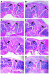
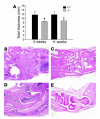
NF-(kappa)B is an important component of both autoimmunity and bone destruction in RA. NF-(kappa)B-inducing kinase (NIK) is a key mediator of the alternative arm of the NF-(kappa)B pathway, which is characterized by the nuclear translocation of RelB/p52 complexes. Mice lacking functional NIK have no peripheral lymph nodes, defective B and T cells, and impaired receptor activator of NF-kappaB ligand-stimulated osteoclastogenesis. We investigated the role of NIK in murine models of inflammatory arthritis using Nik-/- mice. The serum transfer arthritis model is initiated by preformed antibodies and required only intact neutrophil and complement systems in recipients. While Nik-/- mice had inflammation equivalent to that of Nik+/+ controls, they showed significantly less periarticular osteoclastogenesis and less bone erosion. In contrast, Nik-/- mice were completely resistant to antigen-induced arthritis (AIA), which requires intact antigen presentation and lymphocyte function but not lymph nodes. Additionally, transfer of Nik+/+ splenocytes or T cells to Rag2-/- mice conferred susceptibility to AIA, while transfer of Nik-/- cells did not. Nik-/- mice were also resistant to a genetic, spontaneous form of arthritis, generated in mice expressing both the KRN T cell receptor and H-2. Thus, NIK is important in the immune and bone-destructive components of inflammatory arthritis and represents a possible therapeutic target for these diseases.
Figures


Similar articles
-
Inhibition of nuclear factor kappa B inducing kinase suppresses inflammatory responses and the symptoms of chronic periodontitis in a mouse model.Int J Biochem Cell Biol. 2021 Oct;139:106052. doi: 10.1016/j.biocel.2021.106052. Epub 2021 Aug 5. Int J Biochem Cell Biol. 2021. PMID: 34364989
-
Defective nuclear factor-κB-inducing kinase in aly/aly mice prevents bone resorption induced by local injection of lipopolysaccharide.J Periodontal Res. 2011 Apr;46(2):280-4. doi: 10.1111/j.1600-0765.2010.01333.x. Epub 2010 Dec 29. J Periodontal Res. 2011. PMID: 21348872
-
The IkappaB function of NF-kappaB2 p100 controls stimulated osteoclastogenesis.J Exp Med. 2003 Sep 1;198(5):771-81. doi: 10.1084/jem.20030116. Epub 2003 Aug 25. J Exp Med. 2003. PMID: 12939342 Free PMC article.
-
NF-κB–inducing kinase plays an essential T cell–intrinsic role in graft-versus-host disease and lethal autoimmunity in mice.J Clin Invest. 2011 Dec;121(12):4775-86. doi: 10.1172/JCI44943. J Clin Invest. 2011. PMID: 22045568 Free PMC article.
-
Non-canonical NF-κB signaling pathway.Cell Res. 2011 Jan;21(1):71-85. doi: 10.1038/cr.2010.177. Epub 2010 Dec 21. Cell Res. 2011. PMID: 21173796 Free PMC article. Review.
Cited by
-
Effects of testosterone on lean mass gain in elderly men: systematic review with meta-analysis of controlled and randomized studies.Age (Dordr). 2015 Feb;37(1):9742. doi: 10.1007/s11357-014-9742-0. Epub 2015 Feb 1. Age (Dordr). 2015. PMID: 25637335 Free PMC article. Review.
-
Intra-articular nuclear factor-κB blockade ameliorates collagen-induced arthritis in mice by eliciting regulatory T cells and macrophages.Clin Exp Immunol. 2013 May;172(2):217-27. doi: 10.1111/cei.12054. Clin Exp Immunol. 2013. PMID: 23574318 Free PMC article.
-
Functions of nuclear factor kappaB in bone.Ann N Y Acad Sci. 2010 Mar;1192:367-75. doi: 10.1111/j.1749-6632.2009.05315.x. Ann N Y Acad Sci. 2010. PMID: 20392262 Free PMC article. Review.
-
Editorial: Non-canonical NF-κB signaling in immune-mediated inflammatory diseases and malignancies.Front Immunol. 2023 Jul 26;14:1252939. doi: 10.3389/fimmu.2023.1252939. eCollection 2023. Front Immunol. 2023. PMID: 37564643 Free PMC article. No abstract available.
-
NF-κB-inducing kinase increases renal tubule epithelial inflammation associated with diabetes.Exp Diabetes Res. 2011;2011:192564. doi: 10.1155/2011/192564. Epub 2011 Aug 18. Exp Diabetes Res. 2011. PMID: 21869881 Free PMC article.
References
-
- Pope, R., and Perlman, H. 2000. Rheumatoid arthritis. In Principles of molecular rheumatology. G. Tsokos, editor. Humana Press Inc. Totowa, New Jersey, USA. 325–361.
-
- Ruderman EM, Tambar S. Psoriatic arthritis: prevalence, diagnosis, and review of therapy for the dermatologist. Dermatol. Clin. 2004;22:477–486. - PubMed
-
- Smith J, Haynes M. Rheumatoid arthritis — a molecular understanding [review] Ann. Intern. Med. 2002;136:908–922. - PubMed
-
- Goldring S. Pathogenesis of bone erosions in rheumatoid arthritis. Curr. Opin. Rheumatol. 2002;14:406–410. - PubMed
Publication types
MeSH terms
Substances
Grants and funding
LinkOut - more resources
Full Text Sources
Other Literature Sources
Molecular Biology Databases
Miscellaneous

