Comparative Study
. 2005 Jun 5;336(2):184-97.
doi: 10.1016/j.virol.2005.03.032.

Thymic pathogenicity of an HIV-1 envelope is associated with increased CXCR4 binding efficiency and V5-gp41-dependent activity, but not V1/V2-associated CD4 binding efficiency and viral entry

Affiliations
Comparative Study

Thymic pathogenicity of an HIV-1 envelope is associated with increased CXCR4 binding efficiency and V5-gp41-dependent activity, but not V1/V2-associated CD4 binding efficiency and viral entry

Eric G Meissner et al. Virology. .

Abstract

We previously described a thymus-tropic HIV-1 envelope (R3A Env) from a rapid progressor obtained at the time of transmission. An HIV-1 molecular recombinant with the R3A Env supported extensive replication and pathogenesis in the thymus and did not require Nef. Another Env from the same patient did not display the same thymus-tropic pathogenesis (R3B Env). Here, we show that relative to R3B Env, R3A Env enhances viral entry of T cells, increases fusion-induced cytopathicity, and shows elevated binding efficiency for both CD4 and CXCR4, but not CCR5, in vitro. We created chimeric envelopes to determine the region(s) responsible for each in vitro phenotype and for thymic pathogenesis. Surprisingly, while V1/V2 contributed to enhanced viral entry, CD4 binding efficiency, and cytopathicity in vitro, it made no contribution to thymic pathogenesis. Rather, CXCR4 binding efficiency and V5-gp41-associated activity appear to independently contribute to thymic pathogenesis of the R3A Env. These data highlight the contribution of unique HIV pathogenic factors in the thymic microenvironment and suggest that novel mechanisms may be involved in Env pathogenic activity in vivo.


Figures

Fig. 1
Fig. 1
The R3A Env mediates elevated viral fusion with CD4+ T cells. Blam-vpr-containing virions were used to infect Sup-T1 cells by spinoculation. After 2 h of incubation at 37 °C, cells were incubated with flurogenic beta-lactamase substrate for 8 h. The amount of entry was calculated by measuring the ratio of cleaved to uncleaved flurogenic substrate. Shown is a representative of eight independent experiments with error bars derived from triplicate samples and input virus determined by p24 ELISA. (*P <0.05 for R3A vs. either NL4-3 or R3B).
Fig. 2
Fig. 2
The R3A Env shows enhanced sensitivity to sCD4 and reduced sensitivity to Leu3a. (A) R3A Env is sensitive to sCD4 relative to R3B Env. TZM-bl cells were infected with virus incubated with sCD4 for 2 h prior to infection. Infection was quantitated by luciferase assay 48 h post-infection and values were normalized to infection in the absence of sCD4. Shown is a representative of 3 experiments with error bars derived from duplicate samples. (B) R3A Env is resistant to Leu-3a relative to R3B Env. Sup-T1 cells were preincubated with the indicated dose of Leu-3a for 1 h prior to infection with pseudotyped NL4-luc. Infection achieved by spinoculation was quantitated by luciferase assay 48 h post-infection and values were normalized to infection in the absence of Leu-3a. Shown is a representative of 3 experiments with error bars derived from quadruplicate samples (*P <0.05 for R3B vs. either NL4-3 or R3A).
Fig. 3
Fig. 3
The R3A Env shows enhanced binding efficiency for CXCR4, but similar sensitivity to Tak-779 and T20. (A) R3A and R3B Envs show equal sensitivity to a CCR5 antagonist. Infection of U373-MAGI-CCR5E cells was performed in the presence of the CCR5 antagonist TAK-779. Infection was quantitated by counting blue colonies and was normalized to the number of colonies observed with no TAK-779. Shown is a representative of two independent experiments with error bars derived from quadruplicate samples. (B) R3A shows enhanced affinity for CXCR4 relative to R3B. Infection of Sup-T1 cells was performed with NL4-luc-pseudotyped virus in the presence of AMD-3100. Infection was quantitated by luciferase assay after 48 h and was normalized to levels obtained with no AMD-3100. Shown is a representative of seven independent experiments with error bars derived from triplicate samples (*P <0.05 for R3A vs. either NL4-3 or R3B). (C) Sup-T1 cells transduced with vector or X4h shRNA, which reduces CXCR4 surface levels by ~97%, were infected with NL4-luc pseudotyped with the indicated Env. Infection was quantitated by luciferase assay after 48 h and was normalized to levels obtained on vector-transduced cells. Shown is a representative of three independent experiments with error bars derived from triplicate samples (*P <0.05 in comparison to R3A Env or to infection of parental cells). (D) R3A and R3B Envs show equal sensitivity to the fusion inhibitor T20. TZM-bl cells were infected with virus in the presence of the indicated dose of T20. Infection was quantitated and normalized as in A. Shown is a representative of 2 experiments with error bars derived from duplicate samples (*P <0.05 for NL4-R3A vs. either NL4-3 or NL4-R3B).
Fig. 4
Fig. 4
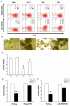
The R3A Env causes cytopathicity through CXCR4-dependent fusion when expressed in T cells. (A) 1G5 cells were transduced with a retroviral vector expressing HIV Env. Env surface levels were detected 3 days post-transduction using the 2G12 monoclonal antibody or human IgG1 as an isotype control. Shown is a representative stain 3 days post-transduction. The percentage of cells in each quadrant is indicated. (B) R3A Env induces extensive syncytia formation. Syncytia were observed by light microscopy 3 days post-transduction. Shown are representative photos of Sup-T1 cells 3 days post-transduction. (C) The number of live cells surviving 3 days post-transduction was quantitated by trypan blue exclusion. Shown is a representative of 8 independent experiments for Sup-T1 cells. (D and E) R3A-induced cytopathicity is dependent on fusion through CXCR4. Vector or R3A-transduced Sup-T1 cells were incubated with no drug, with T20 (D), or with AMD-3100 (E), and cytopathicity was quantitated as in C. Shown is a representative of 2 independent experiments (*P <0.05 for R3A and R3B vs. vector transduced cells).
Fig. 5
Fig. 5
Generation of recombinant env genes to map the pathogenic determinants in the R3A Env. Schematic map of the eight recombinant env genes made using conserved restriction sites. Vertical bars indicate specific amino acid differences between R3A and R3B Env proteins. The total number of differences in each region of Env is indicated. The asterisk indicates the additional putative glycosylation site in the R3B Env.
Fig. 6
Fig. 6
The V1/V2 region is largely responsible for the enhanced entry and CD4 affinity of R3A Env, while CXCR4 affinity cannot be mapped to one particular region. (A) V1/V2 contributes to enhanced virus fusion of T cells. Blam-vpr-containing virions were used to infect Sup-T1 cells at the dose of p24 indicated. Shown is a representative of 5 independent experiments with error bars derived from quadruplicate samples (*P <0.05 for the indicated recombinant relative to the parental envelope at the highest two doses of p24). (B) Soluble CD4 sensitivity also maps to the V1/V2 region. NL4-luc-pseudotyped virus was pretreated with 4 μg/ml sCD4 prior to infection of GHOST-CXCR4 cells. Infection in the presence of sCD4 was compared to infection without sCD4 treatment. Shown is a representative of 2 experiments with error bars derived from quadruplicate samples. (C) Resistance to AMD-3100 is achieved by multiple determinants. Sup-T1 cells were infected with pseudotyped NL4-luc in the presence of 200 nM AMD-3100. Resistance to inhibition observed with the R3A Env was assigned a value of 1 for each of four experiments, and the relative resistance of each recombinant Env was normalized to this value. Shown are the combined results from four independent experiments with standard error bars indicated.
Fig. 7
Fig. 7
V1/V2 and V5-gp41 independently contribute to enhanced fusion-induced cytopathicity of R3A Env. Sup-T1 cells were transduced with Env expression vectors. Cytopathicity was quantitated as in Fig. 2. *Significantly different from the parental envelope, **significantly different from both R3A and R3B Env (P <0.05). Shown are the combined results of 3–6 independent experiments with normalization to vector-transduced cells within each experiment.
Fig. 8
Fig. 8
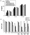
V5-gp41, but not V1/V2, of the R3A Env is uniquely involved in HIV replication and pathogenesis in the thymus model. (A) NL4-3 recombinant viruses expressing the indicated Env were used to infect thymus fragments in the HF-TOC system. Replication was monitored by detection of p24 in the TOC supernatant at the indicated times post-infection. Shown are the combined data from five independent infections. (B) Pathogenesis was assessed by depletion of total CD4+ thymocytes using FACS. Shown are the combined results from five independent experiments. *Indicates significant differences in depletion relative to both NL4-R3B and NL4-R3A at days 11–12 ( P < 0.05).

Similar articles

Cited by

References

    1. Arthos J, Cicala C, Selig SM, White AA, Ravindranath HM, Van Ryk D, Steenbeke TD, Machado E, Khazanie P, Hanback MS, Hanback DB, Rabin RL, Fauci AS. The role of the CD4 receptor versus HIV coreceptors in envelope-mediated apoptosis in peripheral blood mononuclear cells. Virology. 2002;292(1):98–106. - PubMed
    1. Baba M, Nishimura O, Kanzaki N, Okamoto M, Sawada H, Iizawa Y, Shiraishi M, Aramaki Y, Okonogi K, Ogawa Y, Meguro K, Fujino M. A small-molecule, nonpeptide CCR5 antagonist with highly potent and selective anti-HIV-1 activity. Proc. Natl. Acad. Sci. U.S.A. 1999;96(10):5698–5703. - PMC - PubMed
    1. Beaumont T, Quakkelaar E, van Nuenen A, Pantophlet R, Schuitemaker H. Increased sensitivity to CD4 binding site-directed neutralization following in vitro propagation on primary lymphocytes of a neutralization-resistant human immunodeficiency virus IIIB strain isolated from an accidentally infected laboratory worker. J. Virol. 2004;78(11):5651–5657. - PMC - PubMed
    1. Berkowitz RD, Beckerman KP, Schall TJ, McCune JM. CXCR4 and CCR5 expression delineates targets for HIV-1 disruption of T cell differentiation. J. Immunol. 1998;161(7):3702–3710. - PubMed
    1. Bonyhadi ML, Su L, Auten J, McCune JM, Kaneshima H. Development of a human thymic organ culture model for the study of HIV pathogenesis. AIDS Res. Hum. Retroviruses. 1995;11(9):1073–1080. - PubMed

Publication types