. 2005 Jan 12;24(1):138-48.
doi: 10.1038/sj.emboj.7600491. Epub 2004 Nov 25.

Recognition and cleavage of primary microRNA precursors by the nuclear processing enzyme Drosha

Affiliations

Recognition and cleavage of primary microRNA precursors by the nuclear processing enzyme Drosha

Yan Zeng et al. EMBO J. .

Abstract

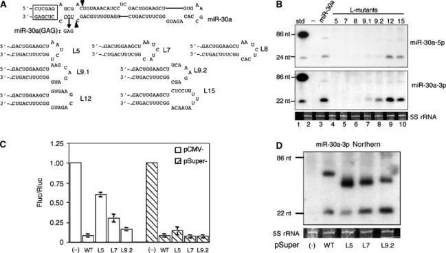
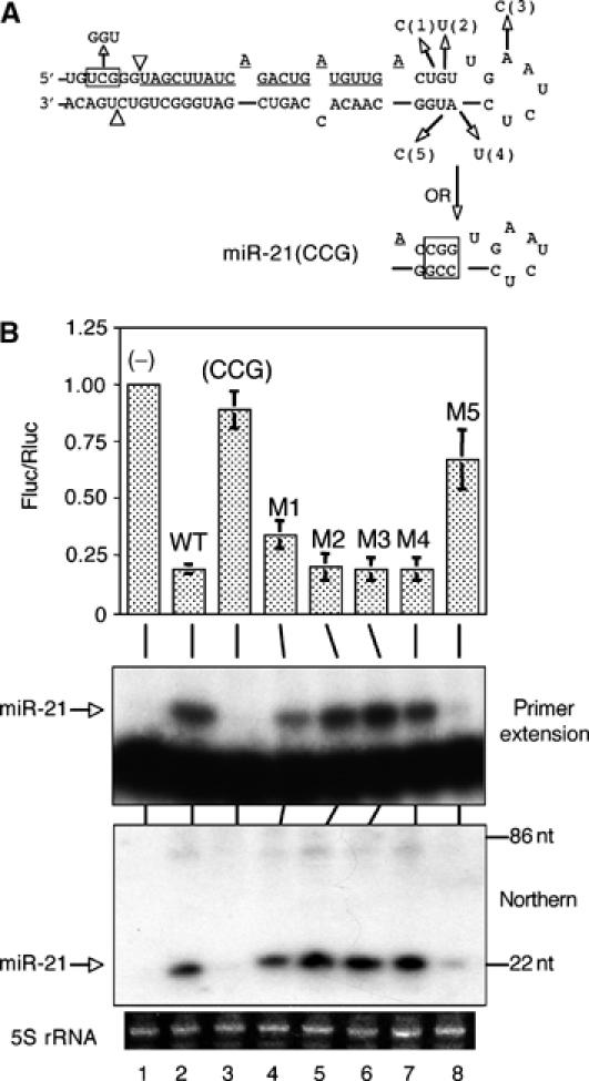
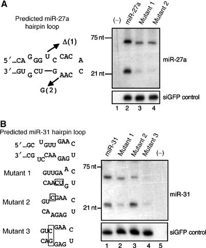
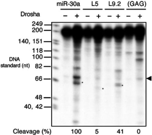
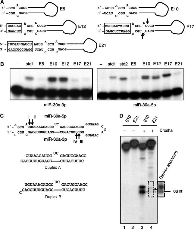
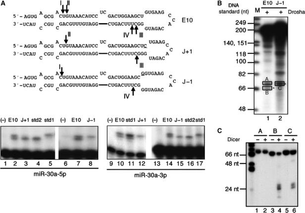
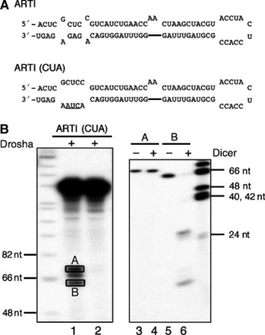
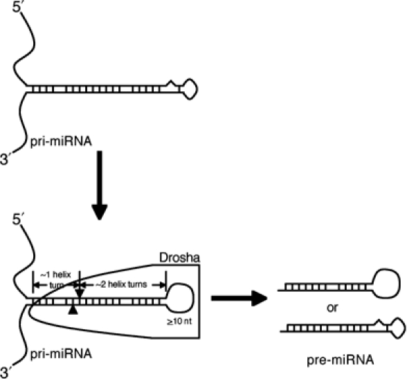
A critical step during human microRNA maturation is the processing of the primary microRNA transcript by the nuclear RNaseIII enzyme Drosha to generate the approximately 60-nucleotide precursor microRNA hairpin. How Drosha recognizes primary RNA substrates and selects its cleavage sites has remained a mystery, especially given that the known targets for Drosha processing show no discernable sequence homology. Here, we show that human Drosha selectively cleaves RNA hairpins bearing a large (>/=10 nucleotides) terminal loop. From the junction of the loop and the adjacent stem, Drosha then cleaves approximately two helical RNA turns into the stem to produce the precursor microRNA. Beyond the precursor microRNA cleavage sites, approximately one helix turn of stem extension is also essential for efficient processing. While the sites of Drosha cleavage are determined largely by the distance from the terminal loop, variations in stem structure and sequence around the cleavage site can fine-tune the actual cleavage sites chosen.


Figures

Figure 1
Figure 1
Analysis of pri-miR-30a hairpin terminal loop mutants. (A) Computer-predicted secondary structure of the pri-miR-30a hairpin (Lagos-Quintana et al, 2001) and sequences of the miR-30a terminal loop mutants. The mutation introduced into miR-30a(GAG), a miR-30a null mutant, is indicated. The boxed sequence derives from appended restriction sites and facilitates miR-30a expression. The arrowheads identify the major cleavage sites for endogenous pre-miR-30a. (B) Northern analyses of miR-30a-5p (upper panel) and miR-30a-3p (lower panel) expression in 293T cells transfected with the indicated pri-miR-30a expression plasmids (lanes 3–10). Lane 1; DNA size markers; lane 2; RNA from nontransfected 293T cells. 5S rRNA was used as a loading control. (C) A reporter assay to compare miR-30a-3p function when expressed from pCMV- or pSuper-based vectors. The firefly luciferase-based reporter, pCMV-luc-8xmiR-30a(P), was cotransfected into 293T cells along with pRL-CMV (Promega), an internal control Renilla luciferase expression plasmid, and pCMV-miR-30a or pSuper-miR-30a variants. The ratio of firefly luciferase activity relative to Renilla luciferase activity from cells transfected with an empty pCMV or pSuper vector (−) is set at 1.00. Average of three experiments with standard deviation is shown. (D) Pre-miR-30a and mature miR-30a-3p expression in 293T cells transfected with the pSuper-miR-30a plasmids was determined by Northern blotting, as shown in panel B. miRNA expression levels induced by pSuper-miR-30a and pCMV-miR-30a are comparable (data not shown).
Figure 2
Figure 2
Analysis of pri-miR-21 loop mutants. (A) Schematic of the computer-predicted miR-21 secondary structure, point mutations (1, 2, 3, 4, and 5), and the miR-21(GGU) and miR-21(CCG) mutants. Mature miR-21 is underlined, and the arrowheads indicate the cleavage sites used to produce the predicted pre-miR-21. (B) Analyses of miR-21 loop mutants. Luciferase assays were performed as in Figure 1C and the similar indicator construct pCMV-luc-8xmiR-21(P) was used as the reporter. Primer extension and Northern blotting assays were performed using oligonucleotide probes described in Supplementary Table 1. 5S rRNA was used as a loading control in the Northern analysis.
Figure 3
Figure 3
Alternative structures for miR-30a, miR-21, miR-27a, and miR-31. Comparison of the published, computer-predicted secondary structures (Lagos-Quintana et al, 2001) with possible new secondary structures proposing larger terminal loops for these miRNA precursors. These proposed structures are based on the mutational analyses, and transfections and in vitro assays, performed in this paper. These larger loops may form as indicated or may be opened by Drosha binding. Mature miRNA sequences are underlined, and predicted or confirmed Drosha cleavage sites are shown by arrows.
Figure 4
Figure 4
Analysis of loop mutants of human pri-miR-27a and pri-miR-31. (A) miR-27a expression. Plasmids encoding wild-type pri-miR-27a, or one of two mutants (1 and 2), were cotransfected with pH1-GFP into 293T cells. Northern blotting was performed to detect the expression of pre- and mature miR-27a (upper panel) and the control GFP siRNA (lower panel). (B) miR-31 expression. Three pri-miR-31 mutants were constructed (boxed sequences), and their miRNA expression levels compared to wild-type miR-31 in transfected 293T cells.
Figure 5
Figure 5
In vitro Drosha cleavage of pri-miR-30a transcripts. FLAG immunoprecipitates from mock-transfected 293T cells (−) or cells transfected with pCK-Drosha-FLAG(+) were incubated with a 32P-labeled ∼202 nt RNA probe encoding the indicated miR-30a variants. RNA cleavage products were recovered and resolved on a denaturing 10% polyacrylamide gel. The pre-miRNA band is indicated by an asterisk, and the background band running slightly above by an arrowhead. Cleavage efficiency was calculated as the intensity of the pre-miRNA band divided by that of the remaining substrate, and set as 100% for the wild-type transcript. DNA size standards are shown next to the autoradiograph.
Figure 6
Figure 6
Analysis of the effects of changes in pri-miR-30a stem length. (A) Predicted hairpin RNA structures adopted by pri-miR-30a variants with different stem extensions beyond the pre-miR-30a structure. Boxed sequences represent added restriction sites. The positions of the 5′ and 3′ ends of the endogenous pre-miR-30a are indicated by arrows in E17. (B) Primer extension experiments to determine the expression level and 5′ ends of mature miR-30a-3p (left panel) or miR-30a-5p (right panel) variants. The sequences of the miR-30a-3p std1 and miR-30a-5p std1 and 2 size standards are given in Supplementary Table 1. (C) The 5′ cleavage sites mapped in panel B are indicated as I, II, III, and IV on the predicted pri-miR-30a RNA structure. Two potential duplex intermediates formed after cleavage are indicated (3′ ends are inferred). (D) In vitro Drosha processing of transcripts encoding E10 and E21. The asterisk indicates the pre-miRNA band. The position of a 66 nt DNA size marker is indicated.
Figure 7
Figure 7
Movement of the loop/stem junction affects Drosha cleavage site selection. (A) Schematic of the predicted RNA secondary structures adopted by the J+1 and J−1 mutants of pCMV-miR-30a(E10). Mutated residues are underlined and the arrows indicate the 5′ ends of the miRNAs, as defined in the primer extension shown in the lower panel. Sequences of the size standards used are given in Supplementary Table 1. (B) Drosha processing of miR-30a transcripts in vitro. M: DNA markers. Pre-miRNA bands are indicated by asterisks. (C) To test the identities of Drosha cleavage products, RNAs from bands A, B, and C in panel B were isolated, digested with recombinant human Dicer, and resolved on a 15% denaturing gel.
Figure 8
Figure 8
Drosha cleavage of an artificial pri-miRNA transcript. (A) Predicted secondary structures adopted by an artificial RNA hairpin, ARTI, and its mutant, ARTI(CUA). (B) In vitro processing of ARTI transcripts. Lanes 1 and 2: Drosha cleavage of the primary pri-miRNA transcripts; lanes 3–6: Dicer cleavage of the two bands, A and B, recovered from lane 1. DNA size makers are indicated.
Figure 9
Figure 9
A model of how a pri-miRNA is processed to produce a pre-miRNA. In this model, Drosha, or a holoenzyme with Drosha providing the catalytic activity, selects an RNA hairpin bearing a terminal loop that is ⩾10 nt long, and cuts ∼22 nt, or ∼2 helix turns, from the terminal loop/stem junction to produce a pre-miRNA. Efficient processing, and possibly recognition, also requires an extended (∼10 bp), mostly double-stranded region located beyond the pre-miRNA stem. See text for detailed discussion.

Similar articles

Cited by

References

    1. Bartel DP (2004) MicroRNAs: genomics, biogenesis, mechanism, and function. Cell 116: 281–297 - PubMed
    1. Bohnsack MT, Czaplinski K, Görlich D (2004) Exportin 5 is a RanGTP-dependent dsRNA-binding protein that mediates nuclear export of pre-miRNAs. RNA 10: 185–191 - PMC - PubMed
    1. Brummelkamp TR, Bernards R, Agami R (2002) A system for stable expression of short interfering RNA in mammalian cells. Science 296: 550–553 - PubMed
    1. Carrington JC, Ambros V (2003) Role of microRNAs in plant and animal development. Science 301: 336–338 - PubMed
    1. Chanfreau G, Buckle M, Jacquier A (2000) Recognition of a conserved class of RNA tetraloops by Saccharomyces cerevisiae RNase III. Proc Natl Acad Sci USA 97: 3142–3147 - PMC - PubMed

Publication types