Identification of a SUMO-binding motif that recognizes SUMO-modified proteins
- PMID: 15388847
- PMCID: PMC521952
- DOI: 10.1073/pnas.0403498101
Identification of a SUMO-binding motif that recognizes SUMO-modified proteins
Abstract
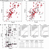
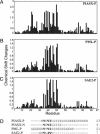
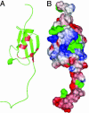
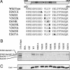
Posttranslational modification by the ubiquitin homologue, small ubiquitin-like modifier 1 (SUMO-1), has been established as an important regulatory mechanism. However, in most cases it is not clear how sumoylation regulates various cellular functions. Emerging evidence suggests that sumoylation may play a general role in regulating protein-protein interactions, as shown in RanBP2/Nup358 and RanGAP1 interaction. In this study, we have defined an amino acid sequence motif that binds SUMO. This motif, V/I-X-V/I-V/I, was identified by NMR spectroscopic characterization of interactions among SUMO-1 and peptides derived from proteins that are known to bind SUMO or sumoylated proteins. This motif binds all SUMO paralogues (SUMO-1-3). Using site-directed mutagenesis, we also show that this SUMO-binding motif in RanBP2/Nup358 is responsible for the interaction between RanBP2/Nup358 and sumoylated RanGAP1. The SUMO-binding motif exists in nearly all proteins known to be involved in SUMO-dependent processes, suggesting its general role in sumoylation-dependent cellular functions.
Figures


Similar articles
-
Insights into E3 ligase activity revealed by a SUMO-RanGAP1-Ubc9-Nup358 complex.Nature. 2005 Jun 2;435(7042):687-92. doi: 10.1038/nature03588. Nature. 2005. PMID: 15931224 Free PMC article.
-
Reconstitution of the Recombinant RanBP2 SUMO E3 Ligase Complex.Methods Mol Biol. 2016;1475:41-54. doi: 10.1007/978-1-4939-6358-4_3. Methods Mol Biol. 2016. PMID: 27631796
-
Protection from isopeptidase-mediated deconjugation regulates paralog-selective sumoylation of RanGAP1.Mol Cell. 2009 Mar 13;33(5):570-80. doi: 10.1016/j.molcel.2009.02.008. Mol Cell. 2009. PMID: 19285941 Free PMC article.
-
Identification of SUMO-binding motifs by NMR.Methods Mol Biol. 2009;497:121-38. doi: 10.1007/978-1-59745-566-4_8. Methods Mol Biol. 2009. PMID: 19107414 Free PMC article. Review.
-
Protein interactions in the sumoylation cascade: lessons from X-ray structures.FEBS J. 2008 Jun;275(12):3003-15. doi: 10.1111/j.1742-4658.2008.06459.x. Epub 2008 May 17. FEBS J. 2008. PMID: 18492068 Review.
Cited by
-
Crossing epigenetic frontiers: the intersection of novel histone modifications and diseases.Signal Transduct Target Ther. 2024 Sep 16;9(1):232. doi: 10.1038/s41392-024-01918-w. Signal Transduct Target Ther. 2024. PMID: 39278916 Free PMC article. Review.
-
Protein SUMOylation promotes cAMP-independent EPAC1 activation.Cell Mol Life Sci. 2024 Jul 4;81(1):283. doi: 10.1007/s00018-024-05315-y. Cell Mol Life Sci. 2024. PMID: 38963422 Free PMC article.
-
SUMOylation regulation of ribosome biogenesis: Emerging roles for USP36.Front RNA Res. 2024;2:1389104. doi: 10.3389/frnar.2024.1389104. Epub 2024 Apr 3. Front RNA Res. 2024. PMID: 38764604 Free PMC article.
-
Regulation of Enhancers by SUMOylation Through TFAP2C Binding and Recruitment of HDAC Complex to the Chromatin.Res Sq [Preprint]. 2024 Apr 2:rs.3.rs-4201913. doi: 10.21203/rs.3.rs-4201913/v1. Res Sq. 2024. PMID: 38645262 Free PMC article. Preprint.
-
Sumoylation of SAP130 regulates its interaction with FAF1 as well as its protein stability and transcriptional repressor function.BMC Mol Cell Biol. 2024 Jan 4;25(1):2. doi: 10.1186/s12860-023-00498-x. BMC Mol Cell Biol. 2024. PMID: 38172660 Free PMC article.
References
-
- Poukka, H., Aarnisalo, P., Karvonen, U., Palvimo, J. J. & Janne, O. A. (1999) J. Biol. Chem. 274, 19441-19446. - PubMed
-
- Muller, S., Ledl, A. & Schmidt, D. (2004) Oncogene 23, 1998-2008. - PubMed
-
- Seeler, J. S. & Dejean, A. (2003) Nat. Rev. Mol. Cell. Biol. 4, 690-699. - PubMed
-
- Kotaja, N., Karvonen, U., Janne, O. A. & Palvimo, J. J. (2002) J. Biol. Chem. 277, 30283-30288. - PubMed
-
- Chauchereau, A., Amazit, L., Quesne, M., Guiochon-Mantel, A. & Milgrom, E. (2003) J. Biol. Chem. 278, 12335-12343. - PubMed
Publication types
MeSH terms
Substances
Grants and funding
LinkOut - more resources
Full Text Sources
Other Literature Sources
Molecular Biology Databases
Miscellaneous

