. 2004 Jul 21;23(14):2789-99.
doi: 10.1038/sj.emboj.7600282. Epub 2004 Jul 1.

The Fos-related antigen Fra-1 is an activator of bone matrix formation

Affiliations

The Fos-related antigen Fra-1 is an activator of bone matrix formation

Robert Eferl et al. EMBO J. .

Abstract

Ectopic expression of the transcription factor Fra-1 in transgenic mice leads to osteosclerosis, a bone disorder characterized by increased bone mass. The molecular basis for this phenotype is unknown and Fra-1 functions cannot be studied by a conventional loss-of-function approach, since fra-1-knockout mice die in utero likely due to placental defects. Here we show that the lethality of fra-1-knockout mice can be rescued by specific deletion of Fra-1 only in the mouse embryo and not in the placenta. Mice lacking Fra-1 (fra-1(delta/delta)) are viable and develop osteopenia, a low bone mass disease. Long bones of fra-1(delta/delta) mice appear to have normal osteoclasts but express reduced amounts of bone matrix components produced by osteoblasts and chondrocytes such as osteocalcin, collagen1a2 and matrix Gla protein. The gene for matrix Gla protein seems to be a specific target of Fra-1 since its expression was markedly increased in the long bones of fra-1-transgenic mice. These results uncover a novel function of Fra-1 in regulating bone mass through bone matrix production by osteoblasts and chondrocytes.


Figures
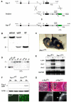
Figure 1
Figure 1
Strategy to generate mice with conditional alleles of fra-1. (A) Scheme of the gene targeting. Exons 3 and 4 of fra-1 were flanked by loxP sites (triangles). These exons contain the dimerization and the DNA-binding domains of Fra-1, and deletion of these exons renders Fra-1 inactive. A GFP reporter gene (GFP) is spliced to the residual N-terminal part of fra-1 upon deletion and is thereby activated. The relevant restriction sites used for Southern blot analysis and the location of the probe are indicated. fra-1: genomic locus of fra-1; vector: targeting vector for fra-1; fra-1f: floxed allele of fra-1; E1–4: exons 1–4 of fra-1; Neo: neomycin phosphotransferase gene; asterisk: splice acceptor of exon 2 of fra-1; DT-A: gene for diphtheria toxin; P1–3: PCR primers used for genotyping. (B) Southern blot analysis of offsprings demonstrating successful targeting of fra-1. For the Southern blot analysis, DNA was digested with HindIII/EcoRV and the bands were detected with the probe indicated in (A). wt: 12.5 kb wild-type allele; f: 4.5 kb floxed allele. (C) The embryonic lethality of fra-1-knockout mice is rescued by conditional deletion with the MORE-cre knock-in allele. The MORE-cre deletes the floxed fra-1 alleles in the whole mouse embryo but not in the placenta. MORE-cre fra-1f/f mice are therefore fra-1Δ/Δ mice that were viable and born at Mendelian frequency. (D) Deletion of fra-1 was confirmed in different tissues of 8-week-old mice by Southern blot analysis and in primary osteoblasts from newborn mice. li: liver; cal: calvaria; l.b.: long bones; b.m.: bone marrow; ob: osteoblasts differentiated in vitro; oc: osteoclasts differentiated in vitro; f: 4.5 kb floxed allele; Δ: 11.5 kb deleted allele. (E) RPA of c-jun, fra-1 and fra-2 in primary E12.5 MEFs. MEFs of the indicated genotypes were starved in 0.5% FCS (−) and then stimulated for 2 h with 10% FCS (+). Note that fra-1 mRNA is not detectable in stimulated fra-1Δ/Δ MEFs. The amount of GAPDH mRNA was used as a loading control. (F) A fusion protein between the residual N-terminal part of Fra-1 and GFP (43 kDa) is generated after deletion and was detected by Western blot analysis using an anti-GFP antibody (upper panel) and by direct immunofluorescence (lower panel). Note that only cells with at least one Δ allele contain the fusion protein. The amount of actin was used as a loading control in the Western blot analysis. (G) c-fosΔ/Δ mice (MORE-cre c-fosf/f) develop osteopetrosis as judged by H&E staining (upper panel, arrows) and X-ray analysis (lower panel, arrows) of long bones (6-week-old mice).
Figure 2
Figure 2
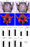
fra-1Δ/Δ mice develop osteopenia. (A–D) Decreased bone mass was detected in the vertebrae of fra-1Δ/Δ mice (3-month-old) by von Kossa staining (A, B) and μCT analysis (C, D). (E–M) Bone parameters measured by dynamic bone histomorphometry after calcein labeling demonstrated decreased bone mass (E), a decrease in absolute numbers of osteoblasts (F) and reduced trabecular thickness (G) in fra-1Δ/Δ mice, which is due to decreased bone formation (I). Numbers of trabeculae (H), osteoblasts per bone perimeter (J) and osteoclasts per bone perimeter (K) were not affected. The surface of osteoblasts (L) and osteoclasts (M) per bone surface was unchanged. BV/TV: trabecular bone volume per tissue volume; Ob.N/TV: osteoblast number per tissue volume; Tb/Th: trabecular thickness; Tb.N: trabecular number; BFR/BS: bone formation rate per bone surface; Ob.N/BPm: osteoblast number per bone perimeter; Oc.N/BPm: osteoclast number per bone perimeter; Ob.S/BS: osteoblast surface per bone surface; Oc.S/BS: osteoclast surface per bone surface. n=18 male mice; P<0.001.
Figure 3
Figure 3
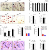
No overt osteoclast phenotype in fra-1Δ/Δ mice. (A, B) TRAP staining of osteoclasts (arrows) on the long bones of fra-1f/Δ and fra-1Δ/Δ mice (6-week-old) showed no major difference. (C) Real-time PCR analysis of mRNAs that are expressed in osteoclasts. Long bones of 6-week-old mice were used for RNA isolation (n=3). Relative expression levels normalized for tubulin with standard deviations indicated are shown. The broken line indicates the amount of bone mass reduction in the long bones of 6-week-old fra-1Δ/Δ mice as judged by bone histomorphometry. trap: tartrate-resistant acidic phosphatase; cathK: cathepsin K; ca II: carbonic anhydrase II; mmp-9: matrix metalloproteinase 9; rank: receptor and activator of NF-κB; tub: tubulin. (D, E) Osteoclast precursor cells derived from the bone marrow of adult mice were differentiated into mature multinucleated osteoclasts on plastic dishes and stained for the osteoclast marker protein TRAP (n=5). Differentiation of fra-1Δ/Δ precursors into osteoclasts was decreased on plastic dishes (E, F). (G) Plating of the precursors on bone slices rescued the osteoclast differentiation defect (n=5). (H) Addition of 2% mouse serum (MS) or 2% ApoE-Mgp serum (Mgp) rescued the osteoclast differentiation defect (n=4). A reciprocal co-culture assay on fra-1Δ/Δ osteoblasts (I) or fra-1-transgenic osteoblasts (J) could not rescue the osteoclast differentiation defect (n=4). Ob: osteoblast; BM: bone marrow; wt: wild type; Δ/Δ: fra-1Δ/Δ; tg: fra-1-transgenic. (K, L) Osteoclast precursor cells derived from the bone marrow of adult mice were differentiated into mature multinucleated osteoclasts on bone slices and the resorption pits (arrows) were stained with toluidine blue. (M) The resorption activity of fra-1Δ/Δ osteoclasts in vitro was analyzed by quantification of resorption pit areas on bone slices (n=5). (N) Deoxypyridinoline (DPD) crosslinks in the urine were measured as an osteoclast activity parameter in vivo. The values were normalized for muscle creatinine that is also excreted in the urine to account for urine concentration (n=9, 6-week-old mice).
Figure 4
Figure 4
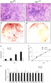
Formation of bone nodules is impaired in cultures of fra-1Δ/Δ osteoblasts. (A, B) Osteoblast precursor cells from the calvariae of newborn mice were differentiated for 21 days in vitro and stained for ALP activity (ALP-positive cells in blue). (C, D) Deposition of extracellular matrix material and bone nodule formation was analyzed by staining of osteoblast cultures (21 days) with alizarin red (bone nodules in red). (E) Time course of bone nodule formation by fra-1f/Δ and fra-1Δ/Δ osteoblasts (n=5). (F) Cumulative cell number (proliferation) of fra-1f/Δ and fra-1Δ/Δ osteoblasts. (G) Real-time PCR analysis of mRNAs for osteoblast differentiation factors and extracellular matrix components in osteoblast cultures (day 9 of differentiation). One representative example out of three independent primary culture experiments is shown. The relative expression levels were normalized for tubulin expression. alp: ALP, bsp: bone sialoprotein; oc: osteocalcin; rankl: receptor and activator of NF-κB; opg: osteoprotegerin; c1a1: collagen1a1; c1a2: collagen1a2; mgp: matrix Gla protein; bgn: biglycan; dec: decorin; tub: tubulin.
Figure 5
Figure 5
Reduced expression of bone matrix components in the long bones of fra-1Δ/Δ mice. (A, B) Real-time PCR analysis of mRNAs for osteoblast differentiation and activity factors (A) and bone matrix genes (B) in long bones (n=3, 6-week-old mice). The relative expression levels were normalized for tubulin expression with the standard deviations indicated. The broken line indicates the amount of bone mass reduction (and reduction in osteoblast numbers) in the long bones of 6-week-old fra-1Δ/Δ mice. Therefore, only the expression levels of osteocalcin and collagen1a2 are reduced in fra-1Δ/Δ osteoblasts. The reduced expression of matrix Gla protein needs no correction for reduced osteoblast numbers since it is mainly expressed in chondrocytes. alp: ALP, bsp: bone sialoprotein; oc: osteocalcin; rankl: receptor and activator of NF-κB; opg: osteoprotegerin; mt-1: membrane-bound matrix metalloproteinase mt-1-mmp; irs1,2: insulin receptor substrate 1,2; tub: tubulin; c1a1: collagen1a1; c1a2: collagen1a2; mgp: matrix Gla protein; bgn: biglycan; dec: decorin; tub: tubulin. (C–J) In situ hybridization experiments for expression of runx2 (C, D), osteocalcin (E, F), collagen1a2 (G, H), and matrix Gla protein (I, J, arrows show expression in hypertrophic chondrocytes) on long bones (n=3, 6-week-old mice).
Figure 6
Figure 6
Increased expression of bone matrix components in the long bones of fra-1-transgenic (fra-1tg) mice. (A) Northern blot analysis of mgp expression in the long bones of fra-1f/Δ, fra-1Δ/Δ and fra-1tg mice. The corresponding littermate controls for the fra-1tg mice expressed mgp at similar levels compared with fra-1f/Δ mice (not shown). Expression of tubulin was used as loading control. (B) In situ hybridization for mgp in the long bones of fra-1+/+ (wild type) and fra-1tg mice. Note the increased blue staining indicative for increased mgp expression in hypertrophic chondrocytes (arrows) of fra-1tg mice. (C) Real-time PCR analysis of mRNAs for osteoblast differentiation markers and genes for bone matrix components in fra-1+/+ and fra-1tg mice (n=3, 3-week-old fra-1tg mice were used before first histological signs of increased bone mass became apparent). The relative expression levels were normalized for tubulin expression and standard deviations are indicated. oc: osteocalcin; rankl: receptor and activator of NF-κB; opg: osteoprotegerin; c1a1: collagen1a1; c1a2: collagen1a2; mgp: matrix Gla protein; bgn: biglycan; dec: decorin; tub: tubulin.

Similar articles

Cited by

References

    1. Akune T, Ogata N, Hoshi K, Kubota N, Terauchi Y, Tobe K, Takagi H, Azuma Y, Kadowaki T, Nakamura K, Kawaguchi H (2002) Insulin receptor substrate-2 maintains predominance of anabolic function over catabolic function of osteoblasts. J Cell Biol 159: 147–156 - PMC - PubMed
    1. Amling M, Priemel M, Holzmann T, Chapin K, Rueger JM, Baron R, Demay MB (1999) Rescue of the skeletal phenotype of vitamin D receptor-ablated mice in the setting of normal mineral ion homeostasis: formal histomorphometric and biomechanical analyses. Endocrinology 140: 4982–4987 - PubMed
    1. Behrens A, Haigh J, Mechta-Grigoriou F, Nagy A, Yaniv M, Wagner EF (2003) Impaired intervertebral disc formation in the absence of Jun. Development 130: 103–109 - PubMed
    1. Boyle WJ, Simonet WS, Lacey DL (2003) Osteoclast differentiation and activation. Nature 423: 337–342 - PubMed
    1. Chung KY, Agarwal A, Uitto J, Mauviel A (1996) An AP-1 binding sequence is essential for regulation of the human alpha2(I) collagen (COL1A2) promoter activity by transforming growth factor-beta. J Biol Chem 271: 3272–3278 - PubMed

Publication types

MeSH terms

LinkOut - more resources