Acute infection with Epstein-Barr virus targets and overwhelms the peripheral memory B-cell compartment with resting, latently infected cells
- PMID: 15113901
- PMCID: PMC400374
- DOI: 10.1128/jvi.78.10.5194-5204.2004
Acute infection with Epstein-Barr virus targets and overwhelms the peripheral memory B-cell compartment with resting, latently infected cells
Abstract
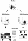
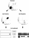
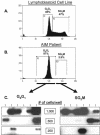
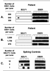
In this paper we demonstrate that during acute infection with Epstein-Barr virus (EBV), the peripheral blood fills up with latently infected, resting memory B cells to the point where up to 50% of all the memory cells may carry EBV. Despite this massive invasion of the memory compartment, the virus remains tightly restricted to memory cells, such that, in one donor, fewer than 1 in 10(4) infected cells were found in the naive compartment. We conclude that, even during acute infection, EBV persistence is tightly regulated. This result confirms the prediction that during the early phase of infection, before cellular immunity is effective, there is nothing to prevent amplification of the viral cycle of infection, differentiation, and reactivation, causing the peripheral memory compartment to fill up with latently infected cells. Subsequently, there is a rapid decline in infected cells for the first few weeks that approximates the decay in the cytotoxic-T-cell responses to viral replicative antigens. This phase is followed by a slower decline that, even by 1 year, had not reached a steady state. Therefore, EBV may approach but never reach a stable equilibrium.
Figures


Similar articles
-
Morphology, immunophenotype, and distribution of latently and/or productively Epstein-Barr virus-infected cells in acute infectious mononucleosis: implications for the interindividual infection route of Epstein-Barr virus.Blood. 1995 Feb 1;85(3):744-50. Blood. 1995. PMID: 7530505
-
EBV-infected B cells in infectious mononucleosis: viral strategies for spreading in the B cell compartment and establishing latency.Immunity. 2000 Oct;13(4):485-95. doi: 10.1016/s1074-7613(00)00048-0. Immunity. 2000. PMID: 11070167
-
Peripheral B cells latently infected with Epstein-Barr virus display molecular hallmarks of classical antigen-selected memory B cells.Proc Natl Acad Sci U S A. 2005 Dec 13;102(50):18093-8. doi: 10.1073/pnas.0509311102. Epub 2005 Dec 5. Proc Natl Acad Sci U S A. 2005. PMID: 16330748 Free PMC article.
-
Regulation and dysregulation of Epstein-Barr virus latency: implications for the development of autoimmune diseases.Autoimmunity. 2008 May;41(4):298-328. doi: 10.1080/08916930802024772. Autoimmunity. 2008. PMID: 18432410 Review.
-
Cell-mediated immunity against Epstein-Barr virus infected B lymphocytes.Springer Semin Immunopathol. 1982;5(1):63-73. doi: 10.1007/BF00201957. Springer Semin Immunopathol. 1982. PMID: 6314570 Review. No abstract available.
Cited by
-
CD8+ T-Cell Deficiency, Epstein-Barr Virus Infection, Vitamin D Deficiency, and Steps to Autoimmunity: A Unifying Hypothesis.Autoimmune Dis. 2012;2012:189096. doi: 10.1155/2012/189096. Epub 2012 Jan 24. Autoimmune Dis. 2012. PMID: 22312480 Free PMC article.
-
EBNA1 Inhibitors Block Proliferation of Spontaneous Lymphoblastoid Cell Lines From Patients With Multiple Sclerosis and Healthy Controls.Neurol Neuroimmunol Neuroinflamm. 2023 Aug 10;10(5):e200149. doi: 10.1212/NXI.0000000000200149. Print 2023 Sep. Neurol Neuroimmunol Neuroinflamm. 2023. PMID: 37562974 Free PMC article.
-
Early age at time of primary Epstein-Barr virus infection results in poorly controlled viral infection in infants from Western Kenya: clues to the etiology of endemic Burkitt lymphoma.J Infect Dis. 2012 Mar 15;205(6):906-13. doi: 10.1093/infdis/jir872. Epub 2012 Feb 1. J Infect Dis. 2012. PMID: 22301635 Free PMC article.
-
Persistence of Epstein-Barr virus in self-reactive memory B cells.J Virol. 2012 Nov;86(22):12330-40. doi: 10.1128/JVI.01699-12. Epub 2012 Sep 5. J Virol. 2012. PMID: 22951828 Free PMC article.
-
Epstein-Barr virus but not cytomegalovirus is associated with reduced vaccine antibody responses in Gambian infants.PLoS One. 2010 Nov 17;5(11):e14013. doi: 10.1371/journal.pone.0014013. PLoS One. 2010. PMID: 21103338 Free PMC article.
References
-
- Babcock, G. J., L. L. Decker, M. Volk, and D. A. Thorley-Lawson. 1998. EBV persistence in memory B cells in vivo. Immunity 9:395-404. - PubMed
-
- Babcock, G. J., D. Hochberg, and A. D. Thorley-Lawson. 2000. The expression pattern of Epstein-Barr virus latent genes in vivo is dependent upon the differentiation stage of the infected B cell. Immunity 13:497-506. - PubMed
Publication types
MeSH terms
Grants and funding
LinkOut - more resources
Full Text Sources
Other Literature Sources
Medical

