. 2004 Apr;15(4):1609-22.
doi: 10.1091/mbc.e03-11-0837. Epub 2004 Jan 23.

Interdependency of fission yeast Alp14/TOG and coiled coil protein Alp7 in microtubule localization and bipolar spindle formation

Affiliations

Interdependency of fission yeast Alp14/TOG and coiled coil protein Alp7 in microtubule localization and bipolar spindle formation

Masamitsu Sato et al. Mol Biol Cell. 2004 Apr.

Abstract

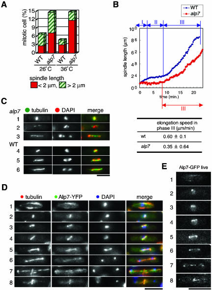
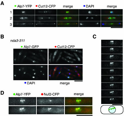
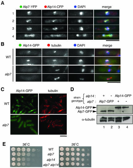
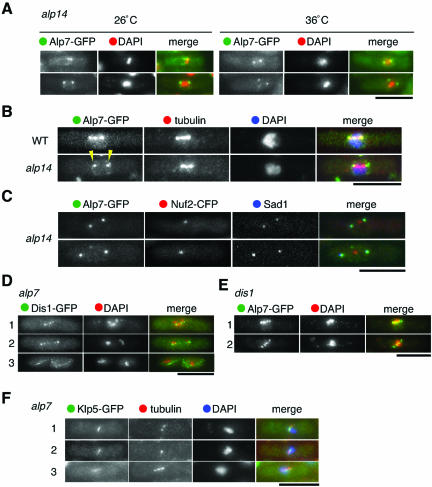
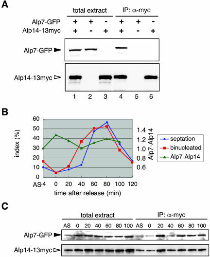
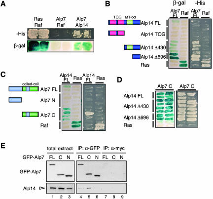
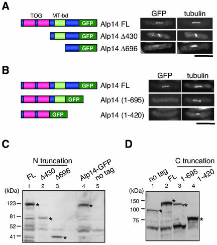
The Dis1/TOG family plays a pivotal role in microtubule organization. In fission yeast, Alp14 and Dis1 share an essential function in bipolar spindle formation. Here, we characterize Alp7, a novel coiled-coil protein that is required for organization of bipolar spindles. Both Alp7 and Alp14 colocalize to the spindle pole body (SPB) and mitotic spindles. Alp14 localization to these sites is fully dependent upon Alp7. Conversely, in the absence of Alp14, Alp7 localizes to the SPBs, but not mitotic spindles. Alp7 forms a complex with Alp14, where the C-terminal region of Alp14 interacts with the coiled-coil domain of Alp7. Intriguingly, this Alp14 C terminus is necessary and sufficient for mitotic spindle localization. Overproduction of either full-length or coiled-coil region of Alp7 results in abnormal V-shaped spindles and stabilization of interphase microtubules, which is induced independent of Alp14. Alp7 may be a functional homologue of animal TACC. Our results shed light on an interdependent relationship between Alp14/TOG and Alp7. We propose a two-step model that accounts for the recruitment of Alp7 and Alp14 to the SPB and microtubules.


Figures

Figure 1.
Figure 1.
Alp7 is a coiled-coil protein that is required for spindle organization and localizes to microtubules. (A) Frequency of mitotic cells. Wild-type and alp7 mutant cells grown at 26 or 36°C (4 h) were fixed and processed for immunofluorescence microscopy with anti-α-tubulin antibody. Frequency of cells with mitotic spindles was counted (at least 200 cells per each sample). (B) Visualization of spindle dynamics in wild-type and alp7 mutants. Strains (wild-type in blue line and alp7 in red line) containing plasmids carrying nmt1-GFP-atb2+ (encoding α2-tubulin) were grown at room temperature (25°C) in the presence of thiamine, and time-lapse live analysis of mitotic spindles was performed. The velocity (μm/min) of spindle elongation at phase 3 is shown with standard deviations. Ten independent samples were observed with live imaging analysis in each strain. (C) Abnormal spindles in alp7 mutants. Representative images displaying mitotic spindles in alp7 mutants (rows 1–3) and wild-type cells (rows 4–6) incubated at 36°C were shown together with DAPI staining. Merged images in which tubulin (green) and DAPI (red) are shown in the right-hand panels. (D) Cellular localization of Alp7. A strain containing Alp7-YFP was grown, fixed with methanol, and processed for immunofluorescence microscopy. Merged images (red for tubulin, green for Alp7-YFP and blue for DAPI) are also shown. Representative images that exhibit Alp7 localization during the cell cycle are presented (1, interphase; 2 and 3, early mitosis; 4, mid-mitosis; 5–7, late mitosis; and 8, postanaphase). (E) Alp7-GFP localization in live cells. A strain containing Alp7-GFP was grown in liquid rich medium at 26°C, and pictures were taken from unfixed live samples. Alp7-GFP images corresponding to stages shown in D (rows 1–8) are shown. Bar, 10 μm.
Figure 2.
Figure 2.
Mitotic localization of Alp7 to the SPB and kinetochore periphery. (A) Colocalization between Alp7-YFP and Cut12-CFP. A doubly tagged strain was fixed with formaldehyde and signals were observed under fluorescence microscopy. Merged images (green for Alp7-YFP, red for Cut12-CFP and blue for DAPI) are also shown. (B) Localization of Alp7 to the mitotic SPB. An nda3-311 strain containing Alp7-GFP and Cut12-CFP was incubated at 20°C for 6 h and fixed with formaldehyde. (C) Time-lapse live analysis of mitotic localization of Alp7-GFP. Alp7 localization during mid-mitosis is depicted in the bottom row. (D) Overlapping localization between Alp7 and a kinetochore marker in mitotic cells. A strain containing Alp7-YFP and Nuf2-CFP was grown, fixed with formaldehyde, and mitotic cells were observed with YFP (green) and CFP (red). Bar, 10 μm.
Figure 3.
Figure 3.
Alp7 colocalizes with Alp14 and is required for the specific localization of Alp14 to the microtubules. (A) Colocalization of Alp7 and Alp14. A strain containing Alp7-YFP and Alp14-CFP was grown, fixed, and observed with YFP, cyan fluorescent protein (CFP), and DAPI staining. Representative pictures of mitotic cells are shown (1; early mitosis; 2 and 3, mid-mitosis; and 4, late mitosis). Merged images for Alp7-YFP (green), Alp14-CFP (red), and DAPI (blue) are shown in the right-hand panels. (B and C) Requirement of Alp7 for Alp14 localization to the microtubules. Wild-type (top rows) or alp7-deleted cells (bottom rows) containing Alp14-GFP was cultured at 26°C, fixed, and processed for immunofluorescence microscopy. In the far-right panels in B, merged images (Alp14-GFP in green, tubulin in red, and DAPI in blue) are shown. Bar, 10 μm. (D) Protein levels of Alp7 and Alp14. Immunoblot against total cell extracts prepared from strains containing Alp7-GFP (lane 1), Alp7-GFP in alp14 mutants (lane 2), Alp14-GFP (lane 3), and Alp14-GFP in alp7 mutants (lane 4) was performed with anti-GFP and anti-α-tubulin antibodies. (E) Nonadditive ts phenotype of alp7alp14 double mutants. Wild-type (top row), alp7-deleted (second), alp14-deleted (third), or alp7alp14-deleted cells (bottom) were spotted on rich plates (106 cells in the far-left spots for each plate and then diluted 10-fold in each subsequent spot rightwards) at 36°C (left) or 26°C (right) and incubated for 3 d.
Figure 4.
Figure 4.
Alp7 localizes to only the SPBs, but not the spindles, in the absence of Alp14, and the requirement of Alp7 for Alp14 localization is specific. (A) Alp7 localization to the SPB in alp14 mutants. A strain containing Alp7-GFP in alp14 mutants was constructed, and GFP signals were observed at 26°C (left) or at 36°C (right). Merged images (green for Alp7-GFP and red for DAPI) are also shown. (B and C) Lack of spindle localization of Alp7 in alp14 mutants. Wild-type and alp14 mutant cells containing Alp7-GFP (B) or Alp14-GFP and Nuf2-CFP (C) were grown at 26°C, fixed, and processed for immunofluorescence microscopy. Merged images are shown with green for Alp7-GFP, red for tubulin, and blue for DAPI (B) or blue for anti-Sad1 (C). Alp7 localization to the two SPBs was marked with arrowheads in B. (D) Dis1 localization in the absence of Alp7. Mitotic cells (rows 1 and 2) and interphase cell (row 3) are shown. Merged images are shown with green in Dis1-GFP and red in DAPI. (E) Alp7 localization in the absence of Dis1. dis1 mutants containing Alp7-GFP were grown at 32°C. Alp7-GFP localization in mitotic cells is shown. Merged images are shown as in A. (F) Klp5 localization in alp7 mutants. Merged images are shown with green in Klp5-GFP, red in tubulin, and blue in DAPI. Bar, 10 μm.
Figure 5.
Figure 5.
Physical interaction between Alp7 and Alp14 during the cell cycle. (A) Coimmunoprecipitation between Alp7 and Alp14. Total cell extracts were prepared from exponentially growing strains containing Alp7-GFP and Alp14–13myc (lane 1 and 4), Alp7-GFP (lanes 2 and 5), or Alp14–13myc (lanes 3 and 6), and immunoprecipitation was performed. Immunoprecipitates were then blotted with mouse monoclonal anti-GFP (top lanes 4–6) or anti-myc antibody (bottom lanes 4–6). Cell extracts used for immunoprecipitation were also run (50 μg, lanes 1–3). (B) A cdc25-22 strain containing Alp7-GFP and Alp14–13myc was shifted to 36°C for 4 h and 15 min (0 min) and then shifted down to 26°C. Samples were taken every 20-min interval for immunoprecipitation and staining with Calcofluor and DAPI. The percentage of septated cells (blue circles) and binucleated cells (red squares) is plotted at each time point. Also shown is quantification of Alp7–Alp14 complex levels (triangles in green). Image J (National Institutes of Health) was used to compare the level of the Alp7–Alp14 complex at each time point. The ratio of precipitated Alp7 and Alp14 was normalized. (C) Coimmunoprecipitation of Alp7 and Alp14 upon cdc25-22 arrest-release. AS means asynchronous culture at 26°C before shift up.
Figure 6.
Figure 6.
Identification of interacting domains of Alp7 and Alp14. (A) Two-hybrid assay. Budding yeast transformants expressing the indicated gene products were streaked on minimal medium lacking histidine (-His) or plates containing X-Gal (β-gal). As a positive control, plasmids expressing Ras and Raf were used. (B) Two-hybrid interaction between full-length Alp7 and various region of Alp14. Schematically shown (left) are regions subcloned (FL, full-length). Full-length Alp7 was used as bait. (C) Interaction between full-length Alp14 and N-terminal or C-terminal half of Alp7. (D) Interaction between subregions of Alp7 and Alp14. (E) Interaction between Alp14 and the C-terminal Alp7 in fission yeast cells. Immunoprecipitation was performed in three different strains, in which full-length (lanes 1, 4, and 7), the C-terminal region (219–474, lanes 2, 5, and 8), and the N-terminal region of Alp7 (1–218, lanes 3, 6, and 9) were expressed with GFP. Cells were grown in rich medium and immunoprecipitation was performed with anti-GFP (lanes 4–6) or anti-myc antibody (lanes 7–9), followed by immunoblotting with anti-GFP (top) or anti-Alp14 antibody (bottom).
Figure 8
Figure 8
Defects in microtubule organization by induced overproduction of Alp7. (A) Schematic representation of strains constructed to dissect functional domains of Alp7. The nmtP3-GFP cassette (Bähler et al., 1998) was integrated in the indicated position of the alp7 locus. (B) Expression of GFP-Alp7 proteins. Strains shown in A (FL, Alp7FL; C, Alp7C; and N, Alp7N) were grown in the presence (+) or absence (-) of thiamine and cell extracts were prepared. Immunoblotting was performed with anti-GFP antibody. Anti-α-tubulin antibody was used as a loading control. (C) Localization of the N-terminal and C-terminal Alp7. Strains containing GFP-tagged Alp7 (full-length, the C-terminal or N-terminal region) were grown in the presence of thiamine (repressed) and GFP signals were observed in individual cells. (D) Toxicity of Alp7 overproduction. Strains containing individual GFP-tagged Alp7 constructs were grown and spotted on rich plates (left) or minimal medium lacking thiamine (right, 106 cells in the far-left spots for each plate and then diluted 10-fold in each subsequent spots rightwards) at 27°C and incubated for 4 d. (E) Structure and organization of microtubules and the SPB in Alp7 overproducing cells. Cells expressing nmtP3-GFP-alp7+FL (rows 1–4) or nmtP3-GFP-alp7C (rows 5 and 6), or alp14-deleted cells containing nmtP3-GFP-alp7+FL (rows 7 and 8) were grown in the absence of thiamine at 27°C for 18 h, and immunofluorescence microscopy was performed with anti-tubulin and anti-Sad1 antibodies. Merged images are shown in the right-hand side (anti-Sad1, green; anti-tubulin, red; and DAPI, blue). Bar, 10 μm.
Figure 7.
Figure 7.
Determination of the minimal region of Alp14 required for in vivo localization to the spindle. (A) N-terminal truncation. Various subclones (depicted in the left-hand side) are constructed with C-terminally tagged GFP (integrated in the chromosome with nmt81 promoter) were tested for mitotic spindles in vivo. GFP signals were observed under derepressed conditions. Bar, 10 μm. (B) C-terminal truncation. GFP was integrated at various positions (left) and GFP signals were observed (right). (C and D) Immunoblotting with anti-GFP (C) or anti-Alp14 antibody (D) against cell extracts used in A (C, N-terminal truncation) and B (D, C-terminal truncation). Bands corresponding to Alp14 or its derivatives in each construct are marked with asterisks.
Figure 9.
Figure 9.
Proposed manner of Alp7-TAC and Alp14-TOG localization to the SPB and microtubules and how this complex regulates microtubule organization. A model depicts a functional dependency between Alp7 and Alp14. Alp7 and Alp14 form a complex, by which Alp7 targets this complex to the SPB. Once localizing to the SPB, Alp14 is responsible for localizing to mitotic spindles and kinetochores. The short C-terminal domain of Alp14 (C in yellow) and the coiled-coil region of Alp7 (C in green) bind each other and are responsible for the specific localization.

Similar articles

Cited by

References

    1. Bähler, J., Wu, J., Longtine, M.S., Shah, N.G., McKenzie III, A., Steever, A.B., Wach, A., Philippsen, P., and Pringle, J.R. (1998). Heterologous modules for efficient and versatile PCR-based gene targeting in Schizosaccharomyces pombe. Yeast 14, 943-951. - PubMed
    1. Bellanger, J.-M., and Gönczy, P. (2003). TAC-1 and ZYG-9 form a complex that promotes microtubule assembly in C. elegans embryos. Curr. Biol. 13, 1488-1498. - PubMed
    1. Bridge, A.J., Morphew, M., Bartlett, R., and Hagan, I.M. (1998). The fission yeast SPB component Cut12 links bipolar spindle formation to mitotic control. Genes Dev. 12, 927-942. - PMC - PubMed
    1. Chen, X.P., Yin, H., and Huffaker, T.C. (1998). The yeast spindle pole body component Spc72p interacts with Stu2p and is required for proper microtubule assembly. J. Cell Biol. 141, 1169-1179. - PMC - PubMed
    1. Cullen, C.F., and Ohkura, H. (2001). Msps protein is localized to acentrosomal poles to ensure bipolarity of Drosophila meiotic spindles. Nat. Cell Biol. 3, 637-642. - PubMed

Publication types

MeSH terms

Substances