A small-molecule antagonist of CXCR4 inhibits intracranial growth of primary brain tumors
- PMID: 14595012
- PMCID: PMC263845
- DOI: 10.1073/pnas.2235846100
A small-molecule antagonist of CXCR4 inhibits intracranial growth of primary brain tumors
Abstract
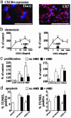
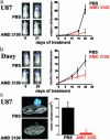
The vast majority of brain tumors in adults exhibit glial characteristics. Brain tumors in children are diverse: Many have neuronal characteristics, whereas others have glial features. Here we show that activation of the Gi protein-coupled receptor CXCR4 is critical for the growth of both malignant neuronal and glial tumors. Systemic administration of CXCR4 antagonist AMD 3100 inhibits growth of intracranial glioblastoma and medulloblastoma xenografts by increasing apoptosis and decreasing the proliferation of tumor cells. This reflects the ability of AMD 3100 to reduce the activation of extracellular signal-regulated kinases 1 and 2 and Akt, all of which are pathways downstream of CXCR4 that promote survival, proliferation, and migration. These studies (i) demonstrate that CXCR4 is critical to the progression of diverse brain malignances and (ii) provide a scientific rationale for clinical evaluation of AMD 3100 in treating both adults and children with malignant brain tumors.
Figures


Similar articles
-
Multiple actions of the chemokine CXCL12 on epithelial tumor cells in human ovarian cancer.Cancer Res. 2002 Oct 15;62(20):5930-8. Cancer Res. 2002. PMID: 12384559
-
Stromal cell-derived factor 1alpha stimulates human glioblastoma cell growth through the activation of both extracellular signal-regulated kinases 1/2 and Akt.Cancer Res. 2003 Apr 15;63(8):1969-74. Cancer Res. 2003. PMID: 12702590
-
G protein-coupled chemokine receptors induce both survival and apoptotic signaling pathways.J Immunol. 2002 Nov 15;169(10):5546-54. doi: 10.4049/jimmunol.169.10.5546. J Immunol. 2002. PMID: 12421931
-
VEGFR inhibitors upregulate CXCR4 in VEGF receptor-expressing glioblastoma in a TGFβR signaling-dependent manner.Cancer Lett. 2015 Apr 28;360(1):60-7. doi: 10.1016/j.canlet.2015.02.005. Epub 2015 Feb 9. Cancer Lett. 2015. PMID: 25676691 Free PMC article.
-
CXCL12/CXCR4 signaling in malignant brain tumors: a potential pharmacological therapeutic target.Brain Tumor Pathol. 2011 Apr;28(2):89-97. doi: 10.1007/s10014-010-0013-1. Epub 2011 Jan 6. Brain Tumor Pathol. 2011. PMID: 21210239 Review.
Cited by
-
Survival and Proliferation of Neural Progenitor-Derived Glioblastomas Under Hypoxic Stress is Controlled by a CXCL12/CXCR4 Autocrine-Positive Feedback Mechanism.Clin Cancer Res. 2017 Mar 1;23(5):1250-1262. doi: 10.1158/1078-0432.CCR-15-2888. Epub 2016 Aug 19. Clin Cancer Res. 2017. PMID: 27542769 Free PMC article.
-
Al[18F]NOTA-T140 Peptide for Noninvasive Visualization of CXCR4 Expression.Mol Imaging Biol. 2016 Feb;18(1):135-42. doi: 10.1007/s11307-015-0872-2. Mol Imaging Biol. 2016. PMID: 26126597 Free PMC article.
-
Identification of anti-malarial compounds as novel antagonists to chemokine receptor CXCR4 in pancreatic cancer cells.PLoS One. 2012;7(2):e31004. doi: 10.1371/journal.pone.0031004. Epub 2012 Feb 3. PLoS One. 2012. PMID: 22319600 Free PMC article.
-
The mesenchymal tumor microenvironment: a drug-resistant niche.Cell Adh Migr. 2012 May-Jun;6(3):285-96. doi: 10.4161/cam.20210. Epub 2012 May 1. Cell Adh Migr. 2012. PMID: 22568991 Free PMC article. Review.
-
The SDF-1-CXCR4 signaling pathway: a molecular hub modulating neo-angiogenesis.Trends Immunol. 2007 Jul;28(7):299-307. doi: 10.1016/j.it.2007.05.007. Epub 2007 Jun 7. Trends Immunol. 2007. PMID: 17560169 Free PMC article.
References
-
- Ries, L. A. G., Eisner, M. P., Kosary, C. L., Hankey, B. F., Miller, B. A., Clegg, L. & Edwards, B. K., eds. (2001) SEER Cancer Statistics Review (Natl. Cancer Inst., Bethesda).
-
- Kleihues, P., Burger, P. C., Collins, V. P., Newcomb, E. W., Ohgaki, H. & Cavenee, W. K. (2000) in World Health Organization Classification of Tumours: Pathology and Genetics of Tumours of the Central Nervous System, eds. Kleihues, P. C. & Cavenee, W. K. (Int. Agency for Res. on Cancer, Lyon, France), pp. 29-39.
-
- Giangaspero, F., Bigner, S. H., Kleihues, P., Pietsch, T. & Trojanowski, J. Q. (2000) in World Health Organization Classification of Tumours: Pathology and Genetics of Tumours of the Central Nervous System, eds. Kleihues, P. C. & Cavenee, W. K. (Int. Agency for Res. on Cancer, Lyon, France), pp. 129-137.
-
- Luster, A. D. (1998) N. Engl. J. Med. 338, 436-445. - PubMed
Publication types
MeSH terms
Substances
LinkOut - more resources
Full Text Sources
Other Literature Sources
Medical
Miscellaneous

