Functional genomic analysis of remyelination reveals importance of inflammation in oligodendrocyte regeneration
- PMID: 14586011
- PMCID: PMC6740899
- DOI: 10.1523/JNEUROSCI.23-30-09824.2003
Functional genomic analysis of remyelination reveals importance of inflammation in oligodendrocyte regeneration
Abstract
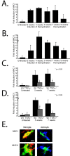
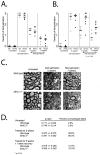
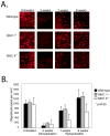
Tumor necrosis factor alpha (TNFalpha), a proinflammatory cytokine, was shown previously to promote remyelination and oligodendrocyte precursor proliferation in a murine model for demyelination and remyelination. We used Affymetrix microarrays in this study to identify (1) changes in gene expression that accompany demyelination versus remyelination and (2) changes in gene expression during the successful remyelination of wild-type mice versus the unsuccessful attempts in mice lacking TNFalpha. Alterations in inflammatory genes represented the most prominent changes, with major histocompatibility complex (MHC) genes dramatically enhanced in microglia and astrocytes during demyelination, remyelination, and as a consequence of TNFalpha stimulation. Studies to examine the roles of these genes in remyelination were then performed using mice lacking specific genes identified by the microarray. Analysis of MHC-II-null mice showed delayed remyelination and regeneration of oligodendrocytes, whereas removal of MHC-I had little effect. These data point to the induction of MHC-II by TNFalpha as an important regulatory event in remyelination and emphasize the active inflammatory response in regeneration after pathology in the brain.
Figures


Similar articles
-
The Role of Galectin-3: From Oligodendroglial Differentiation and Myelination to Demyelination and Remyelination Processes in a Cuprizone-Induced Demyelination Model.Adv Exp Med Biol. 2016;949:311-332. doi: 10.1007/978-3-319-40764-7_15. Adv Exp Med Biol. 2016. PMID: 27714696
-
Deletion of Voltage-Gated Calcium Channels in Astrocytes during Demyelination Reduces Brain Inflammation and Promotes Myelin Regeneration in Mice.J Neurosci. 2020 Apr 22;40(17):3332-3347. doi: 10.1523/JNEUROSCI.1644-19.2020. Epub 2020 Mar 13. J Neurosci. 2020. PMID: 32169969 Free PMC article.
-
Astroglial-derived lymphotoxin-alpha exacerbates inflammation and demyelination, but not remyelination.Glia. 2005 Jan 1;49(1):1-14. doi: 10.1002/glia.20089. Glia. 2005. PMID: 15382206
-
The mechanistic target of rapamycin as a regulator of metabolic function in oligodendroglia during remyelination.Curr Opin Pharmacol. 2022 Apr;63:102193. doi: 10.1016/j.coph.2022.102193. Epub 2022 Mar 1. Curr Opin Pharmacol. 2022. PMID: 35245799 Free PMC article. Review.
-
Possibilities and results in the wide-scale genomic analysis of inflammation. Looking for the child among many midwives.Inflamm Res. 2004 Oct;53(10):517-22. doi: 10.1007/s00011-004-1293-x. Inflamm Res. 2004. PMID: 15597145 Review.
Cited by
-
Astrocytes: Lessons Learned from the Cuprizone Model.Int J Mol Sci. 2023 Nov 16;24(22):16420. doi: 10.3390/ijms242216420. Int J Mol Sci. 2023. PMID: 38003609 Free PMC article. Review.
-
Cuprizone-induced demyelination as a tool to study remyelination and axonal protection.J Mol Neurosci. 2013 Oct;51(2):567-72. doi: 10.1007/s12031-013-0026-4. Epub 2013 May 12. J Mol Neurosci. 2013. PMID: 23666824 Review.
-
Comparative prion disease gene expression profiling using the prion disease mimetic, cuprizone.Prion. 2009 Apr-Jun;3(2):99-109. doi: 10.4161/pri.3.2.9059. Epub 2009 Apr 19. Prion. 2009. PMID: 19535908 Free PMC article.
-
Interferon-triggered transcriptional cascades in the oligodendroglial lineage: a comparison of induction of MHC class II antigen between oligodendroglial progenitor cells and mature oligodendrocytes.J Neuroimmunol. 2009 Jul 25;212(1-2):53-64. doi: 10.1016/j.jneuroim.2009.04.021. Epub 2009 May 20. J Neuroimmunol. 2009. PMID: 19467717 Free PMC article.
-
Heterozygous carriers of galactocerebrosidase mutations that cause Krabbe disease have impaired microglial function and defective repair of myelin damage.Neural Regen Res. 2018 Mar;13(3):393-401. doi: 10.4103/1673-5374.228712. Neural Regen Res. 2018. PMID: 29623914 Free PMC article. Review.
References
-
- Arnett HA, Mason J, Marino M, Suzuki K, Matsushima GK, Ting JP ( 2001) TNF alpha promotes proliferation of oligodendrocyte progenitors and remyelination. Nat Neurosci 4: 1116–1122. - PubMed
-
- Arnett HA, Hellendall RP, Matsushima GK, Suzuki K, Laubach VE, Sherman P, Ting JP ( 2002) The protective role of nitric oxide in a neurotoxicant-induced demyelinating model. J Immunol 168: 427–433. - PubMed
-
- Brosnan CF, Raine CS ( 1996) Mechanisms of immune injury in multiple sclerosis. Brain Pathol 6: 243–257. - PubMed
Publication types
MeSH terms
Substances
Grants and funding
LinkOut - more resources
Full Text Sources
Other Literature Sources
Molecular Biology Databases
Research Materials
