. 2003 Nov;23(21):7488-97.
doi: 10.1128/MCB.23.21.7488-7497.2003.

Chk1 kinase negatively regulates mitotic function of Cdc25A phosphatase through 14-3-3 binding

Affiliations

Chk1 kinase negatively regulates mitotic function of Cdc25A phosphatase through 14-3-3 binding

Mei-Shya Chen et al. Mol Cell Biol. 2003 Nov.

Abstract

The order and fidelity of cell cycle events in mammals is intimately linked to the integrity of the Chk1 kinase-Cdc25A phosphatase pathway. Chk1 phosphorylation targets Cdc25A for destruction and, as shown here, inhibits interactions between Cdc25A and its mitotic substrate cyclin B1-Cdk1. Phosphorylation of Cdc25A on serine 178 and threonine 507 facilitates 14-3-3 binding, and Chk1 phosphorylates both residues in vitro. Mutation of T507 to alanine (T507A) enhanced the biological activity of Cdc25A. Cdc25A(T507A) was more efficient in binding to cyclin B1, activating cyclin B1-Cdk1, and promoting premature entry into mitosis. We propose that the Chk1/Cdc25A/14-3-3 pathway functions to prevent cells from entering into mitosis prior to replicating their genomes to ensure the fidelity of the cell division process.


Figures
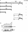
FIG. 1.
FIG. 1.
Dependency of 14-3-3 binding to Cdc25A on S178 and T507. (A) Cosm6 cells transfected with vector alone (mock) or with plasmids encoding the indicated forms of Myc-Cdc25A were lysed and incubated with anti-c-Myc-agarose. Precipitates were resolved by SDS-PAGE and monitored for Cdc25A and 14-3-3 proteins by immunoblotting. +, coprecipitation of 14-3-3 with Cdc25A; −, absence of 14-3-3 in Cdc25A precipitates. (B) 293 cells mock infected or infected with recombinant adenovirus encoding the indicated forms of Cdc25A were lysed and then incubated with anti-c-Myc-agarose. Precipitates were resolved by SDS-PAGE and monitored for Cdc25A and 14-3-3 proteins by immunoblotting.
FIG. 2.
FIG. 2.
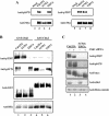
Phosphorylation of Cdc25A at S178 and T507 in vivo and in vitro. (A) Lysates from HeLa cells infected with adenoviruses encoding the indicated Myc-Cdc25A proteins were resolved by SDS-PAGE, and proteins were detected by immunoblotting with phospho-specific antibodies followed by antibodies specific for the Myc epitope (anti-Myc). (B) Kinase assays were performed in vitro in the presence of His6Cdc25A proteins and either GST-Chk1 or GST-Chk2. Reaction products were resolved by SDS-PAGE and subjected to Western blotting to monitor phosphorylation of Cdc25A at T507 (anti-pT507) and S178 (anti-pS178) and to monitor levels of His6Cdc25A (anti-His), GST-Chk1, and GST-Chk2 (anti-GST). (C) HeLa cells were treated with control or Chk1-specific siRNAs followed by infection with recombinant adenovirus encoding the indicated forms of Cdc25A. Lysates were prepared and analyzed by Western blotting with the indicated antibodies.
FIG. 3.
FIG. 3.
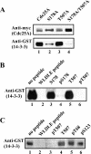
Interactions between Cdc25A and 14-3-3 proteins are direct. (A) Far Western analysis was performed to monitor the binding of 14-3-3 protein to wt and mutant forms of Cdc25A. Membranes containing the indicated form of Myc-Cdc25A were incubated with purified GST-14-3-3ɛ, and 14-3-3 bound to Cdc25A was detected by Western blotting with GST antibodies. (B and C) Specific phosphopeptides compete with 14-3-3 proteins for binding to Cdc25A. Membranes containing the indicated forms of Myc-Cdc25A were incubated with GST-14-3-3ɛ in the presence of the indicated phospho- and nonphosphopeptides. 14-3-3 bound to Cdc25A was detected by Western blotting with GST antibodies.
FIG. 4.
FIG. 4.
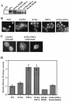
14-3-3 binding mutants activate cyclin B1/Cdk1 and induce a mitotic-like state when expressed in S phase. S-phase HeLa cells expressing GFP and various forms of Myc-Cdc25A were analyzed for Cdc25A levels by Western blotting (A) and for chromosomal integrity by examining DAPI-stained nuclei (B and C). Magnification, ×40 (B) and ×100 (C). Cells were also analyzed for cyclin B1-associated histone H1 kinase activity (D). Standard errors of the means (n = 4) are shown as error bars along the y axis.
FIG. 5.
FIG. 5.
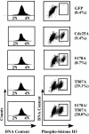
14-3-3 binding mutants induce histone H3 phosphorylation in S-phase cells. S-phase HeLa cells expressing GFP and various forms of Myc-Cdc25A (same cells as for Fig. 4) were costained for DNA content and histone H3 phosphorylation and analyzed by flow cytometry (50). Percentages of mitotic cells are indicated.
FIG. 6.
FIG. 6.
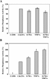
Threonine 507 negatively regulates phosphatase activity of Cdc25A. wt and mutant forms of Cdc25A were purified as GST fusion proteins from bacteria (A) or as Myc fusion proteins from HeLa cells (B). Phosphatase assays were carried out in the presence of Cdc25A and purified cyclin B1/Cdk1 containing Cdk1 phosphorylated on both T14 and Y15. Phosphatase reactions were terminated and histone H1 kinase assays were performed to measure the activation of cyclin B1/Cdk1. 32P incorporation into histone H1 was determined by scintillation counting. Cdc25A precipitates were immunoblotted and quantified by use of Myc antibodies, ECF reagents, and the Storm 860 imaging system (Amersham). Histone H1 kinase activities were normalized for levels of Cdc25A in each reaction. Values obtained for wt Cdc25A were set at 100, and the mutant values are shown relative to this. Standard errors of the means (n ≥ 3) are shown as error bars along the y axis.
FIG. 7.
FIG. 7.
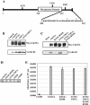
The C terminus of Cdc25A contains a cyclin B1 docking site. (A) Schematic representation of Cdc25A and amino acids comprising the C-terminal cyclin B1 docking site. Asterisks denote K514 and R520. (B) Loss of 14-3-3 binding promotes interactions between Cdc25A and cyclin B1. HeLa cells synchronized at the G1/S border were infected with recombinant adenoviruses encoding GFP (control) or with recombinant viruses encoding various forms of Myc-Cdc25A. Three hours after release from the block, S-phase cells were lysed and incubated with anti-c-Myc-agarose. Myc-Cdc25A precipitates were resolved by SDS-PAGE, and Cdc25A and cyclin B1 were visualized by immunoblotting. (C) The C terminus of Cdc25A is required for interactions between Cdc25A and cyclin B1. HeLa cells synchronized at the G1/S border were infected with recombinant adenoviruses encoding GFP (control) or with recombinant viruses encoding various forms of Myc-Cdc25A. Three hours after release from the block, S-phase cells were lysed and incubated with anti-c-Myc-agarose. Myc-Cdc25A precipitates were resolved by SDS-PAGE, and Cdc25A and cyclin B1 were visualized by immunoblotting. (D and E) Mutation of the cyclin B1 docking site does not impair phosphatase activity of Cdc25A. wt and mutant forms of Cdc25A were purified from bacteria as GST fusion proteins, resolved by SDS-PAGE, and visualized by Western blotting (D). Purified proteins were tested for the ability to dephosphorylate mFP in vitro (E). Reactions were done in triplicate and followed by spectroscopy at 477 nm. Standard errors of the means are shown as error bars along the y axis.
FIG. 8.
FIG. 8.
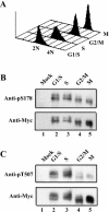
Phosphorylation of Cdc25A at S178 and T507 is cell cycle regulated. HeLa cells expressing Myc-Cdc25A were synchronized at the G1/S border by a double thymidine block protocol. Cells at the G1/S border were collected prior to release from the block, and S- and G2/M-phase cells were harvested at 3 and 7 h after release, respectively. Mitotic cells were obtained by incubation in nocodazole for 10 h after infection and release. Some cells were harvested and analyzed by FACS to determine DNA content (A). The remaining cells were lysed and incubated with anti-c-Myc-agarose. Precipitates were resolved by SDS-PAGE and incubated with phospho-specific antibodies followed by antibodies specific for the Myc epitope to monitor Cdc25A levels (B and C).
FIG. 9.
FIG. 9.
Regulation of Cdc25A by Chk1. Chk1 phosphorylates Cdc25A on several N-terminal serines to promote its ubiquitin-mediated proteolysis (11, 16, 20, 46, 57). Chk1 also phosphorylates Cdc25A on S178 and T507 to facilitate 14-3-3 binding. 14-3-3 binding to the C-terminal cyclin B1 binding domain (BD) of Cdc25A blocks functional interactions between Cdc25A and cyclin B1/Cdk1 throughout interphase. These regulatory pathways prevent cells from entering into mitosis at inappropriate times during the cell cycle and following checkpoint activation. Solid arrows originating from Chk1 indicate phosphorylation sites that mediate 14-3-3 binding, whereas hatched lines represent sites that mediate Cdc25A turnover. S178 is the primary 14-3-3 binding site and S75 is the primary site regulating Cdc25A turnover, as indicated by thicker arrows.

Similar articles

Cited by

References

    1. Atherton-Fessler, S., L. L. Parker, R. L. Geahlen, and H. Piwnica-Worms. 1993. Mechanism of p34cdc2 regulation. Mol. Cell. Biol. 13:1675-1685. - PMC - PubMed
    1. Bartek, J., and J. Lukas. 2001. Pathways governing G1/S transition and their response to DNA damage. FEBS Lett. 490:117-122. - PubMed
    1. Bernardi, R., D. A. Lieberman, and B. Hoffman. 2000. Cdc25A stability is controlled by the ubiquitin-proteasome pathway during cell cycle progression and terminal differentiation. Oncogene 19:2447-2454. - PubMed
    1. Blomberg, I., and I. Hoffman. 1999. Ectopic expression of Cdc25A accelerates the G1/S transition and leads to premature activation of cyclin E- and cyclin A-dependent kinases. Mol. Cell. Biol. 19:6183-6194. - PMC - PubMed
    1. Broggini, M., G. Buraggi, A. Brenna, L. Riva, A. Codegoni, V. Torri, A. Lissoni, C. Mangioni, and M. D'Incalci. 2000. Cell cycle-related phosphatases Cdc25A and B expression correlates with survival in ovarian cancer patients. Anticancer Res. 20:4835-4840. - PubMed

Publication types

MeSH terms