. 2003 Oct;23(20):7256-70.
doi: 10.1128/MCB.23.20.7256-7270.2003.

c-Myc augments gamma irradiation-induced apoptosis by suppressing Bcl-XL

Affiliations

c-Myc augments gamma irradiation-induced apoptosis by suppressing Bcl-XL

Kirsteen H Maclean et al. Mol Cell Biol. 2003 Oct.

Abstract

Alterations in MYC and p53 are hallmarks of cancer. p53 coordinates the response to gamma irradiation (gamma-IR) by either triggering apoptosis or cell cycle arrest. c-Myc activates the p53 apoptotic checkpoint, and thus tumors overexpressing MYC often harbor p53 mutations. Nonetheless, many of these cancers are responsive to therapy, suggesting that Myc may sensitize cells to gamma-IR independent of p53. In mouse embryo fibroblasts (MEFs) and in E micro -myc transgenic B cells in vivo, c-Myc acts in synergy with gamma-IR to trigger apoptosis, but alone, when cultured in growth medium, it does not induce a DNA damage response. Surprisingly, c-Myc also sensitizes p53-deficient MEFs to gamma-IR-induced apoptosis. In normal cells, and in precancerous B cells of E micro -myc transgenic mice, this apoptotic response is associated with the suppression of the antiapoptotic regulators Bcl-2 and Bcl-X(L) and with the concomitant induction of Puma, a proapoptotic BH3-only protein. However, in p53-null MEFs only Bcl-X(L) expression was suppressed, suggesting levels of Bcl-X(L) regulate the response to gamma-IR. Indeed, Bcl-X(L) overexpression blocked this apoptotic response, whereas bcl-X-deficient MEFs were inherently and selectively sensitive to gamma-IR-induced apoptosis. Therefore, MYC may sensitize tumor cells to DNA damage by suppressing Bcl-X.


Figures
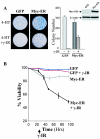
FIG. 1.
FIG. 1.
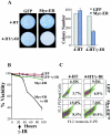
The survival of Myc-expressing MEFs is compromised. (A) GFP-only- or Myc-ER-expressing MEFs (right panel inset) were treated with 4-HT (1 μM) for 24 h and irradiated with 5 Gy, and after 24 h 5 × 10³ cells were plated in fresh growth medium with 4-HT. Colonies were stained with Wright-Giemsa after 10 days. The right panel shows the mean colony numbers from three separate experiments. (B) γ-IR triggers cell death in Myc-expressing MEFs. GFP-only- and Myc-ER-expressing MEFs were treated with 4-HT (1 μM) for 24 h and then left untreated or were irradiated with 5 Gy. At the indicated intervals, viability was determined by trypan blue dye exclusion.
FIG. 2.
FIG. 2.
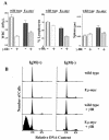
γ-IR selectively compromises B-lymphocyte survival in Eμ-myc transgenic mice. Wild-type and Eμ-myc transgenic littermates were left untreated or were treated with 2 Gy of irradiation (n = 8 for each group). (A) After 4 h the WBC number (left panel) and the percent lymphocytes (middle panel) were evaluated in the peripheral blood of these mice. In addition, the weights of the spleens of these mice were determined (right panel). Although WBC numbers were reduced upon γ-IR treatment in both wild-type and Eμ-myc mice, there were marked reductions in lymphocyte numbers and spleen weights evident in Eμ-myc mice. (B) Bone marrow was harvested from these same animals, and B220+ cells were stained with propidium iodide and analyzed by FACS. Note the marked increase in the numbers of sub-G1 IgM+ and IgM Eμ-myc cells when exposed to γ-IR.
FIG. 3.
FIG. 3.
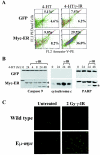
γ-IR triggers apoptosis in Myc-expressing MEFs and in Eμ-myc transgenic B cells. (A) FACS histograms of PE-annexin V binding. GFP-only- or Myc-ER-expressing MEFs were treated with 4-HT (1 μM) for 24 h and irradiated with 5 Gy, and after 24 h cells were stained with annexin-V-PE and propidium iodide (PI). The percentages of annexin V- and PI-negative and -positive cells are given in each quadrant. Results shown are representative of three independent experiments performed with two different MEF cell cultures. (B) γ-IR triggers caspase-9 and PARP cleavage, and the release of cytochrome c into the cytosol, in Myc-expressing MEFs. The indicated cells were treated for 24 h with 4-HT (1 μM) and treated with γ-IR, and lysates were analyzed at the indicated intervals by Western blotting for caspase-9 (left panel), the cytosolic fraction of cytochrome c (29) (middle panel), and PARP (right panel). The asterisk indicates the cleaved forms of caspase-9 and PARP. (C) γ-IR triggers apoptosis in B cells of 6-week-old (precancerous) Eμ-myc transgenic mice. Wild-type and Eμ-myc transgenic littermates were treated with 2 Gy of γ-IR. After 4 h bone marrow was harvested and sorted for B220+ cells by FACS, and cytospins were assessed for apoptotic cells by immunofluorescence using TUNEL assays. TUNEL-positive apoptotic cells appear bright green. Representative fields are shown.
FIG. 4.
FIG. 4.
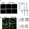
Myc alone does not induce the DNA damage response in MEFs. (A) Detection of Ser139-H2AX foci in GFP-only- or Myc-ER-expressing MEFs treated with 4-HT with or without γ-IR. MEFs were treated with 4-HT (1 μM) for 24 h and irradiated with 5 Gy, and cells were harvested at the indicated intervals. Following fixation and blocking procedures, cells were incubated with anti-Ser139-H2AX antibody and the respective fluorophore-conjugated secondary antibody (Cy3 anti-rabbit). A representative figure from three separate experiments is shown (upper panels), and at right the data are plotted as the number of fluorescent Ser139-H2AX foci per cell. A minimum of 100 counts for three independent fields is shown. (B) Comet assays of DNA damage in GFP-only- or Myc-ER-expressing MEFs. Cells were treated with 4-HT (1 μM) for 24 h, irradiated with 5 Gy, and exposed to an electric field (migration of the DNA in the electric field is from left to right). Arrows indicate GFP-positive cells with obvious comets. (C) p53 phosphorylation on Ser15 does not occur following Myc activation in MEFs. Cells were treated with 4-HT (1 μM) for 24 h and irradiated with 5 Gy, and whole-cell lysates were evaluated for levels of Ser15-phosphorylated p53 and compared to steady-state levels of total p53. Note the robust induction of Ser15-phosphorylated p53 in GFP-only-expressing MEFs treated with γ-IR; this was not detectable in Myc-ER-expressing MEFs treated with 4-HT, despite the robust induction of total p53. The blots shown were exposed for the same length of time. (D) Serum suppresses Myc's ability to provoke Ser15 phosphorylation of p53. The indicated MEFs were treated with 4-HT and cultured in replete growth medium (lanes 1, 3, 5, and 7) or in 0.1% FCS medium (lanes 2, 4, 6, and 8). Cells were then left untreated (lanes 1, 2, 5, and 6) or were treated with 5 Gy of γ-IR (lanes 3, 4, 7, and 8). After 24 h cells were harvested and levels of phospho-p53Ser15 were determined by immunoblot analysis.
FIG.5.
FIG.5.
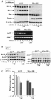
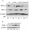
Myc selectively targets the expression of Bcl-2, Bcl-XL, and Puma in MEFs. (A) GFP-only- or Myc-ER-expressing wild-type MEFs were treated for 24 h with 4-HT, irradiated with 5 Gy, and at the indicated intervals harvested for immunoblot analyses (top panels) or RT-PCR analysis of bcl-X and HPRT RNA (lower panels). (B) Only Bcl-XL is suppressed by Myc in p53-defcient MEFs. GFP-only- or Myc-ER-expressing p53-deficent MEFs were treated for 24 h with 4-HT, irradiated with 5 Gy, and at the indicated intervals harvested for immunoblot analyses (left panels) or Northern blot analysis of bcl-X RNA (right panels). Puma expression was not detected in Myc-expressing p53-deficient MEFs (negative data not shown). To control for loading, the levels of 28S and 18S RNA are shown at right. (C) Real-time PCR analysis of bcl-X levels in p53-deficient MEFs engineered to express Myc-ER. The indicated cells were treated for 24 h with 4-HT and irradiated with 5 Gy, and at the indicated intervals RNA was harvested for real-time PCR analysis. The internal control in the real-time reactions is arpp0.
FIG. 6.
FIG. 6.
Myc targets the expression of Bcl-2, Bcl-XL, and Puma in bone marrow (BM) and splenic (S) B cells of Eμ-myc transgenic mice. Wild-type (lanes 1 to 4 and 9 to 12) and precancerous Eμ-myc transgenic littermates (lanes 5 to 8 and 13 to 16) (n = 8 for each group) were left untreated (lanes 1 to 8) or were treated with 2 Gy of γ-IR (lanes 9 to 16). Four hours later, bone marrow and spleens from each of these four groups of mice were pooled and FACS sorted for B220 and IgM, and 20 μg of extracts was assessed by immunoblotting with antibodies specific for Bcl-2, Bcl-XL, and β-actin (A), or 60 μg of extract from IgM+ B cells was analyzed for Puma levels (B).
FIG. 7.
FIG. 7.
Myc sensitizes p53−/− MEFs to γ-IR-induced apoptosis. (A) GFP-only- or Myc-ER-expressing p53−/− MEFs were treated with 4-HT (1 μM) for 24 h and left untreated or were irradiated with 5 Gy. Twenty-four hours later, 5 × 10³ cells were plated in growth medium containing 4-HT. Left panels, colonies were stained with Wright-Giemsa after 10 days. Right panel, the mean colony numbers from three separate experiments is shown. (B) Cells treated as for panel A were examined for apoptosis by trypan blue dye exclusion. (C) Cells treated as for panel A were examined for apoptosis by FACS analyses of propidium iodide (PI)-annexin V-stained cells. The percentages of annexin V- and PI-negative and -positive cells are given in each quadrant. Results shown are representative of three independent experiments performed with two different MEF cell cultures.
FIG. 8.
FIG. 8.
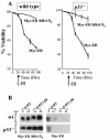
bcl-X suppression is necessary for Myc to trigger apoptosis following γ-IR. Wild-type and p53−/− MEFs were transduced with MSCV-Myc-ER-IRES-GFP- and MSCV-hBcl-XL-IRES-YFP-expressing viruses, and doubly transduced MEFs were compared to those expressing Myc-ER alone. (A) GFP-only- or Myc-ER-expressing MEFs were treated with 4-HT (1 μM) for 24 h and irradiated with 5 Gy, and viability was determined by trypan blue dye exclusion at the indicated intervals. Viability experiments shown are representative of three independent experiments performed in triplicate. (B) Immunoblot analysis with an antibody specific for human Bcl-XL (left panels) demonstrated that overexpression of human Bcl-XL protein was sustained in Myc-expressing cells exposed to γ-IR, contrary to results with endogenous mouse Bcl-XL (right panels).
FIG. 9.
FIG. 9.
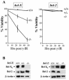
bcl-X loss, but not loss of bcl-2, radiosensitizes MEFs. (A) MEFs prepared from the indicated embryos were cultured in growth medium and exposed to 5 Gy of γ-IR, and their viability was assessed by trypan blue dye exclusion. Viability experiments shown are representative of three independent experiments with three independent MEF cell cultures, performed in triplicate. (B) Immunoblot analysis of Bcl-XL and Bcl-2 protein levels in bcl-X+/+, bcl-X+/−, and bcl-X−/− MEFs (left panels) and in bcl-2+/+, bcl-2+/−, and bcl-2−/− MEFs (right panels).

Similar articles

Cited by

References

    1. Adams, J. M., A. W. Harris, C. A. Pinkert, L. M. Corcoran, W. S. Alexander, S. Cory, R. D. Palmiter, and R. L. Brinster. 1985. The c-myc oncogene driven by immunoglobulin enhancers induces lymphoid malignancy in transgenic mice. Nature 318:533-538. - PubMed
    1. Askew, D. S., R. A. Ashmun, B. C. Simmons, and J. L. Cleveland. 1991. Constitutive c-myc expression in an IL-3-dependent myeloid cell line suppresses cell cycle arrest and accelerates apoptosis. Oncogene 6:1915-1922. - PubMed
    1. Banin, S., L. Moyal, S. Shieh, Y. Taya, C. W. Anderson, L. Chessa, N. I. Smorodinsky, C. Prives, Y. Reiss, Y. Shiloh, and Y. Ziv. 1998. Enhanced phosphorylation of p53 by ATM in response to DNA damage. Science 281:1674-1677. - PubMed
    1. Baudino, T. A., and J. L. Cleveland. 2001. The Max network gone mad. Mol. Cell. Biol. 21:691-702. - PMC - PubMed
    1. Baudino, T. A., C. McKay, H. Pendeville-Samain, J. A. Nilsson, K. H. Maclean, E. L. White, A. C. Davis, J. N. Ihle, and J. L. Cleveland. 2002. c-Myc is essential for vasculogenesis and angiogenesis during development and tumor progression. Genes Dev. 16:2530-2543. - PMC - PubMed

Publication types

MeSH terms