

Replication complex of human parechovirus 1
- PMID: 12857920
- PMCID: PMC165257
- DOI: 10.1128/jvi.77.15.8512-8523.2003
Replication complex of human parechovirus 1
Abstract
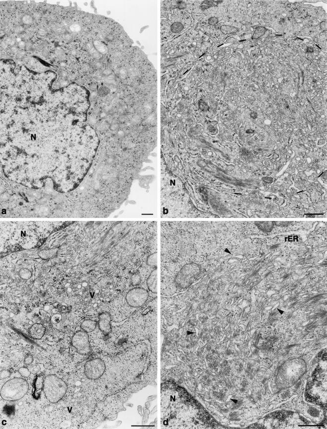
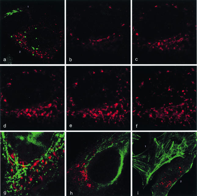
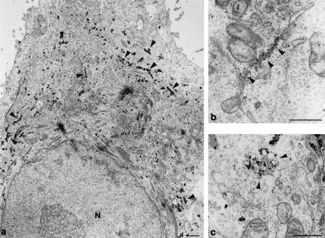
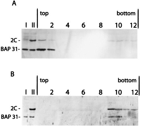
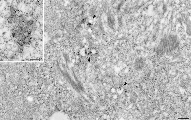
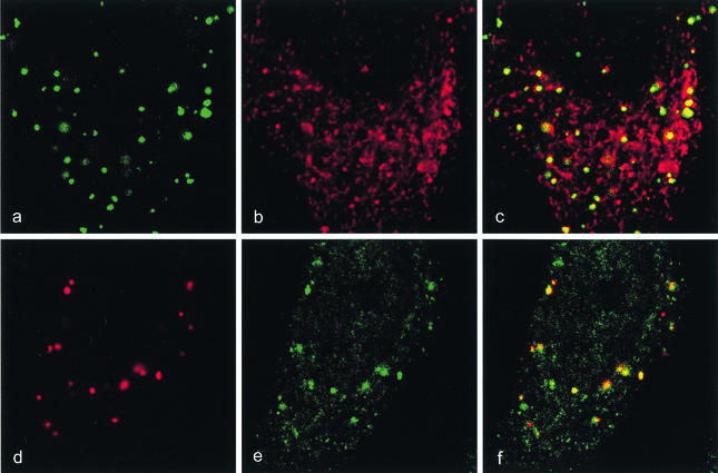
The parechoviruses differ in many biological properties from other picornaviruses, and their replication strategy is largely unknown. In order to identify the viral RNA replication complex in human parechovirus type 1 (HPEV-1)-infected cells, we located viral protein and RNA in correlation to virus-induced membrane alterations. Structural changes in the infected cells included a disintegrated Golgi apparatus and disorganized, dilated endoplasmic reticulum (ER) which had lost its ribosomes. Viral plus-strand RNA, located by electron microscopic (EM) in situ hybridization, and the viral protein 2C, located by EM immunocytochemistry were found on clusters of small vesicles. Nascent viral RNA, visualized by 5-bromo-UTP incorporation, localized to compartments which were immunocytochemically found to contain the viral protein 2C and the trans-Golgi marker 1,4-galactosyltransferase. Protein 2C was immunodetected additionally on altered ER membranes which displayed a complex network-like structure devoid of cytoskeletal elements and with no apparent involvement in viral RNA replication. This protein also exhibited membrane binding properties in an in vitro assay. Our data suggest that the HPEV-1 replication complex is built up from vesicles carrying a Golgi marker and forming a structure different from that of replication complexes induced by other picornaviruses.
Figures


Similar articles
-
Intracellular localization and effects of individually expressed human parechovirus 1 non-structural proteins.J Gen Virol. 2007 Mar;88(Pt 3):831-841. doi: 10.1099/vir.0.82201-0. J Gen Virol. 2007. PMID: 17325355
-
Membrane rearrangement and vesicle induction by recombinant poliovirus 2C and 2BC in human cells.Virology. 1994 Jul;202(1):129-45. doi: 10.1006/viro.1994.1329. Virology. 1994. PMID: 8009827
-
ATP hydrolysis and AMP kinase activities of nonstructural protein 2C of human parechovirus 1.J Virol. 2006 Jan;80(2):1053-8. doi: 10.1128/JVI.80.2.1053-1058.2006. J Virol. 2006. PMID: 16379008 Free PMC article.
-
Functions of alphavirus nonstructural proteins in RNA replication.Prog Nucleic Acid Res Mol Biol. 2002;71:187-222. doi: 10.1016/s0079-6603(02)71044-1. Prog Nucleic Acid Res Mol Biol. 2002. PMID: 12102555 Free PMC article. Review.
-
Flaviviral Replication Complex: Coordination between RNA Synthesis and 5'-RNA Capping.Viruses. 2015 Aug 13;7(8):4640-56. doi: 10.3390/v7082837. Viruses. 2015. PMID: 26287232 Free PMC article. Review.
Cited by
-
Human Parechovirus: an Increasingly Recognized Cause of Sepsis-Like Illness in Young Infants.Clin Microbiol Rev. 2017 Nov 15;31(1):e00047-17. doi: 10.1128/CMR.00047-17. Print 2018 Jan. Clin Microbiol Rev. 2017. PMID: 29142080 Free PMC article. Review.
-
Parechovirus A Pathogenesis and the Enigma of Genotype A-3.Viruses. 2019 Nov 14;11(11):1062. doi: 10.3390/v11111062. Viruses. 2019. PMID: 31739613 Free PMC article. Review.
-
Wrapping things up about virus RNA replication.Traffic. 2005 Nov;6(11):967-77. doi: 10.1111/j.1600-0854.2005.00339.x. Traffic. 2005. PMID: 16190978 Free PMC article. Review.
-
The nonstructural protein 2C of a Picorna-like virus displays nucleic acid helix destabilizing activity that can be functionally separated from its ATPase activity.J Virol. 2013 May;87(9):5205-18. doi: 10.1128/JVI.00245-13. Epub 2013 Feb 28. J Virol. 2013. PMID: 23449794 Free PMC article.
-
The open reading frame 3a protein of severe acute respiratory syndrome-associated coronavirus promotes membrane rearrangement and cell death.J Virol. 2010 Jan;84(2):1097-109. doi: 10.1128/JVI.01662-09. Epub 2009 Nov 4. J Virol. 2010. PMID: 19889773 Free PMC article.
References
-
- Aldabe, R., and L. Carrasco. 1995. Induction of membrane proliferation by poliovirus proteins 2C and 2BC. Biochem. Biophys. Res. Commun. 206:664-676. - PubMed
-
- Bienz, K., and D. Egger. 1995. Immunocytochemistry and in situ hybridization in the electron microscope: combined application in the study of virus-infected cells. Histochem. Cell Biol. 103:325-338. - PubMed
-
- Bienz, K., D. Egger, and L. Pasamontes. 1987. Association of polioviral proteins of the P2 genomic region with the viral replication complex and virus-induced membrane synthesis as visualized by electron microscopic immunocytochemistry and autoradiography. Virology 160:220-226. - PubMed
Publication types
MeSH terms
Substances
LinkOut - more resources
Full Text Sources
