. 2002 Aug 15;21(16):4338-48.
doi: 10.1093/emboj/cdf433.

Different telomere damage signaling pathways in human and mouse cells

Affiliations

Different telomere damage signaling pathways in human and mouse cells

Agata Smogorzewska et al. EMBO J. .

Abstract

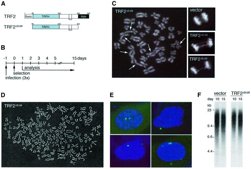
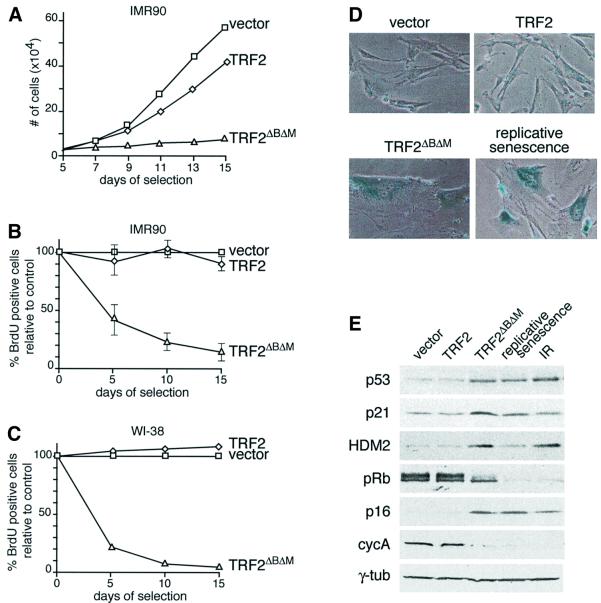
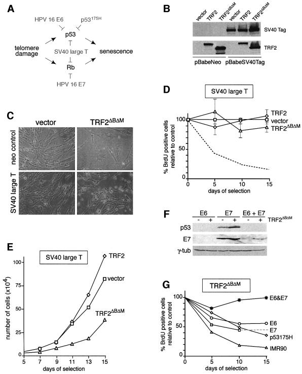
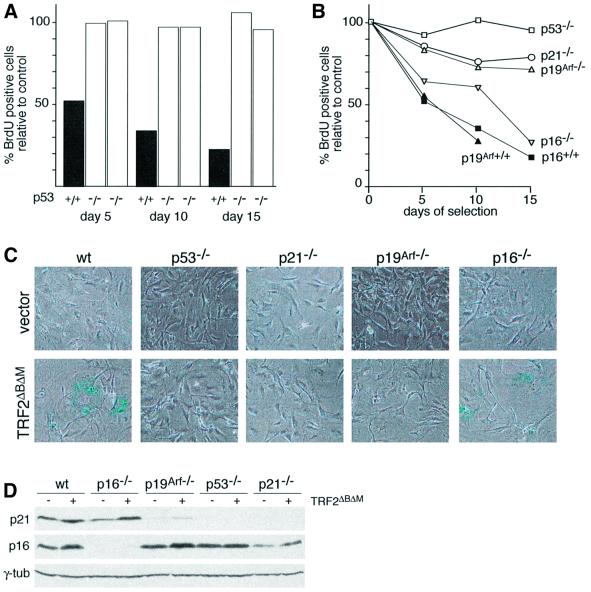
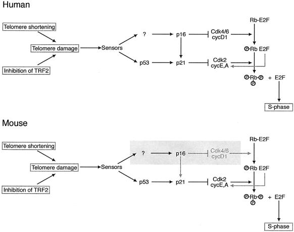
Programmed telomere shortening in human somatic cells is thought to act as a tumor suppressor pathway, limiting the replicative potential of developing tumor cells. Critically short human telomeres induce senescence either by activating p53 or by inducing the p16/RB pathway, and suppression of both pathways is required to suppress senescence of aged human cells. Here we report that removal of TRF2 from human telomeres and the ensuing de-protection of chromosome ends induced immediate premature senescence. Although the telomeric tracts remained intact, the TRF2(DeltaBDeltaM)-induced premature senescence was indistinguishable from replicative senescence and could be mediated by either the p53 or the p16/RB pathway. Telomere de-protection also induced a growth arrest and senescent morphology in mouse cells. However, in this setting the loss of p53 function was sufficient to completely abrogate the arrest, indicating that the p16/RB response to telomere dysfunction is not active in mouse cells. These findings reveal a fundamental difference in telomere damage signaling in human and mouse cells that bears on the use of mouse models for the telomere tumor suppressor pathway.


Figures

None
Fig. 1. TRF2ΔBΔM induces telomere de-protection and karyotypic abnormalities in IMR90 cells without loss of telomeric DNA. (A) Schematic of TRF2 and TRF2ΔBΔM. (B) Experimental timeline. (C) Chromosome end fusions. Large panel, metaphase spread with multiple chromosome end fusions (arrows). Small panels, anaphase cells (top, control; bottom, TRF2ΔBΔM). (D) TRF2ΔBΔM-induced tetraploidy in IMR90. Diplochromosomes were observed in a subset of tetraploid metaphases. (E) TRF2ΔBΔM-induced centrosome amplification in IMR90. IF for γ-tubulin (FITC, green). (F) Persistence of telomeric DNA in TRF2ΔBΔM-expressing cells. Genomic blot of telomeric HinfI–RsaI fragments detected with a TTAGGG repeat probe. DNA was isolated from IMR90 cells on day 10 and 15 of selection, after infection with pWZLvector or pWZLTRF2ΔBΔM. (C–E) represent analysis on day 7 of selection and DAPI DNA staining.
None
Fig. 2. Premature senescence in primary human fibroblasts expressing TRF2ΔBΔM. (A) Growth curve of IMR90 cells expressing TRF2, or TRF2ΔBΔM and vector control cells. Cells were infected at passage 23 and analyzed according to the schematic in Figure 1B. After 3 weeks, TRF2-expressing cells proliferated at the same rate as vector control cells. (B) S-phase index in TRF2ΔBΔM-expressing IMR90 cells. The percentage of cells in S phase was measured using BrdU incorporation on day 5, 10 and 15 (see Materials and methods). Values represent the mean of 4–5 experiments and the SD is given. (C) S-phase index in TRF2ΔBΔM-expressing WI-38 cells. Method as in (B) but using WI-38 fibroblasts at passage 26. (D) TRF2ΔBΔM-induced senescent morphology and SA-β-gal expression. IMR90 cells (passage 22) infected with the indicated viruses as in (A) were stained for SA-β-gal on day 16. The bottom right panel shows IMR90 cells that have undergone replicative senescence (passage 42). (E) Immunoblot analysis of TRF2ΔBΔM-expressing fibroblasts, senescent fibroblasts and γ-irradiated fibroblasts. IMR90 cells (passage 22) infected with the indicated viruses and analyzed on day 15. Naturally senescent cells were collected at passage 42. Irradiated cells (10 Gray from a 137Cs source) were maintained for 15 days with daily feeding before preparation of the lysate. Lysates of 1.5 × 10⁵ cell equivalents were loaded in each lane. Immunoblotting for the indicated proteins was performed as detailed in Materials and methods.
None
Fig. 3. TRF2ΔBΔM-induced premature senescence in human cells mediated by p53 and p16/RB. (A) Oncoproteins used to inhibit the p53 and p16/RB pathways. (B) Immunoblot analysis of expression of SV40 large T antigen and retroviral TRF2 proteins. IMR90 cells infected with pBabeNeo or pBabeSV40Tag were superinfected with the indicated viruses and SV40 Tag and TRF2 (Ab 647) were detected as described in Materials and methods. (C) Morphology of SV40 Tag-expressing cells infected with the TRF2ΔBΔM virus. Cells were infected as in (B) and photographed on day 13 after selection. (D) S-phase index in cells expressing SV40 Tag and TRF2ΔBΔM. Procedure as in Figure 2B. Mean values and SDs were derived from three experiments. (E) Growth curve of IMR90 cells expressing SV40 Tag and TRF2ΔBΔM or control viruses (vector or TRF2). (F) Immunoblot analysis of IMR90 cells expressing HPV16 E6 and/or E7. IMR90 cells were infected with viruses for HPV16 E6, E7, or both, and superinfected with a control retrovirus (– lanes) or the TRF2ΔBΔM retrovirus (+ lanes). WCL (equal cell number equivalents) were analyzed on day 8 of selection for the expression of p53 and E7, as described in Materials and methods. Reduction of the basal level of p53 (– lanes) served as a marker for the activity of E6. γ-tubulin served as a loading control. (G) Effects of separate inhibition of the p53 and p16/RB pathways in TRF2ΔBΔM-induced premature senescence. Young (passage 19) IMR90 cells were first infected with viruses carrying HPV16 E6, HPV16 E7, HPV16 E6 and E7, p53175H, or with the pBabeNeo control virus (graph labeled IMR90). Subsequently, cultures were superinfected with the TRF2ΔBΔM virus or the vector control. The percentage BrdU-positive cells was determined in triplicate, as described in Materials and methods. The SDs were 1–5%. The dotted line extrapolates the data point for E7-expressing cells on day 10 to a day 15 data point from a different experiment.
None
Fig. 4. TRF2ΔBΔM-induced growth arrest in murine cells depends on p53. (A) S-phase index of TRF2ΔBΔM-expressing p53+/+ and p53–/– MEFs. Cells of the indicated genotypes were infected with the TRF2ΔBΔM virus or the vector control, and the incorporation of BrdU in the parallel cultures was measured as described in Figure 2B. The bar graph shows the percentage of TRF2ΔBΔM-expressing cells in S phase compared with controls (control virus infection set at 100%) as measured using BrdU incorporation. Two experiments are shown for two independent batches of MEFs deficient in p53. (B) Effects of deficiency for p53, p21, p19Arf or p16 on the TRF2ΔBΔM-induced growth arrest in MEFs. Cells of the indicated genotypes were infected with TRF2ΔBΔM or a control virus. BrdU incorporation in TRF2ΔBΔM-expressing cells was normalized to control infected cells as described in Figure 2B. Values are derived from triplicate measurements. SDs were 1–5%. (C) Effects of p53, p21, p19Arf and p16 status on the TRF2ΔBΔM-induced morphology and SA-β-gal expression. Cells used in the experiment shown in (B) were plated at equal cell density on day 10 and assayed for β-gal activity 2 days later. (D) Levels of p21 and p16 in mouse cells expressing TRF2ΔBΔM. Cells of the indicated genotypes were infected with the TRF2ΔBΔM virus or the vector control. Lysates (104 cells/lane) were prepared on day 10.
None
Fig. 5. Schematic illustrating the difference in the telomere damage signaling pathways in human and mouse cells. Telomere damage created by telomere shortening or removal of TRF2 is proposed to activate the same signaling pathway. The sensor(s) upstream of p16 and p53 have not been identified. In human cells, cell cycle arrest in response to telomere damage can be transduced through either the p53 or the p16/Rb pathways. In mouse cells, the telomere damage signal is transduced only through the p53 pathway. In the absence of p53, the p16/Rb pathway alone is insufficient to induce cell cycle arrest in response to telomere damage (shading). The arrow from p16 to p21 illustrates the ability of p16 to displace p21 from the CDK4/6 complex, resulting in a release of a latent pool of p21 that can inhibit cycE/A-dependent CDK2.

Similar articles

Cited by

References

    1. Artandi S.E., Chang,S., Lee,S.L., Alson,S., Gottlieb,G.J., Chin,L. and DePinho,R.A. (2000) Telomere dysfunction promotes non-reciprocal translocations and epithelial cancers in mice. Nature, 406, 641–645. - PubMed
    1. Benn P.A. (1976) Specific chromosome aberrations in senescent fibroblast cell lines derived from human embryos. Am. J. Hum. Genet., 28, 465–473. - PMC - PubMed
    1. Blasco M.A., Lee,H.W., Hande,M.P., Samper,E., Lansdorp,P.M., DePinho,R.A. and Greider,C.W. (1997) Telomere shortening and tumor formation by mouse cells lacking telomerase RNA. Cell, 91, 25–34. - PubMed
    1. Bodnar A.G. et al. (1998) Extension of life-span by introduction of telomerase into normal human cells. Science, 279, 349–352. - PubMed
    1. Broccoli D., Smogorzewska,A., Chong,L. and de Lange,T. (1997) Human telomeres contain two distinct Myb-related proteins, TRF1 and TRF2. Nat. Genet., 17, 231–235. - PubMed

Publication types

Substances