. 2002 Sep;22(17):6158-69.
doi: 10.1128/MCB.22.17.6158-6169.2002.

c-Myc functionally cooperates with Bax to induce apoptosis

Affiliations

c-Myc functionally cooperates with Bax to induce apoptosis

Philippe Juin et al. Mol Cell Biol. 2002 Sep.

Abstract

c-Myc promotes apoptosis by destabilizing mitochondrial integrity, leading to the release of proapoptotic effectors including holocytochrome c. Candidate mediators of c-Myc in this process are the proapoptotic members of the Bcl-2 family. We show here that fibroblasts lacking Bak remain susceptible to c-Myc-induced apoptosis whereas bax-deficient fibroblasts are resistant. However, despite this requirement for Bax, c-Myc activation exerts no detectable effects on Bax expression, localization, or conformation. Moreover, susceptibility to c-Myc-induced apoptosis can be restored in bax-deficient cells by ectopic expression of Bax or by microinjection of a peptide comprising a minimal BH3 domain. Microinjection of BH3 peptide also restores sensitivity to c-Myc-induced apoptosis in p53-deficient primary fibroblasts that are otherwise resistant. By contrast, there is no synergy between BH3 peptide and c-Myc in fibroblasts deficient in both Bax and Bak. We conclude that c-Myc triggers a proapoptotic mitochondrial destabilizing activity that cooperates with proapoptotic members of the Bcl-2 family.


Figures
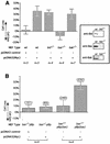
FIG. 1.
FIG. 1.
c-Myc-induced apoptosis in fibroblasts requires Bax but not Bid or Bak. (A) MEFs of the indicated genotypes were grown in 20% heat-inactivated FCS and were microinjected with either empty pcDNA3 or pcDNA3(Myc) (100 ng/ml) mixed in RNase-free water with pEGFP-N1 (20 ng/ml) indicator as described in Materials and Methods. Eighteen hours after microinjection, the number of GFP-expressing cells was evaluated by fluorescence microscopy; cells were then washed twice in PBS and cultured in the absence of FCS for an additional 24 h at 37°. The number of GFP-expressing cells still viable was then determined. Cell loss or cell gain was expressed as a percentage of the initial number of GFP-positive cells prior to serum deprivation. Data presented are mean values ± standard errors of the means (SEM) from the indicated number of independent experiments. wt, wild type. Insert: MEFs with genes knocked out were validated for loss of relevant BH3 protein by immunoblot analysis. Lysates of wild-type MEFs and bid−/−, bax−/−, or bak−/− MEFs were fractionated by SDS-PAGE (105 cells per lane) and immunoblotted with anti-Bid, anti-Bax, or anti-Bak antibodies as indicated. (B) bax−/− MEFs were infected with either pBp(Bax) or retrovirus vector alone, and their sensitivities to c-Myc-induced apoptosis were assayed by microinjection with either empty pcDNA3 or pcDNA3(Myc) mixed with pEGFP-N1 indicator as described above. Data presented are mean values ± SEM from the indicated number of independent experiments. For each type of infected cell, each experiment comprised two paired observations, with either pcDNA3 or pcDNA3(Myc) plasmid DNA being injected within distinct areas of the same cell population seeded on glass coverslips the previous day. For each type of infected cell and each type of microinjected plasmid, the average number of GFP-positive cells prior to serum deprivation is indicated in brackets.
FIG. 2.
FIG. 2.
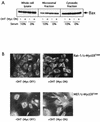
Activation of c-Myc induces apoptosis but has no effect on Bax expression, localization, or conformation. (A) Rat-1/c-MycERTAM cells were either serum deprived for 24 h or maintained in 10% FCS prior to stimulation with either 100 nM OHT or ethanol vehicle control for 6 h. Cells were then harvested and either lysed directly or biochemically fractionated into cytosolic and heavy membrane fractions prior to solubilization in SDS sample buffer, as described in Materials and Methods. Forty micrograms of heavy membrane fraction, 20 μg of cytosolic fraction, or 20 μg of whole-cell lysate was thenfractionated by SDS-PAGE and immunoblotted with anti-Bax antibody (see Materials and Methods). (B) Rat-1/c-MycERTAM cells (upper panels) serum deprived for 48 h or MEFs infected with pBp(c-MycERTAM) (lower panels) and serum deprived for 24 h were either left untreated (left panels) or treated with OHT for 8 h (right panels) prior to fixation and immunostaining with polyclonal Bax (57-72) antibody as described in Materials and Methods. Bar = 20 μM. (C) Rat-1/c-MycERTAM cells were serum deprived for 48 h prior to treatment with OHT for an additional 24 h. Cells were then fixed and immunostained by using polyclonal Bax NT antibody (green) and monoclonal anti-cytochrome c antibody (red). As a positive control for altered Bax conformation during apoptosis, Rat-1/c-MycERTAM cells grown in 10% FCS in the absence of OHT were either treated with human recombinant TNF alpha (5 ng/ml) and actinomycin D (2 μg/ml) or left untreated for 20 h prior to fixation and immunostaining with polyclonal Bax NT antibody and monoclonal anti-cytochrome c antibody. Images were collected on a Leica TCS NT confocal microscope with a 63 × 1.4 PL Apo objective (Leica, Rueil-Malmaison, France).
FIG. 2.
FIG. 2.
Activation of c-Myc induces apoptosis but has no effect on Bax expression, localization, or conformation. (A) Rat-1/c-MycERTAM cells were either serum deprived for 24 h or maintained in 10% FCS prior to stimulation with either 100 nM OHT or ethanol vehicle control for 6 h. Cells were then harvested and either lysed directly or biochemically fractionated into cytosolic and heavy membrane fractions prior to solubilization in SDS sample buffer, as described in Materials and Methods. Forty micrograms of heavy membrane fraction, 20 μg of cytosolic fraction, or 20 μg of whole-cell lysate was thenfractionated by SDS-PAGE and immunoblotted with anti-Bax antibody (see Materials and Methods). (B) Rat-1/c-MycERTAM cells (upper panels) serum deprived for 48 h or MEFs infected with pBp(c-MycERTAM) (lower panels) and serum deprived for 24 h were either left untreated (left panels) or treated with OHT for 8 h (right panels) prior to fixation and immunostaining with polyclonal Bax (57-72) antibody as described in Materials and Methods. Bar = 20 μM. (C) Rat-1/c-MycERTAM cells were serum deprived for 48 h prior to treatment with OHT for an additional 24 h. Cells were then fixed and immunostained by using polyclonal Bax NT antibody (green) and monoclonal anti-cytochrome c antibody (red). As a positive control for altered Bax conformation during apoptosis, Rat-1/c-MycERTAM cells grown in 10% FCS in the absence of OHT were either treated with human recombinant TNF alpha (5 ng/ml) and actinomycin D (2 μg/ml) or left untreated for 20 h prior to fixation and immunostaining with polyclonal Bax NT antibody and monoclonal anti-cytochrome c antibody. Images were collected on a Leica TCS NT confocal microscope with a 63 × 1.4 PL Apo objective (Leica, Rueil-Malmaison, France).
FIG. 3.
FIG. 3.
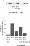
Inhibition of Bcl-xL antiapoptotic function by Bax-BH3 peptide. (A) Bax-BH3 peptide inhibits interaction between GST-Bcl-xL and Bax in vitro. Rat-1/c-MycERTAM fibroblasts grown in 10% FCS in the absence of OHT were lysed as described in Materials and Methods. Cell lysates were incubated with glutathione Sepharose beads prebound with either GST or GST-Bcl-xL(ΔTM) in the presence or absence of 200 μM Bax-BH3 peptide or Bax-BH3L63A mutant peptide as indicated. Glutathione Sepharose beads were then washed, bound polypeptides were eluted with SDS sample buffer and fractionated by SDS-PAGE, and the presence of endogenous Bax was assessed by immunoblotting with anti-Bax antibody. (B) Bax-BH3 peptide inhibits the antiapoptotic action of Bcl-xL in intact cells. Either GST (0.2 mg/ml) or GST-Bcl-xL(ΔTM) (0.2 mg/ml) in PBS were mixed with the fluorescent microinjection marker Oregon Green dextran (0.5%, wt/vol) in the presence or absence of either Bax-BH3 peptide (0.2 mg/ml) or Bax-BH3L63A mutant peptide (0.2 mg/ml) and then microinjected into Rat-1/c-MycERTAM fibroblasts grown in 10% FCS in the absence of OHT. Thirty minutes following microinjection, cells were washed once in PBS and subjected to 25 mJ of UV irradiation (Stratalinker UV cross-linker; Stratagene, La Jolla, Calif.) in the absence of medium, followed by incubation at 37°C for 6 h in normal growth medium. The viability of microinjected cells was then assessed by fluorescence microscopy as described in Materials and Methods. The data presented are from one experiment that is representative of four independent experiments. Each experiment comprised five observations; GST, GST-Bcl-xL, GST-Bcl-xL/Bax-BH3, and GST-Bcl-xL/Bax-BH3L63A solutions were injected within distinct areas of the same cell population seeded on glass coverslips 24 h prior to UV treatment. The same microcapillary loaded with GST-Bcl-xL/Bax-BH3 was used to microinject the cell population that was not treated with UV.
FIG. 4.
FIG. 4.
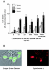
Induction of cell death by Bax-BH3 peptide in intact cells. (A) Induction of cell death by Bax-BH3 peptide is titratable. Bax-BH3 peptide at the indicated concentrations was mixed with 0.5% (wt/vol) Oregon Green dextran in PBS and microinjected into Rat-1/c-MycERTAM fibroblasts, which were grown in 10% FCS in the absence of OHT. Cells were then incubated at 37°C, and the deaths of microinjected cells were assessed morphologically by fluorescence microscopy at the indicated times. Where indicated, zVADfmk was added to a final concentration of 100 μM 2 h prior to microinjection. (B) Bax-BH3 peptide triggers asynchronous release of cytochrome c from mitochondria. Bax-BH3 peptide (1 mg/ml) mixed with 0.5% (wt/vol) Oregon Green dextran in PBS was microinjected into Rat-1/c-MycERTAM fibroblasts cultured in the absence of OHT. Cells were then incubated for 5 h at 37°C prior to fixation and immunostaining with anti-cytochrome c antibody. Cells were analyzed by fluorescence microscopy. Left panel, an overlay of phase contrast and green fluorescence allows identification of microinjected cells; right panel, cyto-chrome c immunostaining of the same cells. Bar = 20 μM. This frame is representative of at least four experiments. (C) Dependence of Bax-BH3 peptide-induced cell death on endogenous Bax/Bak. Wild-type (wt) and knockout MEFs of the indicated genotypes were microinjected with either Bax-BH3 (2 mg/ml) or Bax-BH3L63A (2 mg/ml) as indicated, and cell viability was assessed as described above.
FIG. 4.
FIG. 4.
Induction of cell death by Bax-BH3 peptide in intact cells. (A) Induction of cell death by Bax-BH3 peptide is titratable. Bax-BH3 peptide at the indicated concentrations was mixed with 0.5% (wt/vol) Oregon Green dextran in PBS and microinjected into Rat-1/c-MycERTAM fibroblasts, which were grown in 10% FCS in the absence of OHT. Cells were then incubated at 37°C, and the deaths of microinjected cells were assessed morphologically by fluorescence microscopy at the indicated times. Where indicated, zVADfmk was added to a final concentration of 100 μM 2 h prior to microinjection. (B) Bax-BH3 peptide triggers asynchronous release of cytochrome c from mitochondria. Bax-BH3 peptide (1 mg/ml) mixed with 0.5% (wt/vol) Oregon Green dextran in PBS was microinjected into Rat-1/c-MycERTAM fibroblasts cultured in the absence of OHT. Cells were then incubated for 5 h at 37°C prior to fixation and immunostaining with anti-cytochrome c antibody. Cells were analyzed by fluorescence microscopy. Left panel, an overlay of phase contrast and green fluorescence allows identification of microinjected cells; right panel, cyto-chrome c immunostaining of the same cells. Bar = 20 μM. This frame is representative of at least four experiments. (C) Dependence of Bax-BH3 peptide-induced cell death on endogenous Bax/Bak. Wild-type (wt) and knockout MEFs of the indicated genotypes were microinjected with either Bax-BH3 (2 mg/ml) or Bax-BH3L63A (2 mg/ml) as indicated, and cell viability was assessed as described above.
FIG. 5.
FIG. 5.
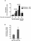
c-Myc and Bax-BH3 peptide cooperate to induce cell death. (A) c-Myc sensitizes cells to Bax-BH3 peptide-induced apoptosis. Rat-1/c-MycERTAM fibroblasts grown in 10% FCS were treated with either ethanol vehicle control or 100 nM OHT to activate c-Myc 2 h prior to microinjection with Bax-BH3 peptide (1 mg/ml) as described in the legend to Fig. 4A. Cells were then incubated at 37°C for the indicated times and analyzed by fluorescence microscopy for cell death. The data are mean values ± SEM from at least five experiments. Each experiment comprised two paired observations, and the same microcapillary was used to microinject cells, which were treated with either OHT or ethanol, with the same peptide solution on the same day. (B) Effect of c-Myc on Bax-BH3 peptide-induced holocytochrome c release. Rat-1/c-MycERTAM fibroblasts grown in 10% FCS were treated with either ethanol vehicle control or 100 nM OHT to activate c-Myc 2 h prior to microinjection with Bax-BH3 peptide (1 mg/ml) as described above. Cells were then incubated for 5 h at 37°C, fixed, and immunostained with anti-cytochrome c antibody as described in the legend to Fig. 4B. The percentage of microinjected cells exhibiting cytosolic cytochrome c was then determined. The data are mean values ± SEM from four independent experiments performed as described above.
FIG. 6.
FIG. 6.
Multiple BH3 peptides cooperate with c-Myc to induce cell death in Bax-deficient cells. (A to C) bax−/− MEFs infected with pBp(c-MycERTAM) were treated with either ethanol vehicle control or 100 nM OHT to activate c-Myc 2 h prior to microinjection with 2 mg of Bax-BH3 (A), Bak-BH3 (B), or Bid-BH3 (C) peptide per ml. Cell death was assessed as described in the legend to Fig. 4A. Data are the mean values ± SEM from at least three independent experiments. Each experiment comprised two paired observations as described in the legend to Fig. 5A. (D) Additional experiments were performed as described for panels A to C by using bax−/−/bak−/− MEFs infected with pBp(c-MycERTAM). (E) bax−/− MEFs infected with pBp(c-MycERTAM) were microinjected with pcDNA3(Bak) (100 ng/ml) and pEGFP-N1 (20 ng/ml). The number of GFP-expressing cells was evaluated by fluorescence microscopy 18 h after microinjection. Cells were then washed twice in PBS and treated with 100 nM OHT or ethanol vehicle control in the absence of FCS. Cell loss was evaluated at the times indicated, as described in the legend to Fig. 1A. The data presented are mean values ± SEM from three independent experiments.
FIG. 7.
FIG. 7.
Role of p53 in c-Myc-induced sensitization to Bax-BH3 peptide and Bax. (A) c-Myc induces apoptosis of p53−/−/bax−/− MEFs ectopically expressing Bax. Experiments were performed as described in the legend to Fig. 1B with p53−/−/bax−/− MEFs. (B) Cooperation between c-Myc and the Bax-BH3 peptide in p53-null cells. Experiments were performed as described in the legend to Fig. 6 with p53+/− and p53−/− MEFs infected with pBp(c-MycERTAM). Microinjection was performed with Bax-BH3 peptide (2 mg/ml). The data are mean values ± SEM from the indicated number of experiments.

Similar articles

Cited by

References

    1. Attardi, L. D., E. E. Reczek, C. Cosmas, E. G. Demicco, M. E. McCurrach, S. W. Lowe, and T. Jacks. 2000. PERP, an apoptosis-associated target of p53, is a novel member of the PMP-22/gas3 family. Genes Dev. 14:704-718. - PMC - PubMed
    1. Bissonnette, R., F. Echeverri, A. Mahboubi, and D. Green. 1992. Apoptotic cell death induced by c-myc is inhibited by bcl-2. Nature 359:552-554. - PubMed
    1. Cheng, E. H., M. C. Wei, S. Weiler, R. A. Flavell, T. W. Mak, T. Lindsten, and S. J. Korsmeyer. 2001. BCL-2, BCL-X(L) sequester BH3 domain-only molecules preventing BAX- and BAK-mediated mitochondrial apoptosis. Mol. Cell 8:705-711. - PubMed
    1. Cosulich, S. C., V. Worrall, P. J. Hedge, S. Green, and P. R. Clarke. 1997. Regulation of apoptosis by BH3 domains in a cell-free system. Curr. Biol. 7:913-920. - PubMed
    1. Datta, S. R., H. Dudek, X. Tao, S. Masters, H. Fu, Y. Gotoh, and M. E. Greenberg. 1997. Akt phosphorylation of BAD couples survival signals to the cell-intrinsic death machinery. Cell 91:231-241. - PubMed

MeSH terms