. 2001 Aug;75(16):7637-50.
doi: 10.1128/JVI.75.16.7637-7650.2001.

Binding of human immunodeficiency virus type 1 gp120 to CXCR4 induces mitochondrial transmembrane depolarization and cytochrome c-mediated apoptosis independently of Fas signaling

Affiliations

Binding of human immunodeficiency virus type 1 gp120 to CXCR4 induces mitochondrial transmembrane depolarization and cytochrome c-mediated apoptosis independently of Fas signaling

R Roggero et al. J Virol. 2001 Aug.

Abstract

Apoptosis of CD4(+) T lymphocytes, induced by contact between human immunodeficiency virus type 1 (HIV-1) envelope glycoprotein (gp120) and its receptors, could contribute to the cell depletion observed in HIV-infected individuals. CXCR4 appears to play an important role in gp120-induced cell death, but the mechanisms involved in this apoptotic process remain poorly understood. To get insight into the signal transduction pathways connecting CXCR4 to apoptosis following gp120 binding, we used different cell lines expressing wild-type CXCR4 and a truncated form of CD4 that binds gp120 but lacks the ability to transduce signals. The present study demonstrates that (i) the interaction of cell-associated gp120 with CXCR4-expressing target cells triggers a rapid dissipation of the mitochondrial transmembrane potential resulting in the cytosolic release of cytochrome c from the mitochondria to cytosol, concurrent with activation of caspase-9 and -3; (ii) this apoptotic process is independent of Fas signaling; and (iii) cooperation with a CD4 signal is not required. In addition, following coculture with cells expressing gp120, a Fas-independent apoptosis involving mitochondria and caspase activation is also observed in primary umbilical cord blood CD4(+) T lymphocytes expressing high levels of CXCR4. Thus, this gp120-mediated apoptotic pathway may contribute to CD4(+) T-cell depletion in AIDS.


Figures
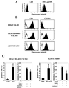
FIG. 1
FIG. 1
Description of the two cellular model systems. (A) Cell surface expression of gp120 at the surface of 8.E5 cells and the stable gp120-transfected HEK cell line (black histograms) are compared to the parental CEM and HEK lines (white histograms), as detected by flow cytometry. Cells were incubated with medium containing anti-gp120 human polyclonal antibodies and bound Ab was detected with a secondary FITC-labeled goat anti-human Ig. (B) Expression of mutated CD4 and CXCR4 molecules at the surface of the HEK/CD4.403 and HEK/CD4.403/CXCR4 clones and the A2.01/CD4.403 T-cell line. Cells were incubated with medium alone (white histograms) or medium containing the anti-CD4 (left) or anti-CXCR4 (right) MAbs at 10 μg/ml (black histograms). Bound MAb was detected with a FITC-labeled goat anti-mouse Ig. The fluorescence intensity was recorded in the log mode on an EPICS XL4 cytofluorometer. (C) Apoptosis of the HEK/CD4.403/CXCR4 and A2.01/CD4.403 cell lines cocultured with cells expressing gp120 (8.E5 and HEK.gp120) in the presence or absence of SDF-1 (500 ng/ml) or the caspase-3 inhibitor (50 μM).
FIG. 2
FIG. 2
The death receptor Fas is not involved in gp120-induced apoptosis of CXCR4+ cells. Expression of extracellular Fas (A) and intracellular FasL (B) in HEK transfected cells and the A2.01/CD4.403 cell line after coculture for 3 days with 8.E5 or CEM cells and HEK.gp120 or HEK cell lines, respectively. These unpermeabilized or permeabilized cells were incubated with medium alone (white histograms) or medium containing anti-Fas and anti-FasL antibodies (grey histograms), respectively. (C) On the left, representative photographs of apoptotic HEK/CD4.403/CXCR4 cells (Hoechst staining) after coculture with CEM or 8.E5 cells for 3 days in the presence or absence of the anti-Fas ZB4 MAb inhibitor or after treatment with the anti-Fas MAb CH11 (1 μg/ml) for 6 h. Apoptotic cells are indicated by arrowheads. On the right, representative data from three independent flow cytometry experiments demonstrating annexin-V and propidium iodide labeling of A2.01/CD4.403 cells cocultured for 3 days with HEK or HEK.gp120 cells in the presence or absence of the anti-Fas ZB4 MAb inhibitor or the anti-Fas CH11 MAb (1 μg/ml) for 6 h. (D) Percentage of apoptotic HEK/CD4.403/CXCR4 (condensed chromatin) and A2.01/CD4.403 (annexin-V positive/propidium iodide negative) cells after coculture with gp120 negative or positive cells for 3 days in the presence or absence of the anti-Fas ZB4 MAb inhibitor or the anti-Fas CH11 MAb (1 μg/ml) for 6 h. Data shown reflect means ± standard deviations from at least three replicates. Statistical analysis was performed as described in Materials and Methods.
FIG. 2
FIG. 2
The death receptor Fas is not involved in gp120-induced apoptosis of CXCR4+ cells. Expression of extracellular Fas (A) and intracellular FasL (B) in HEK transfected cells and the A2.01/CD4.403 cell line after coculture for 3 days with 8.E5 or CEM cells and HEK.gp120 or HEK cell lines, respectively. These unpermeabilized or permeabilized cells were incubated with medium alone (white histograms) or medium containing anti-Fas and anti-FasL antibodies (grey histograms), respectively. (C) On the left, representative photographs of apoptotic HEK/CD4.403/CXCR4 cells (Hoechst staining) after coculture with CEM or 8.E5 cells for 3 days in the presence or absence of the anti-Fas ZB4 MAb inhibitor or after treatment with the anti-Fas MAb CH11 (1 μg/ml) for 6 h. Apoptotic cells are indicated by arrowheads. On the right, representative data from three independent flow cytometry experiments demonstrating annexin-V and propidium iodide labeling of A2.01/CD4.403 cells cocultured for 3 days with HEK or HEK.gp120 cells in the presence or absence of the anti-Fas ZB4 MAb inhibitor or the anti-Fas CH11 MAb (1 μg/ml) for 6 h. (D) Percentage of apoptotic HEK/CD4.403/CXCR4 (condensed chromatin) and A2.01/CD4.403 (annexin-V positive/propidium iodide negative) cells after coculture with gp120 negative or positive cells for 3 days in the presence or absence of the anti-Fas ZB4 MAb inhibitor or the anti-Fas CH11 MAb (1 μg/ml) for 6 h. Data shown reflect means ± standard deviations from at least three replicates. Statistical analysis was performed as described in Materials and Methods.
FIG. 3
FIG. 3
Mitochondrial depolarization occurs during gp120-induced apoptosis. (A) Following a 1-day coculture of HEK/CD4.403 and HEK/CD4.403/CXCR4 cells with CEM (black histograms) or 8.E5 (white histograms) cells, the former cells were stained with DiOC6 (40 nM, 15 min, 37°C), and ΔΨm was analyzed by flow cytometry. HEK/CD4.403/CXCR4 cells treated with the uncoupling reagent mClCCP (5 μM, 15 min) and dexamethazone (100 μM, overnight) were used as controls. Results of data from one of five representative experiments are shown. (B) The same experiments were performed following coculture of the A2.01/CD4.403 cell line with HEK (black histogram) or HEK.gp120 (white histogram) cells. Controls were identical to those described above for panel A.
FIG. 4
FIG. 4
gp120-induced cytochrome c release from cells expressing CXCR4. (A) HEK/CD4.403 and HEK/CD4.403/CXCR4 cell lines were cocultured for 1 day with CEM or 8.E5 cells, and A2.01/CD4.403 cells were cocultured with HEK or HEK.gp120 cell lines. HEK/CD4.403, HEK/CD4.403/CXCR4 and A2.01/CD4.403 cells were dounced, and S100 cytosol was prepared as described in Materials and Methods. Cytosolic fractions (25 μg) were run on an SDS-polyacrylamide gel and Western blotted with an anti-cytochrome c antibody that recognizes a denaturated form of this molecule or an anti-cytochrome oxidase subunit II antibody (12CA-F12). Protein loading was controlled using an anti-actin antibody. Data of three independent experiments are shown. (B) Transfected HEK cell lines were cocultured for 3 days with 8.E5 or CEM cells and then triple stained with Hoechst to detect chromatin condensation (blue, left), an anti-cytochrome c antibody detected with FITC-conjugated secondary antibody (green, center) to detect the presence of cytochrome c in mitochondria, and ΔΨm-sensitive dye MitoTracker Orange to visualize mitochondrial polarization (red, right). Cells treated with dexamethasone (50 μM) were used as a positive control of mitochondrial depolarization. (C) Mitochondrion damage was controlled by triple staining with Hoechst (blue, left), an anti-Hsp60 antibody detected with a FITC-conjugated secondary antibody (green, center), and MitoTracker Orange (red, right).
FIG. 4
FIG. 4
gp120-induced cytochrome c release from cells expressing CXCR4. (A) HEK/CD4.403 and HEK/CD4.403/CXCR4 cell lines were cocultured for 1 day with CEM or 8.E5 cells, and A2.01/CD4.403 cells were cocultured with HEK or HEK.gp120 cell lines. HEK/CD4.403, HEK/CD4.403/CXCR4 and A2.01/CD4.403 cells were dounced, and S100 cytosol was prepared as described in Materials and Methods. Cytosolic fractions (25 μg) were run on an SDS-polyacrylamide gel and Western blotted with an anti-cytochrome c antibody that recognizes a denaturated form of this molecule or an anti-cytochrome oxidase subunit II antibody (12CA-F12). Protein loading was controlled using an anti-actin antibody. Data of three independent experiments are shown. (B) Transfected HEK cell lines were cocultured for 3 days with 8.E5 or CEM cells and then triple stained with Hoechst to detect chromatin condensation (blue, left), an anti-cytochrome c antibody detected with FITC-conjugated secondary antibody (green, center) to detect the presence of cytochrome c in mitochondria, and ΔΨm-sensitive dye MitoTracker Orange to visualize mitochondrial polarization (red, right). Cells treated with dexamethasone (50 μM) were used as a positive control of mitochondrial depolarization. (C) Mitochondrion damage was controlled by triple staining with Hoechst (blue, left), an anti-Hsp60 antibody detected with a FITC-conjugated secondary antibody (green, center), and MitoTracker Orange (red, right).
FIG. 5
FIG. 5
Caspase-9 is activated during the apoptotic process triggered by gp120 binding to CXCR4. HEK/CD4.403 and HEK/CD4.403/CXCR4 cell lines were cocultured for 2 days with CEM or 8.E5 cells, and A2.01/CD4.403 cells were cocultured with HEK or HEK.gp120 cell lines. The cytosolic fraction of these cells was then analyzed by immunoblotting for caspase-9 as described in Materials and Methods. Protein loading was controlled using an anti-actin antibody. Results representative of three independent experiments are shown.
FIG. 6
FIG. 6
Induction of caspase-3 activation after gp120 binding to CXCR4. (A) Activation of caspase-3 was determined by flow cytometry after labeling of HEK/CD4.403 and HEK/CD4.403/CXCR4 cells cocultured with CEM or 8.E5 cells and A2.01/CD4.403 cells cocultured with HEK or HEK.gp120 cells with a phycoerythrin-conjugated anti-caspase-3 antibody that preferentially recognizes activated caspase-3. The anti-Fas CH11 MAb was used as a positive control; HEK/CD4.403/CXCR4 and A201/CD4.403 cells were incubated in the presence or absence of CH11 MAb (1 μg/ml). Results are the means ± standard deviations of three independent experiments. (B) Immunofluorescence detection of activated and cleaved caspase-3 and apoptotic nuclei in HEK transfected cells expressing CD4.403 and/or CXCR4 molecules after coculture for 3 days with CEM or 8.E5 cells or following treatment with the CH11 MAb. Apoptotic cells are indicated by arrowheads.
FIG. 6
FIG. 6
Induction of caspase-3 activation after gp120 binding to CXCR4. (A) Activation of caspase-3 was determined by flow cytometry after labeling of HEK/CD4.403 and HEK/CD4.403/CXCR4 cells cocultured with CEM or 8.E5 cells and A2.01/CD4.403 cells cocultured with HEK or HEK.gp120 cells with a phycoerythrin-conjugated anti-caspase-3 antibody that preferentially recognizes activated caspase-3. The anti-Fas CH11 MAb was used as a positive control; HEK/CD4.403/CXCR4 and A201/CD4.403 cells were incubated in the presence or absence of CH11 MAb (1 μg/ml). Results are the means ± standard deviations of three independent experiments. (B) Immunofluorescence detection of activated and cleaved caspase-3 and apoptotic nuclei in HEK transfected cells expressing CD4.403 and/or CXCR4 molecules after coculture for 3 days with CEM or 8.E5 cells or following treatment with the CH11 MAb. Apoptotic cells are indicated by arrowheads.
FIG. 7
FIG. 7
gp120-induced apoptosis of UC CD4+ T cells. (A) Cell-surface expression of CD4 (black histogram) and CXCR4 (grey histogram) in purified UC CD4+ T cells, detected by flow cytometry as described in the Fig. 1 legend. (B) Percentage of apoptotic CD4+ cells cocultured with gp120+ or gp120 HEK cells for 2 to 4 days and (on the right) inhibition of apoptosis by SDF-1 (500 ng/ml). Results are from at least two independent experiments. (C) Mitochondrial depolarization of CD4+ T cells after coculture for 1 day with HEK (black histogram) or HEK.gp120 (white histogram) cells, analyzed by flow cytometry using DiOC6 as previously described. Data representative of five individual experiments are shown. (D) gp120-induced caspase-3 activation in CD4+ T cells after coculture with HEK.gp120 cells for 2 to 4 days and (on the right) inhibition of apoptosis by the caspase-3 inhibitor (50 μM). Results are the means ± standard deviations of two independent experiments. (E) The death receptor Fas is not involved in gp120-mediated UC CD4+ T-cell apoptosis. The expression of extracellular Fas and intracellular FasL was analyzed by flow cytometry after 4, 16, and 24 h of coculture with HEK or HEK.gp120 cells. Cells were incubated with medium alone (white histograms) or medium containing anti-Fas or anti-FasL (black histograms). Representative data from one of two independent experiments are shown. The percentage of inhibition of gp120-induced UC apoptosis by the anti-Fas antibody ZB4 is shown on the right. Error bar reflects means ± standard deviations from three replicates.

Similar articles

Cited by

References

    1. Adachi S, Gottlieb R A, Babior B M. Lack of release of cytochrome C from mitochondria into cytosol early in the course of Fas-mediated apoptosis of Jurkat cells. J Biol Chem. 1998;273:19892–19894. - PubMed
    1. Badley A D, Dockrell D, Paya C V. Apoptosis in AIDS. Adv Pharmacol. 1997;41:271–294. - PubMed
    1. Badley A D, Dockrell D, Simpson M, Schut R, Lynch D H, Leibson P, Paya C V. Macrophage-dependent apoptosis of CD4+ T lymphocytes from HIV-infected individuals is mediated by FasL and tumor necrosis factor. J Exp Med. 1997;185:55–64. - PMC - PubMed
    1. Badley A D, McElhinny J A, Leibson P J, Lynch D H, Alderson M R, Paya C V. Upregulation of Fas ligand expression by human immunodeficiency virus in human macrophages mediates apoptosis of uninfected T lymphocytes. J Virol. 1996;70:199–206. - PMC - PubMed
    1. Badley A D, Parato K, Cameron D W, Kravcik S, Phenix B N, Ashby D, Kumar A, Lynch D H, Tschopp J, Angel J B. Dynamic correlation of apoptosis and immune activation during treatment of HIV infection. Cell Death Differ. 1999;6:420–432. - PubMed

Publication types