. 2001 Jun 1;20(11):2702-14.
doi: 10.1093/emboj/20.11.2702.

The death substrate Gas2 binds m-calpain and increases susceptibility to p53-dependent apoptosis

Affiliations

The death substrate Gas2 binds m-calpain and increases susceptibility to p53-dependent apoptosis

R Benetti et al. EMBO J. .

Abstract

Gas2 is a caspase-3 substrate that plays a role in regulating microfilament and cell shape changes during apoptosis. Here we provide evidence that overexpression of Gas2 efficiently increases cell susceptibility to apoptosis following UV irradiation, etoposide and methyl methanesulfonate treatments, and that these effects are dependent on increased p53 stability and transcription activity. To investigate possible pathways linking Gas2 to p53, a yeast two-hybrid screen swas performed, indicating m-calpain as a strong Gas2- interacting protein. Moreover, we demonstrate that Gas2 physically interacts with m-calpain in vivo and that recombinant Gas2 inhibits calpain-dependent processing of p53. Importantly, the Gas2 dominant-negative form (Gas2171-314) that binds calpain but is unable to inhibit its activity abrogates Gas2's ability to stabilize p53, to enhance p53 transcriptional activity and to induce p53-dependent apoptosis. Finally, we show that Gas2 is able to regulate the levels of p53 independently of Mdm2 status, suggesting that, like calpastatin, it may enhance p53 stability by inhibiting calpain activity.


Figures

None
Fig. 1. Gas2 overexpression increases susceptibility to apoptosis. (A) A confocal-generated overlay showing nuclear morphology in U2OS cells expressing Gas2 and treated further with the indicated apoptotic stimuli. U2OS cells transfected with Gas2wt were treated with the different apoptotic stimuli and 12 h later cells were processed for immunofluorescence to visualize Gas2 (green). Propidium iodide was used to visualize nuclei (red). (B) Diagram showing the percentage of apoptotic nuclei in U2OS cells (p53 wild-type) expressing Gas2 and treated with the indicated apoptotic stimuli. (C) Diagram showing the percentage of apoptotic nuclei in U2OS cells expressing Gas2 and a control protein, and treated with etoposide and MMS. (D) Diagram showing the percentage of apoptotic nuclei in wild-type and p53–/– MEFs, which differ only in their p53 status, expressing Gas2 and treated with UV. (E) Diagram showing the comparison of viability between cells microinjected with different combinations of vectors as indicated. Data represent the means of at least three independent experiments and error bars represent standard deviations.
None
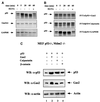
Fig. 2. Gas2 overexpression regulates p53 levels. (A) Mouse Balb/C (10)1 fibroblasts were transfected with wild-type p53, either alone or together with Gas2wt or β-catenin, as indicated. Cells were lysed 24 h after transfection and subjected to western blot analysis with p53-specific DO-1 antibody (upper panel). The same membrane was probed subsequently with anti-Gas2 antibody (middle panel) and anti-actin antibody as a loading control (lower panel). (B) Balb/C (10)1 mouse fibroblasts were transfected with wild-type p53 together with empty vector or with increasing amounts of Gas2wt as indicated. A 100 ng aliquot of pEGFP was co-transfected to monitor transfection efficiency. Western blotting was performed with DO-1 antibody (upper panel), with anti-Gas2 antibody to confirm the expression of transfected Gas2 (middle panel) or with anti-GFP antibody to evaluate transfection efficiency (lower panel). (C) U2OS cells were transfected with empty vector, Gas2 and β-catenin as indicated. Cells were lysed 24 h after transfection and subjected to western blot analysis with DO-1 antibody. The same membrane subsequently was probed with anti-Gas2 antibody and anti-actin antibody to estimate the total amount of protein loaded in each lane. (D) U2OS cells were transfected with the indicated amounts of empty vector or Gas2. A 100 ng aliquot of pGFP was co-transfected to monitor the efficiency of transfection. Western blotting was performed with DO-1, with anti-Gas2 antibody to confirm the expression of transfected Gas2 or with anti-GFP antibody to evaluate transfection efficiency.
None
Fig. 3. Gas2 enhances the transcriptional activity of p53. (A) U2OS cells were transfected with the p21-Luc reporter together with empty vector or Gas2. Luciferase assays were performed 24 h later. (B) Balb/C (10)1 fibroblasts were transfected with p21-Luc reporter and wild-type p53, together with empty vector or Gas2. Luciferase assays were performed 24 h later. Data represent arithmetic means ± SD from three independent experiments. (C) MEFs were transfected with the indicated combinations of plasmids. Protein levels of p53, p21/Waf1, Mdm2 and Gas2 were determined by western blot analysis.
None
Fig. 4. Gas2 interacts with m-calpain. (A) Schematic representation of m-calpain. In the large subunit, the N-terminal region (domains I and II; amino acids 1–355) contains the catalytic domains, while the C-terminal region (domains III and IV; amino acids 355–700) represents the regulatory regions of the protease. EF-hand structures capable of binding calcium are indicated as white bars. (B) Interaction of calpain with Gas2wt in yeast. The LexA–Gas2 fusion employed is represented on the left and the various deletions of calpain fused to B42 are shown on the right. + indicates a positive interaction, as judged by β-galactosidase activity and ability to grow in the absence of leucine; – indicates no detectable interaction. (C) Balb/C (10)1 cells were transfected with Gas2 together with HA-m-calpain (lane 1) or HA-E4FΔ350 (lane 2). After 24 h, lysates were immunoprecipitated with an anti-Gas2 antibody. Total lysates: an aliquot of each lysate was checked for the expression of the transfected plasmid by staining with anti-HA. The same membrane was probed subsequently with an anti-Gas2 antibody. (D) Immunoprecipitation was performed on endogenous Gas2 and endogenous calpain. Balb/C 3T3 cell lysates were immunoprecipitated with an anti-Gas2 antibody (lane 1) or with a pre-immune serum (lane 2). Immuno complexes were resolved on a 10% SDS–polyacrylamide gel and subjected to western blot analysis with anti-calpain antibody. An aliquot of each lysate (total lysates) was checked for the expression of endogenous Gas2wt and calpain. Running positions of molecular weight markers and of the various proteins are indicated.
None
Fig. 5. Gas2 is not a substrate of calpain, inhibits calpain activity in vitro and stabilizes p53 in the absence of Mdm2. (A) Proteolysis experiments were carried out as described in the text. Aliquots were taken at the indicated time points, with time zero corresponding to the addition of calcium for activating calpain. EGTA was added at the concentration of 1 mM. Full-length proteins are indicated by arrows. (B) Equal amounts of in vitro translated Gas2, calpastatin and GAPDH proteins were added separately to in vitro translated p53 (indicated by an arrow). Reactions were incubated at room temperature with purified bovine m-calpain, and aliquots of samples were collected after 5, 20 and 60 min, as indicated. Time 0 corresponds to the addition of calcium. EGTA was added at the concentration of 1 mM. (C) p53–/– Mdm2–/– MEFs were transfected with p53 together with an equal amount of Gas2, calpastatin and β-catenin as indicated. Cells were harvested 24 h after transfection and subjected to western blot analysis using p53-specific antibody (DO-1) (upper panel), Gas2-specific antibody (middle panel) and anti-actin antibody (lower panel).
None
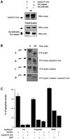
Fig. 6. Characterization of the dominant-negative form of Gas2. (A) Gas2Δ171–314 associates with the large subunit of m-calpain in mammalian cells. Balb/C (10)1 cells were transfected with GFP-tagged Gas2Δ171–314 together with HA-tagged E4FΔ350 (lane 1) and HA-tagged m-calpain (lane 2). Lysates were immunoprecipitated with anti-HA antibody. Immunocomplexes were resolved by SDS–PAGE and subjected to western blot analysis with anti-Gas2 antibody. (B) Gas2Δ171–314 is not able to inhibit calpain activity in vitro and to counteract the Gas2wt effect. (a and b) Equal amounts of in vitro translated Gas2Δ171–314 and GAPDH proteins were added separately to in vitro translated p53 (indicated by an arrow). Reactions were incubated at room temperature with purified bovine m-calpain (final concentration of 50 mg/ml) and aliquots of samples were collected at time 0, which corresponds to addition of calcium required for activating calpains, and after 30 min, as indicated. (c and d) Gas2wt and GAPDH or Gas2wt and Gas2Δ171–314 were added separately to in vitro translated p53 (indicated by an arrow). Reactions were incubated at room temperature with purified bovine m-calpain (50 mg/ml) and aliquots of samples were collected at time 0 and after 30 min, as indicated. (C) Diagram showing the percentage of apoptotic nuclei in U2OS cells expressing Gas2wt either alone or together with Gas2Δ171–314 in comparison with a control after UV irradiation, etoposide or MMS treatments. Data represent arithmetic means ± SD from three independent experiments. (D) Expression of Gas2Δ171–314 efficiently reduces the steady-state levels of p53 as stabilized by the wild-type form and by calpastatin. U2OS cells were co-transfected with Gas2wt or calpastatin alone or together with Gas2Δ171–314. Cells were subjected to western blot analysis using DO-1 antibody (upper panel), Gas2 antibody (middle panel) and anti-actin antibody (lower panel). (E) The presence of Gas2Δ171–314 efficiently reduces the Gas2wt-dependent increased transcription from the p21 promoter. U2OS cells were transfected with the p21-Luc reporter together with Gas2 or calpastatin either alone or together with Gas2Δ171–314. Luciferase assays were performed 24 h later. Data represent arithmetic means ± SD from five independent experiments.
None
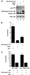
Fig. 7. Gas2Δ171–314 decreases endogenous p53 levels and its transcriptional activity. (A) U2OS cells were transfected with GFP, Mdm2 or Gas2Δ171–314 as indicated. Western blotting of total lysates was performed with DO-1 to monitor the p53 levels, with anti-Gas2 antibody to confirm the expression of transfected Gas2 and polyclonal anti-actin antibody as loading control. (B) U2OS cells were transfected with the p21-Luc reporter together with a control vector, GasΔ171–314 or Mdm2. Luciferase assays were performed 24 h later. Data represent arithmetic means ± SD from three independent experiments. (C) p53–/– or p53–/– Mdm2–/– MEFs were transfected with the p21-Luc reporter together with GFP or GFP-tagged Gas2Δ171–314. Luciferase assays were performed 24 h later. Data represent arithmetic means ± SD from five independent experiments. (D) Diagram showing the viability of Balb/C 3T3 cells microinjected with GFP or GFP-tagged Gas2Δ171–314 after UV damage and in conditions where Gas2wt is physiologic ally induced. After microinjection and Gas2 induction, cells were treated with UV and 12 h later GFP staining was visualized to calculate cell recovery. (E) Diagram showing the apoptotic susceptibility of wild-type or p53–/– MEFs transfected with GFP or GFP-tagged Gas2Δ171–314 after UV damage and in conditions where Gas2wt is physiologically induced. After transfection and Gas2 induction, cells were treated with UV and 12 h later GFP staining was visualized to calculate cell recovery.
None
Fig. 8. A working model for the regulatory network involving Gas2, calpain and p53. Mdm2 and calpain can negatively regulate p53 levels. Overexpression of either Gas2 or calpastatin can lead to p53 accumulation by inhibiting calpain. A rapid increase in p53 concentration with no de novo transcription is particularly advantageous in cells that are more prone to respond to different stimuli. Further modifications or different cellular environments are required to convert p53 into a fully active protein and to trigger the apoptotic program.

Similar articles

Cited by

References

    1. Atencio I., Ramachandra,M., Shabram,P. and Demers,G. (2000) Calpain inhibitor 1 activates p53-dependent apoptosis in tumor cell lines. Cell Growth Differ., 11, 247–253. - PubMed
    1. Brancolini C. and Schneider,C. (1997) Cut and die: proteolytic cascades regulating apoptosis. Adv. Clin. Pathol., 3, 177–189. - PubMed
    1. Brancolini C., Bottega,S. and Schneider,C. (1992) Gas2, a growth arrest-specific protein, is a component of the microfilament network system. J. Cell Biol., 117, 1251–1261. - PMC - PubMed
    1. Brancolini C., Benedetti,M. and Schneider,C. (1995) Microfilament reorganization during apoptosis: the role of Gas2, a possible substrate for ICE-like proteases. EMBO J., 14, 5179–5190. - PMC - PubMed
    1. Brancolini C., Marzinotto,S., Edomi,P., Agostoni,E., Fiorentini,C., Muller,H. and Schneider,C. (1999) Rho-dependent regulation of cell spreading by the tetraspan membrane protein Gas3/PMP22. Mol. Biol. Cell, 10, 2441–2459. - PMC - PubMed

Publication types

MeSH terms