. 2001 May 7;193(9):1005-14.
doi: 10.1084/jem.193.9.1005.

Vascular endothelial growth factor and angiopoietin-1 stimulate postnatal hematopoiesis by recruitment of vasculogenic and hematopoietic stem cells

Affiliations

Vascular endothelial growth factor and angiopoietin-1 stimulate postnatal hematopoiesis by recruitment of vasculogenic and hematopoietic stem cells

K Hattori et al. J Exp Med. .

Abstract

Tyrosine kinase receptors for angiogenic factors vascular endothelial growth factor (VEGF) and angiopoietin-1 (Ang-1) are expressed not only by endothelial cells but also by subsets of hematopoietic stem cells (HSCs). To further define their role in the regulation of postnatal hematopoiesis and vasculogenesis, VEGF and Ang-1 plasma levels were elevated by injecting recombinant protein or adenoviral vectors expressing soluble VEGF(165), matrix-bound VEGF(189), or Ang-1 into mice. VEGF(165), but not VEGF(189), induced a rapid mobilization of HSCs and VEGF receptor (VEGFR)2(+) circulating endothelial precursor cells (CEPs). In contrast, Ang-1 induced delayed mobilization of CEPs and HSCs. Combined sustained elevation of Ang-1 and VEGF(165) was associated with an induction of hematopoiesis and increased marrow cellularity followed by proliferation of capillaries and expansion of sinusoidal space. Concomitant to this vascular remodeling, there was a transient depletion of hematopoietic activity in the marrow, which was compensated by an increase in mobilization and recruitment of HSCs and CEPs to the spleen resulting in splenomegaly. Neutralizing monoclonal antibody to VEGFR2 completely inhibited VEGF(165), but not Ang-1-induced mobilization and splenomegaly. These data suggest that temporal and regional activation of VEGF/VEGFR2 and Ang-1/Tie-2 signaling pathways are critical for mobilization and recruitment of HSCs and CEPs and may play a role in the physiology of postnatal angiogenesis and hematopoiesis.


Figures

Figure 1
Figure 1
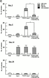
AdVEGF165 and AdAng-1 promote mobilization of mature and immature hematopoietic cells. Immunodeficient SCID mice received AdVEGF165 (1.5 × 10⁸ PFU) and/or AdAng-1 (109 PFU), AdNull vector (109 PFU), or the AdVEGF189 vector (2 × 10⁸ PFU) by a single intravenous administration on day 0. (A) Total WBCs were counted using a Neubauer hematocytometer and stained by crystal violet. n = 6. (B) Morphological characterization of PBMCs was determined by Wright-Giemsa staining. Original magnification: ×200. (C) Differential leukocyte counts were obtained by examining the blood smears from each mouse (200 cells counted/smear). n = 4.
Figure 1
Figure 1
AdVEGF165 and AdAng-1 promote mobilization of mature and immature hematopoietic cells. Immunodeficient SCID mice received AdVEGF165 (1.5 × 10⁸ PFU) and/or AdAng-1 (109 PFU), AdNull vector (109 PFU), or the AdVEGF189 vector (2 × 10⁸ PFU) by a single intravenous administration on day 0. (A) Total WBCs were counted using a Neubauer hematocytometer and stained by crystal violet. n = 6. (B) Morphological characterization of PBMCs was determined by Wright-Giemsa staining. Original magnification: ×200. (C) Differential leukocyte counts were obtained by examining the blood smears from each mouse (200 cells counted/smear). n = 4.
Figure 2
Figure 2
AdVEGF165 and/or AdAng-1 induce peripheral mobilization of hematopoietic progenitor cells and stem cells. (A) The number of mobilized progenitors was determined using standard CFU assays. Large numbers of progenitors were mobilized by VEGF (day 5, predominantly CFU-GM) and Ang-1 (day 14, predominantly CFU-M) (n = 4). The pluripotency of the mobilized cells was determined by CFU-S assay (B) and BM repopulating assay (C and D). Compared with AdVEGF, the combination of AdVEGF plus AdAng-1 induced significant long-term mobilization of CFU-S up to 21 d. Days 3, 7, and 14, *P < 0.01; day 21, **P < 0.05. Ang-1, VEGF, or combined VEGF and Ang-1 promoted mobilization of BM repopulating cells (n = 9). PBMCs (106 cells) from SCID mice (H-2Kd) treated with AdNull, AdVEGF165, AdAng-1, or a combination (AdVEGF165 and AdAng-1) were transplanted into irradiated C57BL/6 (H-2Kb) mice by intravenous injection on day 0. (C) The number of engrafted H-2Kd cells was determined by flow cytometry. Compared with the AdNull group, AdAng-1– and AdVEGF-treated mice showed significant mobilization of cells capable of reconstituting hematopoiesis in lethally irradiated mice. **P < 0.05. In contrast, all the mice transplanted with PBMCs from the peripheral blood of AdNull-treated mice failed to engraft. (D) As a control group, 90% of the mice transplanted with untreated BM (BMT group) were engrafted and survived the effects of lethal irradiation.
Figure 2
Figure 2
AdVEGF165 and/or AdAng-1 induce peripheral mobilization of hematopoietic progenitor cells and stem cells. (A) The number of mobilized progenitors was determined using standard CFU assays. Large numbers of progenitors were mobilized by VEGF (day 5, predominantly CFU-GM) and Ang-1 (day 14, predominantly CFU-M) (n = 4). The pluripotency of the mobilized cells was determined by CFU-S assay (B) and BM repopulating assay (C and D). Compared with AdVEGF, the combination of AdVEGF plus AdAng-1 induced significant long-term mobilization of CFU-S up to 21 d. Days 3, 7, and 14, *P < 0.01; day 21, **P < 0.05. Ang-1, VEGF, or combined VEGF and Ang-1 promoted mobilization of BM repopulating cells (n = 9). PBMCs (106 cells) from SCID mice (H-2Kd) treated with AdNull, AdVEGF165, AdAng-1, or a combination (AdVEGF165 and AdAng-1) were transplanted into irradiated C57BL/6 (H-2Kb) mice by intravenous injection on day 0. (C) The number of engrafted H-2Kd cells was determined by flow cytometry. Compared with the AdNull group, AdAng-1– and AdVEGF-treated mice showed significant mobilization of cells capable of reconstituting hematopoiesis in lethally irradiated mice. **P < 0.05. In contrast, all the mice transplanted with PBMCs from the peripheral blood of AdNull-treated mice failed to engraft. (D) As a control group, 90% of the mice transplanted with untreated BM (BMT group) were engrafted and survived the effects of lethal irradiation.
Figure 3
Figure 3
AdVEGF165 and Ang-1 promote mobilization of CEPs with late outgrowth potential. (A) Mobilized PBMCs were isolated from AdVEGF165- and/or AdAng-1– or AdNull-treated SCID mice on days 0–28 and stained with FITC-conjugated anti-VEGFR2 (clone DC101) mAb and propidium iodide. (A) 104 cells were analyzed on a Beckman Coulter Elite flow cytometer, and representative percentages of positive populations in PBMCs are shown. (B) The percentages of mobilized circulating VEGFR2+CD11b cells from AdVEGF165- and/or AdAng-1– or AdNull-treated SCID mice (n = 3) are shown from days 0 –28. For quantification of CEPs with late outgrowth potential, mobilized PBMCs were obtained from AdVEGF165 and/or AdAng-1 or AdNull-treated SCID mice on days 0–21 and plated in the presence of endothelial growth medium (reference 13) on collagen/fibronectin-coated plastic dishes. Endothelial colonies (CFU-ECs) were identified by vWF immunostaining and metabolic labeling with DiI-Ac-LDL. (C) CFU-ECs proliferated 2 wk after the start of culture (mean ± SEM). Colonies that formed within the first 3 d (early outgrowth) and colonies formed 14 d (late outgrowth) after vector administration were quantified by vWF and DiI-Ac-LDL labeling. (D) The majority of CFU-EC proliferated 3 wk after the start of culture, forming focal DiI-Ac-LDL+ endothe-lial monolayers as seen at 14 (E), 20 (F), and 25 d (G).
Figure 3
Figure 3
AdVEGF165 and Ang-1 promote mobilization of CEPs with late outgrowth potential. (A) Mobilized PBMCs were isolated from AdVEGF165- and/or AdAng-1– or AdNull-treated SCID mice on days 0–28 and stained with FITC-conjugated anti-VEGFR2 (clone DC101) mAb and propidium iodide. (A) 104 cells were analyzed on a Beckman Coulter Elite flow cytometer, and representative percentages of positive populations in PBMCs are shown. (B) The percentages of mobilized circulating VEGFR2+CD11b cells from AdVEGF165- and/or AdAng-1– or AdNull-treated SCID mice (n = 3) are shown from days 0 –28. For quantification of CEPs with late outgrowth potential, mobilized PBMCs were obtained from AdVEGF165 and/or AdAng-1 or AdNull-treated SCID mice on days 0–21 and plated in the presence of endothelial growth medium (reference 13) on collagen/fibronectin-coated plastic dishes. Endothelial colonies (CFU-ECs) were identified by vWF immunostaining and metabolic labeling with DiI-Ac-LDL. (C) CFU-ECs proliferated 2 wk after the start of culture (mean ± SEM). Colonies that formed within the first 3 d (early outgrowth) and colonies formed 14 d (late outgrowth) after vector administration were quantified by vWF and DiI-Ac-LDL labeling. (D) The majority of CFU-EC proliferated 3 wk after the start of culture, forming focal DiI-Ac-LDL+ endothe-lial monolayers as seen at 14 (E), 20 (F), and 25 d (G).
Figure 4
Figure 4
Chronic elevation of AdVEGF and AdAng-1 induces vascular remodeling of BM with concomitant splenomegaly. Administration of AdVEGF165 and/or AdAng-1 or AdNull was performed as described in the legend to Fig. 1. On days 7, 14, and 60 after inoculation three mice in each group were killed, and their organs were collected and processed for histological analysis. Paraffin sections of the spleen and BM were stained by hematoxylin and eosin. Representative BM sections from day 7 after AdNull (A) and day 7 after AdVEGF165 plus AdAng-1 (B) are shown. BM cellularity was increased in AdVEGF165 plus AdAng-1–treated SCID mice at day 7 compared with AdNull-treated mice. (C) On day 14, sinusoidal space and proliferation of capillaries were progressively increased in the BM of AdVEGF165 plus AdAng-1–treated SCID mice. (D) BM cellularity and vascularity returned to baseline levels on day 60. Macroscopic and microscopic observations of spleens from BALB/c mice treated with recombinant VEGF (E and G) or PBS as a control (F and H) on day 10 are shown. These mice were injected subcutaneously with 100 ng of recombinant VEGF or only vehicle, on a daily basis. There was a remarkable increase in the size of the spleen and follicular hyperplasia 10 d after VEGF treatment compared with control. Original magnifications: (A, C, D, G, and H) ×200; (B) ×400; (E and F) ×20.
Figure 4
Figure 4
Chronic elevation of AdVEGF and AdAng-1 induces vascular remodeling of BM with concomitant splenomegaly. Administration of AdVEGF165 and/or AdAng-1 or AdNull was performed as described in the legend to Fig. 1. On days 7, 14, and 60 after inoculation three mice in each group were killed, and their organs were collected and processed for histological analysis. Paraffin sections of the spleen and BM were stained by hematoxylin and eosin. Representative BM sections from day 7 after AdNull (A) and day 7 after AdVEGF165 plus AdAng-1 (B) are shown. BM cellularity was increased in AdVEGF165 plus AdAng-1–treated SCID mice at day 7 compared with AdNull-treated mice. (C) On day 14, sinusoidal space and proliferation of capillaries were progressively increased in the BM of AdVEGF165 plus AdAng-1–treated SCID mice. (D) BM cellularity and vascularity returned to baseline levels on day 60. Macroscopic and microscopic observations of spleens from BALB/c mice treated with recombinant VEGF (E and G) or PBS as a control (F and H) on day 10 are shown. These mice were injected subcutaneously with 100 ng of recombinant VEGF or only vehicle, on a daily basis. There was a remarkable increase in the size of the spleen and follicular hyperplasia 10 d after VEGF treatment compared with control. Original magnifications: (A, C, D, G, and H) ×200; (B) ×400; (E and F) ×20.
Figure 5
Figure 5
Neutralizing mAb to VEGFR2 inhibits mobilization of leukocytes and hematopoietic progenitor cells induced by AdVEGF165 but not AdAng-1. (A) SCID mice were inoculated with AdVEGF165 (1.5 × 10⁸ PFU) or AdNull (109 PFU) on day 0. Half of the AdVEGF165-treated mice received 800 μg of anti-VEGFR2 (DC101) mAb at 2-d intervals starting from day 2. Total WBCs are expressed as the mean ± SEM (n = 8). •, AdVEGF165; ▴, AdVEGF165 + DC101; ▪, AdNull. (B) SCID mice inoculated with AdVEGF165 (1.5 × 10⁸ PFU) and AdAng-1 (109 PFU) received 800 μg of anti-VEGFR2 (DC101) at 2-d intervals starting from day 0. AdAng-1 (109 PFU) inoculated SCID mice were used as a control. Total WBCs were quantified using a Neubauer hematocytometer. All data are expressed as the mean ± SEM (n = 8). ▵, AdAng-1; •, AdVEGF165 + AdAng-1 + DC101; ▪, AdVEGF165 + AdAng-1; ♦, AdNull. (C) PBMCs from AdAng-1 alone, AdVEGF165 + AdAng-1 (treated or untreated with DC101), or AdNull-treated mice were seeded into the colony assays and four types of CFU (CFU-GM, BFU-E, CFU-M, and CFU-GEMM) were scored at 3, 5, 14, and 28 d after the onset of treatment. All data are expressed as the mean ± SEM (n = 4). Data points achieving statistical significance are shown. *P < 0.005.
Figure 5
Figure 5
Neutralizing mAb to VEGFR2 inhibits mobilization of leukocytes and hematopoietic progenitor cells induced by AdVEGF165 but not AdAng-1. (A) SCID mice were inoculated with AdVEGF165 (1.5 × 10⁸ PFU) or AdNull (109 PFU) on day 0. Half of the AdVEGF165-treated mice received 800 μg of anti-VEGFR2 (DC101) mAb at 2-d intervals starting from day 2. Total WBCs are expressed as the mean ± SEM (n = 8). •, AdVEGF165; ▴, AdVEGF165 + DC101; ▪, AdNull. (B) SCID mice inoculated with AdVEGF165 (1.5 × 10⁸ PFU) and AdAng-1 (109 PFU) received 800 μg of anti-VEGFR2 (DC101) at 2-d intervals starting from day 0. AdAng-1 (109 PFU) inoculated SCID mice were used as a control. Total WBCs were quantified using a Neubauer hematocytometer. All data are expressed as the mean ± SEM (n = 8). ▵, AdAng-1; •, AdVEGF165 + AdAng-1 + DC101; ▪, AdVEGF165 + AdAng-1; ♦, AdNull. (C) PBMCs from AdAng-1 alone, AdVEGF165 + AdAng-1 (treated or untreated with DC101), or AdNull-treated mice were seeded into the colony assays and four types of CFU (CFU-GM, BFU-E, CFU-M, and CFU-GEMM) were scored at 3, 5, 14, and 28 d after the onset of treatment. All data are expressed as the mean ± SEM (n = 4). Data points achieving statistical significance are shown. *P < 0.005.

Similar articles

Cited by

References

    1. Moore M.A. Stem cell proliferationex vivo and in vivo observations. Stem Cells. 1997;1:239–248. - PubMed
    1. Rafii S. Circulating endothelial precursorsmystery, reality, and promise. J. Clin. Invest. 2000;105:17–19. - PMC - PubMed
    1. Takahashi T., Kalka C., Masuda H., Chen D., Silver M., Kearney M., Magner M., Isner J.M., Asahara T. Ischemia- and cytokine-induced mobilization of bone marrow-derived endothelial progenitor cells for neovascularization. Nat. Med. 1999;5:434–438. - PubMed
    1. Peichev M., Naiyer A.J., Pereira D., Zhu Z., Lane W.J., Williams M., Oz M.C., Hicklin D.J., Witte L., Moore M.A., Rafii S. Expression of VEGFR-2 and AC133 by circulating human CD34(1) cells identifies a population of functional endothelial precursors. Blood. 2000;95:952–958. - PubMed
    1. Frey B.M., Rafii S., Teterson M., Eaton D., Crystal R.G., Moore M.A. Adenovector-mediated expression of human thrombopoietin cDNA in immune-compromised miceinsights into the pathophysiology of osteomyelofibrosis. J. Immunol. 1998;160:691–699. - PubMed

Publication types

MeSH terms