Gamma interferon expression in CD8(+) T cells is a marker for circulating cytotoxic T lymphocytes that recognize an HLA A2-restricted epitope of human cytomegalovirus phosphoprotein pp65
- PMID: 11329470
- PMCID: PMC96113
- DOI: 10.1128/CDLI.8.3.628-631.2001
Gamma interferon expression in CD8(+) T cells is a marker for circulating cytotoxic T lymphocytes that recognize an HLA A2-restricted epitope of human cytomegalovirus phosphoprotein pp65
Abstract
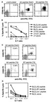
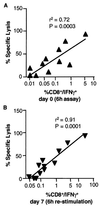
Antigen-specific CD8(+) T cells with cytotoxic activity are often critical in immune responses to infectious pathogens. To determine whether gamma interferon (IFN-gamma) expression is a surrogate marker for cytotoxic T lymphocytes (CTL), human cytomegalovirus-specific CTL responses were correlated with CD8(+) T-cell IFN-gamma expression determined by cytokine flow cytometry. A strong positive correlation was observed between specific lysis of peptide-pulsed targets in a (51)Cr release assay and frequencies of peptide-activated CD8(+) T cells expressing IFN-gamma at 6 h (r(2) = 0.72) or 7 days (r(2) = 0.91). Enumeration of responding cells expressing perforin, another marker associated with CTL, did not improve this correlation. These results demonstrate that IFN-gamma expression can be a functional surrogate for identification of CTL precursor cells.
Figures


Similar articles
-
The human cytotoxic T-lymphocyte (CTL) response to cytomegalovirus is dominated by structural protein pp65: frequency, specificity, and T-cell receptor usage of pp65-specific CTL.J Virol. 1996 Nov;70(11):7569-79. doi: 10.1128/JVI.70.11.7569-7579.1996. J Virol. 1996. PMID: 8892876 Free PMC article.
-
Rapid monitoring of immune reconstitution after allogeneic stem cell transplantation--a comparison of different assays for the detection of cytomegalovirus-specific T cells.Eur J Haematol. 2013 Dec;91(6):534-45. doi: 10.1111/ejh.12187. Epub 2013 Oct 14. Eur J Haematol. 2013. PMID: 23952609
-
Design and synthesis of HLA-A*02-restricted Hantaan virus multiple-antigenic peptide for CD8+ T cells.Virol J. 2020 Jan 31;17(1):15. doi: 10.1186/s12985-020-1290-x. Virol J. 2020. PMID: 32005266 Free PMC article.
-
Use of a lentiviral vector encoding a HCMV-chimeric IE1-pp65 protein for epitope identification in HLA-Transgenic mice and for ex vivo stimulation and expansion of CD8(+) cytotoxic T cells from human peripheral blood cells.Hum Immunol. 2004 May;65(5):514-22. doi: 10.1016/j.humimm.2004.02.018. Hum Immunol. 2004. PMID: 15172452
-
The matrix protein pp65(341-350): a peptide that induces ex vivo stimulation and in vitro expansion of CMV-specific CD8+ T cells in subjects bearing either HLA-A*2402 or A*0101 allele.Transfusion. 2003 Nov;43(11):1567-74. doi: 10.1046/j.1537-2995.2003.00564.x. Transfusion. 2003. PMID: 14617317
Cited by
-
Impact of cryopreservation on tetramer, cytokine flow cytometry, and ELISPOT.BMC Immunol. 2005 Jul 18;6:17. doi: 10.1186/1471-2172-6-17. BMC Immunol. 2005. PMID: 16026627 Free PMC article.
-
Killer cell proteases can target viral immediate-early proteins to control human cytomegalovirus infection in a noncytotoxic manner.PLoS Pathog. 2020 Apr 13;16(4):e1008426. doi: 10.1371/journal.ppat.1008426. eCollection 2020 Apr. PLoS Pathog. 2020. PMID: 32282833 Free PMC article.
-
The combination therapy with EpCAM/CD3 BsAb and MUC-1/CD3 BsAb elicited antitumor immunity by T-cell adoptive immunotherapy in lung cancer.Int J Med Sci. 2021 Jul 31;18(15):3380-3388. doi: 10.7150/ijms.61681. eCollection 2021. Int J Med Sci. 2021. PMID: 34522164 Free PMC article.
-
Direct loading of CTL epitopes onto MHC class I complexes on dendritic cell surface in vivo.Biomaterials. 2018 Nov;182:92-103. doi: 10.1016/j.biomaterials.2018.08.008. Epub 2018 Aug 6. Biomaterials. 2018. PMID: 30107273 Free PMC article.
-
The efficacy of T cell-mediated immune responses is reduced by the envelope protein of the chimeric HIV-1/SIV-KB9 virus in vivo.J Immunol. 2008 Oct 15;181(8):5510-21. doi: 10.4049/jimmunol.181.8.5510. J Immunol. 2008. PMID: 18832708 Free PMC article.
References
-
- Ahmed R, Gray D. Immunological memory and protective immunity: understanding their relation. Science. 1996;272:54–60. - PubMed
-
- Altman J D, Moss P A H, Goulder P J R, Barouch D H, McHeyzer-Williams M G, Bell J I, McMichael A J, Davis M M. Phenotypic analysis of antigen-specific T lymphocytes. Science. 1996;274:94–96. . (Erratum, 280:1821, 1998.) - PubMed
-
- Belz G T, Stevenson P G, Doherty P C. Contemporary analysis of MHC-related immunodominance hierarchies in the CD8+ t cell response to influenza A viruses. J Immunol. 2000;165:2404–2409. - PubMed
-
- Chai J G, Bartok I, Scott D, Dyson J, Lechler R. T:T antigen presentation by activated murine CD8+ T cells induces anergy and apoptosis. J Immunol. 1998;160:3655–3665. - PubMed
MeSH terms
Substances
LinkOut - more resources
Full Text Sources
Other Literature Sources
Research Materials

