Genetic disruption of mineralocorticoid receptor leads to impaired neurogenesis and granule cell degeneration in the hippocampus of adult mice
- PMID: 11258486
- PMCID: PMC1083761
- DOI: 10.1093/embo-reports/kvd088
Genetic disruption of mineralocorticoid receptor leads to impaired neurogenesis and granule cell degeneration in the hippocampus of adult mice
Abstract
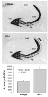
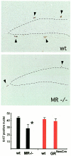
To dissect the effects of corticosteroids mediated by the mineralocorticoid (MR) and the glucocorticoid receptor (GR) in the central nervous system, we compared MR-/- mice, whose salt loss syndrome was corrected by exogenous NaCI administration, with GR-/- mice having a brain-specific disruption of the GR gene generated by the Cre/loxP-recombination system. Neuropathological analyses revealed a decreased density of granule cells in the hippocampus of adult MR-/- mice but not in mice with disruption of GR. Furthermore, adult MR-/- mice exhibited a significant reduction of granule cell neurogenesis to 65% of control levels, possibly mediated by GR due to elevated corticosterone plasma levels. Neurogenesis was unaltered in adult mice with disruption of GR. Thus, we could attribute long-term trophic effects of adrenal steroids on dentate granule cells to MR. These MR-related alterations may participate in the pathogenesis of hippocampal changes observed in ageing, chronic stress and affective disorders.
Figures


Similar articles
-
Age-dependent expression of glucocorticoid- and mineralocorticoid receptors on neural precursor cell populations in the adult murine hippocampus.Aging Cell. 2004 Dec;3(6):363-71. doi: 10.1111/j.1474-9728.2004.00130.x. Aging Cell. 2004. PMID: 15569353
-
The role of the hippocampal mineralocorticoid and glucocorticoid receptors in the hypothalamo-pituitary-adrenal axis of the aged Fisher rat.Mol Cell Neurosci. 1994 Oct;5(5):400-12. doi: 10.1006/mcne.1994.1050. Mol Cell Neurosci. 1994. PMID: 7820364
-
Glucocorticoids and serotonin alter glucocorticoid receptor (GR) but not mineralocorticoid receptor (MR) mRNA levels in fetal mouse hippocampal neurons, in vitro.Brain Res. 2001 Mar 30;896(1-2):130-6. doi: 10.1016/s0006-8993(01)02075-3. Brain Res. 2001. PMID: 11277981
-
[Corticosteroid receptor and stress].Nihon Shinkei Seishin Yakurigaku Zasshi. 2000 Nov;20(5):181-8. Nihon Shinkei Seishin Yakurigaku Zasshi. 2000. PMID: 11326543 Review. Japanese.
-
Brain mineralocorticoid receptor diversity: functional implications.J Steroid Biochem Mol Biol. 1993 Dec;47(1-6):183-90. doi: 10.1016/0960-0760(93)90073-6. J Steroid Biochem Mol Biol. 1993. PMID: 8274434 Review.
Cited by
-
Hippocampal neurogenesis is not enhanced by lifelong reduction of glucocorticoid levels.Hippocampus. 2005;15(4):491-501. doi: 10.1002/hipo.20074. Hippocampus. 2005. PMID: 15744738 Free PMC article.
-
Regulation of mineralocorticoid receptor expression during neuronal differentiation of murine embryonic stem cells.Endocrinology. 2010 May;151(5):2244-54. doi: 10.1210/en.2009-0753. Epub 2010 Mar 5. Endocrinology. 2010. PMID: 20207834 Free PMC article.
-
Role of Mineralocorticoid Receptor in Adipogenesis and Obesity in Male Mice.Endocrinology. 2020 Feb 1;161(2):bqz010. doi: 10.1210/endocr/bqz010. Endocrinology. 2020. PMID: 32036385 Free PMC article.
-
Prenatal alcohol exposure is associated with altered subcellular distribution of glucocorticoid and mineralocorticoid receptors in the adolescent mouse hippocampal formation.Alcohol Clin Exp Res. 2014 Feb;38(2):392-400. doi: 10.1111/acer.12236. Epub 2013 Aug 19. Alcohol Clin Exp Res. 2014. PMID: 23992407 Free PMC article.
-
The multifaceted mineralocorticoid receptor.Compr Physiol. 2014 Jul;4(3):965-94. doi: 10.1002/cphy.c130044. Compr Physiol. 2014. PMID: 24944027 Free PMC article. Review.
References
-
- Almeida O.F.X., Conde, G.L., Crochemore, C., Demeneix, B.A., Fischer, D., Hassan, A.H.S., Meyer, M., Holsboer, F. and Michaelidis, T.M. (2000) Subtle shifts in the ratio between pro- and antiapoptotic molecules after activation of corticosteroid receptors decide neuronal fate. FASEB J., 14, 779–790. - PubMed
-
- Beato M., Herrlich, P. and Schütz, G. (1995) Steroid hormone receptors: many actors in search of a plot. Cell, 83, 851–857. - PubMed
-
- Bleich M. et al. (1999) Rescue of the mineralocorticoid receptor knock-out mouse. Pflugers Arch., 438, 245–254. - PubMed
-
- Cameron H.A. and Gould, E. (1994) Adult neurogenesis is regulated by adrenal steroids in the dentate gyrus. Neuroscience, 61, 203–209. - PubMed
Publication types
MeSH terms
Substances
LinkOut - more resources
Full Text Sources
Molecular Biology Databases

