Insulin/IGF-1 and TNF-alpha stimulate phosphorylation of IRS-1 at inhibitory Ser307 via distinct pathways
- PMID: 11160134
- PMCID: PMC199174
- DOI: 10.1172/JCI10934
Insulin/IGF-1 and TNF-alpha stimulate phosphorylation of IRS-1 at inhibitory Ser307 via distinct pathways
Abstract
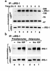
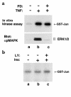
Serine/threonine phosphorylation of IRS-1 might inhibit insulin signaling, but the relevant phosphorylation sites are difficult to identify in cultured cells and to validate in isolated tissues. Recently, we discovered that recombinant NH2-terminal Jun kinase phosphorylates IRS-1 at Ser307, which inhibits insulin-stimulated tyrosine phosphorylation of IRS-1. To monitor phosphorylation of Ser307 in various cell and tissue backgrounds, we prepared a phosphospecific polyclonal antibody designated alphapSer307. This antibody revealed that TNF-alpha, IGF-1, or insulin stimulated phosphorylation of IRS-1 at Ser307 in 3T3-L1 preadipocytes and adipocytes. Insulin injected into mice or rats also stimulated phosphorylation of Ser307 on IRS-1 immunoprecipitated from muscle; moreover, Ser307 was phosphorylated in human muscle during the hyperinsulinemic euglycemic clamp. Experiments in 3T3-L1 preadipocytes and adipocytes revealed that insulin-stimulated phosphorylation of Ser307 was inhibited by LY294002 or wortmannin, whereas TNF-alpha-stimulated phosphorylation was inhibited by PD98059. Thus, distinct kinase pathways might converge at Ser307 to mediate feedback or heterologous inhibition of IRS-1 signaling to counterregulate the insulin response.
Figures


Similar articles
-
Tumor necrosis factor alpha-mediated insulin resistance, but not dedifferentiation, is abrogated by MEK1/2 inhibitors in 3T3-L1 adipocytes.Mol Endocrinol. 2000 Oct;14(10):1557-69. doi: 10.1210/mend.14.10.0542. Mol Endocrinol. 2000. PMID: 11043572
-
Aspirin inhibits serine phosphorylation of insulin receptor substrate 1 in tumor necrosis factor-treated cells through targeting multiple serine kinases.J Biol Chem. 2003 Jul 4;278(27):24944-50. doi: 10.1074/jbc.M300423200. Epub 2003 Apr 24. J Biol Chem. 2003. PMID: 12714600
-
Three mitogen-activated protein kinases inhibit insulin signaling by different mechanisms in 3T3-L1 adipocytes.Mol Endocrinol. 2003 Mar;17(3):487-97. doi: 10.1210/me.2002-0131. Epub 2002 Dec 5. Mol Endocrinol. 2003. PMID: 12554784
-
Hyperosmotic stress inhibits insulin receptor substrate-1 function by distinct mechanisms in 3T3-L1 adipocytes.J Biol Chem. 2003 Jul 18;278(29):26550-7. doi: 10.1074/jbc.M212273200. Epub 2003 May 1. J Biol Chem. 2003. PMID: 12730242
-
Insulin resistance associated to obesity: the link TNF-alpha.Arch Physiol Biochem. 2008 Jul;114(3):183-94. doi: 10.1080/13813450802181047. Arch Physiol Biochem. 2008. PMID: 18629684 Review.
Cited by
-
Insulin receptor substrate-2 is expressed in kidney epithelium and up-regulated in diabetic nephropathy.FEBS J. 2013 Jul;280(14):3232-43. doi: 10.1111/febs.12305. Epub 2013 May 29. FEBS J. 2013. PMID: 23617393 Free PMC article.
-
An anti-diabetes agent protects the mouse brain from defective insulin signaling caused by Alzheimer's disease- associated Aβ oligomers.J Clin Invest. 2012 Apr;122(4):1339-53. doi: 10.1172/JCI57256. J Clin Invest. 2012. PMID: 22476196 Free PMC article.
-
TNFα and SOCS3 regulate IRS-1 to increase retinal endothelial cell apoptosis.Cell Signal. 2012 May;24(5):1086-92. doi: 10.1016/j.cellsig.2012.01.003. Epub 2012 Jan 12. Cell Signal. 2012. PMID: 22266116 Free PMC article.
-
Mechanisms of action of natural products on type 2 diabetes.World J Diabetes. 2023 Nov 15;14(11):1603-1620. doi: 10.4239/wjd.v14.i11.1603. World J Diabetes. 2023. PMID: 38077803 Free PMC article. Review.
-
Inhibition of insulin signaling in endothelial cells by protein kinase C-induced phosphorylation of p85 subunit of phosphatidylinositol 3-kinase (PI3K).J Biol Chem. 2012 Feb 10;287(7):4518-30. doi: 10.1074/jbc.M111.286591. Epub 2011 Dec 12. J Biol Chem. 2012. PMID: 22158866 Free PMC article.
References
-
- Withers DJ, White MF. Insulin action and type 2 diabetes: lessons from knockout mice. Curr Opin Endocrinol Diab. 1999;6:141–145.
-
- DeFronzo RA. Pathogenesis of type 2 diabetes: metabolic and molecular implications for identifying diabetes genes. Diabetes Review. 1997;5:177–269.
-
- DeFronzo RA, Prato SD. Insulin resistance and diabetes mellitus. J Diabetes Complications. 1996;10:243–245. - PubMed
-
- DeFronzo RA, Barzilai N, Simonson DC. Mechanism of metformin action in obese and lean noninsulin-dependent diabetic subjects. J Clin Endocrinol Metab. 1997;73:1294–1301. - PubMed
-
- Kulkarni RN, et al. Tissue-specific knockout of the insulin receptor in pancreatic β cells creates an insulin secretory defect similar to that in type 2 diabetes. Cell. 1999;96:329–339. - PubMed
Publication types
MeSH terms
Substances
Grants and funding
LinkOut - more resources
Full Text Sources
Other Literature Sources
Medical
Molecular Biology Databases
Miscellaneous

