

Tomato spotted wilt virus glycoproteins exhibit trafficking and localization signals that are functional in mammalian cells
- PMID: 11134314
- PMCID: PMC113997
- DOI: 10.1128/JVI.75.2.1004-1012.2001
Tomato spotted wilt virus glycoproteins exhibit trafficking and localization signals that are functional in mammalian cells
Abstract
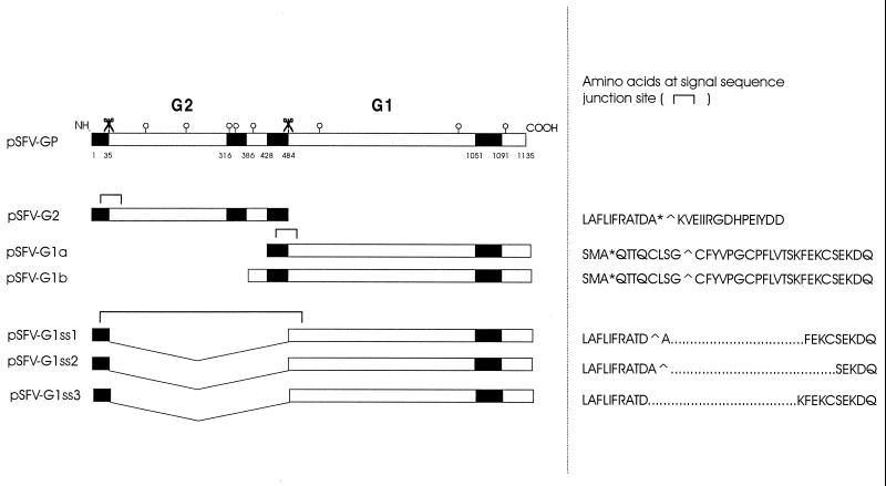
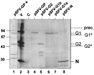
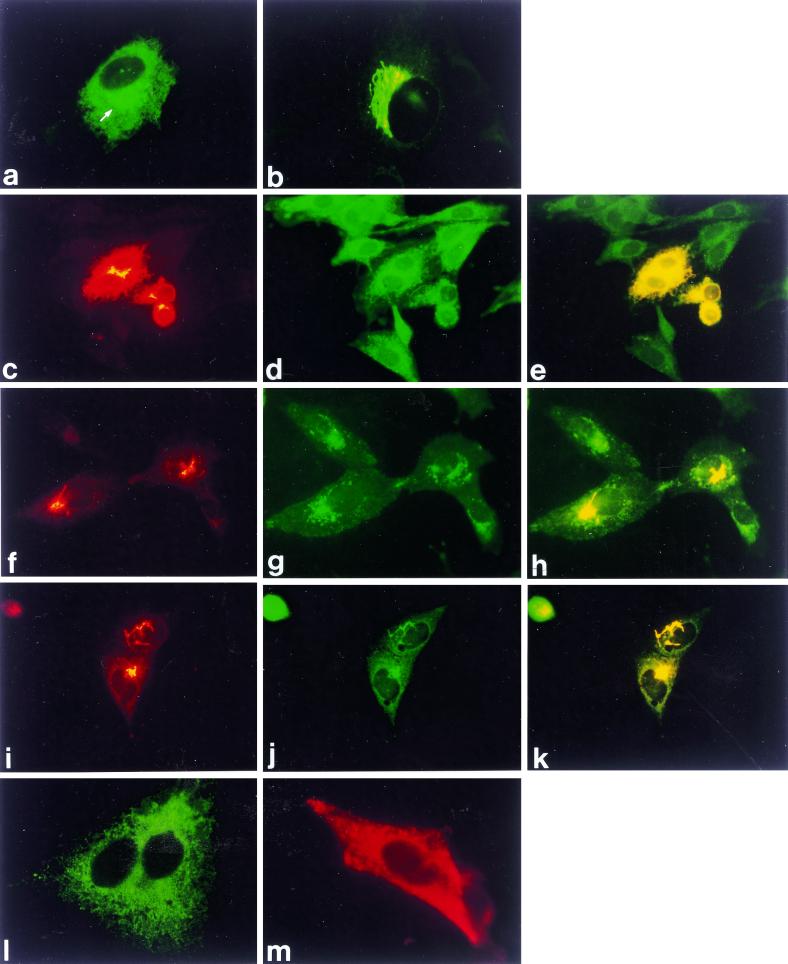
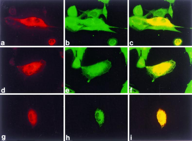
The glycoprotein precursor (G1/G2) gene of tomato spotted wilt virus (TSWV) was expressed in BHK cells using the Semliki Forest virus expression system. The results reveal that in this cell system, the precursor is efficiently cleaved and the resulting G1 and G2 glycoproteins are transported from the endoplasmic reticulum (ER) to the Golgi complex, where they are retained, a process that could be blocked by tunicamycin. Expression of G2 alone resulted in transport to and retention in the Golgi complex, albeit less efficient, suggesting that G2 contains a Golgi retention signal. G1 alone was retained in the ER, irrespective of whether it contained the precursor's signal sequence or its own N-terminal hydrophobic sequence. Coexpression of G1 and G2 from separate gene constructs resulted in rescue of efficient G1 transport, as the proteins coaccumulated in the Golgi complex, indicating that their interaction is essential for proper targeting to this organelle. The results demonstrate that transport and targeting of the plant TSWV glycoproteins in mammalian BHK cells are strikingly similar to those of animal-infecting bunyavirus glycoproteins in mammalian cells. The observations are likely to reflect the dual tropism of TSWV, which replicates both in its plant host and in its animal (thrips) vector.
Figures

Similar articles
-
Tomato spotted wilt virus glycoproteins induce the formation of endoplasmic reticulum- and Golgi-derived pleomorphic membrane structures in plant cells.J Gen Virol. 2008 Aug;89(Pt 8):1811-1818. doi: 10.1099/vir.0.2008/001164-0. J Gen Virol. 2008. PMID: 18632951
-
Requirements for ER-arrest and sequential exit to the golgi of Tomato spotted wilt virus glycoproteins.Traffic. 2009 Jun;10(6):664-72. doi: 10.1111/j.1600-0854.2009.00900.x. Traffic. 2009. PMID: 19302268
-
Distinct Mechanism for the Formation of the Ribonucleoprotein Complex of Tomato Spotted Wilt Virus.J Virol. 2017 Nov 14;91(23):e00892-17. doi: 10.1128/JVI.00892-17. Print 2017 Dec 1. J Virol. 2017. PMID: 28904194 Free PMC article.
-
Natural Resources Resistance to Tomato Spotted Wilt Virus (TSWV) in Tomato (Solanum lycopersicum).Int J Mol Sci. 2021 Oct 12;22(20):10978. doi: 10.3390/ijms222010978. Int J Mol Sci. 2021. PMID: 34681638 Free PMC article. Review.
-
Thrips transmission of tospoviruses.Curr Opin Virol. 2015 Dec;15:80-9. doi: 10.1016/j.coviro.2015.08.003. Epub 2015 Sep 2. Curr Opin Virol. 2015. PMID: 26340723 Review.
Cited by
-
Viral genetic determinants for thrips transmission of Tomato spotted wilt virus.Proc Natl Acad Sci U S A. 2005 Apr 5;102(14):5168-73. doi: 10.1073/pnas.0407354102. Epub 2005 Mar 7. Proc Natl Acad Sci U S A. 2005. PMID: 15753307 Free PMC article.
-
Bunyaviral N Proteins Localize at RNA Processing Bodies and Stress Granules: The Enigma of Cytoplasmic Sources of Capped RNA for Cap Snatching.Viruses. 2022 Jul 29;14(8):1679. doi: 10.3390/v14081679. Viruses. 2022. PMID: 36016301 Free PMC article.
-
Determination of key residues in tospoviral NSm required for Sw-5b recognition, their potential ability to overcome resistance, and the effective resistance provided by improved Sw-5b mutants.Mol Plant Pathol. 2022 May;23(5):622-633. doi: 10.1111/mpp.13182. Epub 2021 Dec 27. Mol Plant Pathol. 2022. PMID: 34962031 Free PMC article.
-
Molecular characterization of the full-length L and M RNAs of Tomato yellow ring virus, a member of the genus Tospovirus.Virus Genes. 2013 Jun;46(3):487-95. doi: 10.1007/s11262-013-0880-8. Epub 2013 Jan 19. Virus Genes. 2013. PMID: 23334441
-
Crystal structure of tomato spotted wilt virus GN reveals a dimer complex formation and evolutionary link to animal-infecting viruses.Proc Natl Acad Sci U S A. 2020 Oct 20;117(42):26237-26244. doi: 10.1073/pnas.2004657117. Epub 2020 Oct 5. Proc Natl Acad Sci U S A. 2020. PMID: 33020295 Free PMC article.
References
-
- Adam G, Peters D, Goldbach R W. Serological comparison of tospovirus isolates using polyclonal and monoclonal antibodies: Proceedings of the International Symposium on Tospoviruses and Thrips of Floral and Vegetable Crops. Acta Hortic (Wageningen) 1996;431:135–158.
-
- Bar-Peled M, Bassham D C, Raikhel N V. Transport of proteins in eucaryotic cells: more questions ahead. Plant Mol Biol. 1996;32:223–249. - PubMed
-
- De Ávila A C, de Haan P, Smeets M L L, de O. Resende R, Kormelink R, Kitajima E W, Goldbach R W, Peters D. Distinct levels of relationships between tospovirus isolates. Arch Virol. 1993;128:211–227. - PubMed
MeSH terms
Substances
LinkOut - more resources
Full Text Sources
