. 2000 Sep 12;97(19):10430-5.
doi: 10.1073/pnas.190332597.

Activation of HIF1alpha ubiquitination by a reconstituted von Hippel-Lindau (VHL) tumor suppressor complex

Affiliations

Activation of HIF1alpha ubiquitination by a reconstituted von Hippel-Lindau (VHL) tumor suppressor complex

T Kamura et al. Proc Natl Acad Sci U S A. .

Abstract

Mutations in the VHL tumor suppressor gene result in constitutive expression of many hypoxia-inducible genes, at least in part because of increases in the cellular level of hypoxia-inducible transcription factor HIF1alpha, which in normal cells is rapidly ubiquitinated and degraded by the proteasome under normoxic conditions. The recent observation that the VHL protein is a subunit of an Skp1-Cul1/Cdc53-F-box (SCF)-like E3 ubiquitin ligase raised the possibility that VHL may be directly responsible for regulating cellular levels of HIF1alpha by targeting it for ubiquitination and proteolysis. In this report, we test this hypothesis directly. We report development of methods for production of the purified recombinant VHL complex and present direct biochemical evidence that it can function with an E1 ubiquitin-activating enzyme and E2 ubiquitin-conjugating enzyme to activate HIF1alpha ubiquitination in vitro. Our findings provide new insight into the function of the VHL tumor suppressor protein, and they provide a foundation for future investigations of the mechanisms underlying VHL regulation of oxygen-dependent gene expression.


Figures
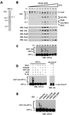
Figure 1
Figure 1
The purified recombinant VHL tumor suppressor complex activates HIF1α ubiquitination in vitro. (A) An aliquot of recombinant His-HPC4-HIF1α used as substrate in the ubiquitination reactions of C and D was subjected to 8% SDS/PAGE, and proteins were visualized by Coomassie staining. His-HPC4-HIF1α was expressed in Sf21 cells and purified by Ni2+-agarose chromatography. (B) The recombinant VHL complex was purified as described in Materials and Methods from lysates of insect cells infected with baculoviruses encoding His-VHL, Cul2, Elongin B, Elongin C, and myc-Rbx1. Aliquots of TSK DEAE-NPR column fractions were subjected to 13% SDS/PAGE, and proteins were visualized by Coomassie staining (Top). Aliquots of TSK DEAE-NPR column fractions were subjected to 8% or 13% SDS/PAGE, and proteins were transferred to Hybond P membranes and visualized by Western blotting as described in Materials and Methods, using the antibodies indicated in the figure (Bottom). EloB, Elongin B; EloC, Elongin C. (C) Aliquots of TSK-DEAE-NPR column fractions indicated in the figure were assayed as described in Materials and Methods for the ability to activate HIF1α ubiquitination. Reactions contained ≈50 ng of the His-HPC4-HIF1α shown in A. Reaction products were subjected to 8% SDS/PAGE, and GST-ubiquitin-HIF1α conjugates (GST-Ub-HIF1α) were transferred to Hybond P membranes and visualized by Western blotting as described in Materials and Methods, using anti-HPC4 antibodies. (D) Aliquots of TSK-DEAE-NPR column fraction 25 were assayed as described in Materials and Methods for the ability to activate HIF1α or Cln2 ubiquitination activity in the presence and absence of Uba1, hUbc5a, GST-ubiquitin (GST-Ub), and ATP. Reactions contained ≈50 ng of either the His-HPC4-HIF1α shown in A or about 50 ng of the phosphorylated Cln2 complex. Reaction products were subjected to 8% SDS/PAGE and transferred to Hybond P membranes and visualized by Western blotting as described in Materials and Methods, using anti-HPC4 or anti-HA antibodies. (E) Aliquots of TSK-DEAE-NPR column fraction 25 were assayed as described in Materials and Methods for the ability to activate HIF1α ubiquitination activity in the presence of Uba1, GST-ubiquitin (GST-Ub), ATP, and approximately equimolar amounts of the E2 ubiquitin-conjugating enzymes shown in the figure (≈100 ng hUbc5a, ≈200 ng hUbc3, ≈100 ng mE2-21K, ≈200 ng mE2-35K, ≈150 ng mE2-24K). Reactions contained ≈50 ng of the His-HPC4-HIF1α shown in A. Reaction products were subjected to 8% SDS/PAGE, and GST-ubiquitin-HIF1α conjugates (GST-Ub-HIF1α) were transferred to Hybond P membranes and visualized by Western blotting as described in Materials and Methods, using anti-HPC4 antibodies.
Figure 2
Figure 2
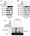
All subunits of the VHL complex are required for maximal activation of HIF1α ubiquitination. (A) Total cell lysates from Sf21 cells infected with the baculoviruses indicated in the figure were subjected to 8% or 13% SDS/PAGE, and proteins were transferred to Hybond P membranes and visualized by Western blotting as described in Materials and Methods, using the antibodies indicated in the figure. (B) The cell lysates of A were subjected to immunoprecipitations with anti-VHL antibodies as described in Materials and Methods. The immunoprecipitates were subjected to 8% or 13% SDS/PAGE, and proteins were transferred to Hybond P membranes and visualized by Western blotting as described in Materials and Methods, using the antibodies indicated in the figure. (C) The cell lysates of A were subjected to immunoprecipitations with anti-VHL antibodies as described in Materials and Methods. The immunoprecipitates were assayed for the ability to activate HIF1α ubiquitination as described in Materials and Methods. Reaction products were subjected to 8% SDS/PAGE, and GST-ubiquitin-HIF1α conjugates (GST-Ub-HIF1α) were transferred to Hybond P membranes and visualized by Western blotting as described in Materials and Methods, using anti-HIF1α antibodies. EloB, Elongin B; EloC, Elongin C.
Figure 3
Figure 3
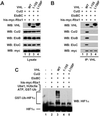
VHL mutants that do not bind stably to Elongins B and C do not assemble into the VHL complex and do not detectably activate HIF1α ubiquitination. (A) Total cell lysates from Sf21 cells infected with the baculoviruses indicated in the figure were subjected to 8% or 13% SDS/PAGE, and proteins were transferred to Hybond P membranes and visualized by Western blotting as described in Materials and Methods, using the antibodies indicated in the figure. (B) The cell lysates of A were subjected to immunoprecipitations with anti-VHL antibodies as described in Materials and Methods. The immunoprecipitates were subjected to 8% or 13% SDS/PAGE, and proteins were transferred to Hybond P membranes and visualized by Western blotting as described in Materials and Methods, using the antibodies indicated in the figure. (C) The cell lysates of A were subjected to immunoprecipitations with anti-VHL antibodies as described in Materials and Methods. The immunoprecipitates were assayed for the ability to activate HIF1α ubiquitination as described in Materials and Methods. Reaction products were subjected to 8% SDS/PAGE, and GST-ubiquitin-HIF1α conjugates (GST-Ub-HIF1α) were transferred to Hybond P membranes and visualized by Western blotting as described in Materials and Methods, using anti-HIF1α antibodies. EloB, Elongin C; EloC, Elongin C.
Figure 4
Figure 4
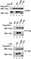
Interaction of HIF1α with VHL mutants VHL[1–155] and VHL[L158P]. (Upper) Total cell lysates from Sf21 cells infected with the baculoviruses indicated in the figure were subjected to 8% or 13% SDS/PAGE, and proteins were transferred to Hybond P membranes and visualized by Western blotting as described in Materials and Methods, using the antibodies indicated in the figure. (Middle and Lower) Cell lysates were subjected to immunoprecipitations with anti-VHL or anti-Flag antibodies as described in Materials and Methods. The immunoprecipitates were subjected to 8% or 13% SDS/PAGE, and proteins were transferred to Hybond P membranes and visualized by Western blotting as described in Materials and Methods, using the antibodies indicated in the figure.
Figure 5
Figure 5
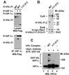
A VHL mutant lacking a portion of the β domain is defective in HIF1α binding and ubiquitination. (A) Total cell lysates or anti-HIF1α immunoprecipitates from the indicated baculovirus-infected Sf21 cells were immunoblotted with anti-Flag antibody. (B) Sf21 cells were coinfected with baculoviruses encoding Cul2, Elongin B, Elongin C, His-myc-Rbx1, and either His-VHL-Flag or His-VHL-Flag[117–213]. Recombinant VHL complexes were purified as described in Materials and Methods. After TSK DEAE-NPR HPLC, aliquots of peak fractions from were subjected to 13% SDS/PAGE, and proteins were visualized by Coomassie staining. (C) Aliquots of the TSK DEAE-NPR fractions shown in B were assayed as described in Materials and Methods for the ability to activate HIF1a ubiquitination. Reactions contained ≈50 ng of the His-HPC4-HIF1a shown in Fig. 1A. The reaction products were subjected to 8% SDS/PAGE, transferred to Hybond P membranes, and visualized by Western blotting using anti-HPC4 antibody. H, His; Fl, Flag.

Similar articles

Cited by

References

    1. Latif F, Tory K, Gnarra J, Yao M, Duh F M, Orcutt M L, Stackhouse T, Kuzmin I, Modi W, Geil L, et al. Science. 1993;260:1317–1320. - PubMed
    1. Gnarra J R, Duan D R, Weng Y, Humphrey J S, Chen D Y, Lee S, Pause A, Dudley C F, Latif F, Kuzmin I, et al. Biochim Biophys Acta. 1996;1242:201–210. - PubMed
    1. Gnarra J R, Zhou S, Merrill M J, Wagner J R, Krumm A, Papavassiliou E, Oldfield E H, Klausner R D, Linehan W M. Proc Natl Acad Sci USA. 1996;93:10589–10594. - PMC - PubMed
    1. Iliopoulos O, Levy A P, Jiang C, Kaelin W G, Goldberg M A. Proc Natl Acad Sci USA. 1996;93:10595–10599. - PMC - PubMed
    1. Siemeister G, Weindel K, Mohrs K, Barleon B, Martiny-Baron G, Marme D. Cancer Res. 1996;56:2299–2301. - PubMed

Publication types