. 2000 Aug 15;19(16):4298-309.
doi: 10.1093/emboj/19.16.4298.

Mechanism of regulation of the hypoxia-inducible factor-1 alpha by the von Hippel-Lindau tumor suppressor protein

Affiliations

Mechanism of regulation of the hypoxia-inducible factor-1 alpha by the von Hippel-Lindau tumor suppressor protein

K Tanimoto et al. EMBO J. .

Abstract

In normoxic cells the hypoxia-inducible factor-1 alpha (HIF-1 alpha) is rapidly degraded by the ubiquitin-proteasome pathway, and activation of HIF-1 alpha to a functional form requires protein stabilization. Here we show that the product of the von Hippel-Lindau (VHL) tumor suppressor gene mediated ubiquitylation and proteasomal degradation of HIF-1 alpha under normoxic conditions via interaction with the core of the oxygen-dependent degradation domain of HIF-1 alpha. The region of VHL mediating interaction with HIF-1 alpha overlapped with a putative macromolecular binding site observed within the crystal structure of VHL. This motif of VHL also represents a mutational hotspot in tumors, and one of these mutations impaired interaction with HIF-1 alpha and subsequent degradation. Interestingly, the VHL binding site within HIF-1 alpha overlapped with one of the minimal transactivation domains. Protection of HIF-1 alpha against degradation by VHL was a multistep mechanism, including hypoxia-induced nuclear translocation of HIF-1 alpha and an intranuclear hypoxia-dependent signal. VHL was not released from HIF-1 alpha during this process. Finally, stabilization of HIF-1 alpha protein levels per se did not totally bypass the need of the hypoxic signal for generating the transactivation response.


Figures

None
Fig. 1. VHL mediates proteasomal degradation of HIF-1α under normoxic conditions. (A) Expression of HIF-1α under normoxic conditions. Increasing amounts [0.2 µg (+), 0.5 µg (++), 1.0 µg (+++)] of pFLAG CMV2/HIF-1α were transiently transfected into COS7 cells, and the cells were incubated for 12 h at normoxia (21% O2) or hypoxia (1% O2), as indicated. Whole-cell extracts were analyzed by immunoblotting using anti-FLAG antibodies. (B) Degradation of HIF-1α in the presence of VHL. pFLAG CMV2/HIF-1α was cotransfected into COS7 cells together with empty vector or wild-type VHL expression vector (pCMX/VHL). Whole-cell extracts were analyzed by immunoblotting using anti-FLAG or anti-VHL antibodies. (C) VHL mediates proteasomal degradation of HIF-1α. Cells were transfected with pFLAG CMV2/HIF-1α and pCMX/VHL, incubated in the absence or presence of 5 µM MG-132 for 6 h before harvesting, and cellular extracts were analyzed as in (B). (D) VHL does not affect dioxin receptor (DR) protein levels. COS7 cells were transfected with a FLAG-tagged dioxin receptor expression vector (pCMV/DR/FLAG) in the absence or presence of pCMX/VHL, and analyzed as in (B). The mobilities of molecular weight (kDa) markers are shown on the right hand side of the blots.
None
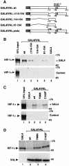
Fig. 2. VHL requires two functional domains to induce HIF-1α degradation. (A) Schematic representation of GAL4-fused wild-type (wt), deletion or single amino acid point mutant (pmt) forms of VHL. (B) VHL directly interacts with HIF-1α. Equal concentrations of in vitro translated 35S-labeled full-length HIF-1α were incubated with in vitro translated wild-type GAL4/VHL or VHL deletion mutants or GAL4-DBD spanning the GAL4 DNA binding domain alone. Co-immunoprecipitation assays with anti-GAL4 antibody (upper panel) or control preimmune serum (lower panel) were carried out as described in Materials and methods. The precipitated material was analysed by SDS–PAGE and autoradiography. For loading controls, 10% of input 35S-labeled HIF-1α is shown in lane 1. (C) A tumor-derived point mutation impairs the interaction between VHL and HIF-1α. In vitro translated proteins were incubated and analyzed by co-immunoprecipitation as in (B). (D) Tumor-derived point-mutated forms of VHL fail to induce HIF-1α degradation. pFLAG CMV2/HIF-1α was transiently coexpressed in COS7 cells in the absence or presence of GAL4-fused wild-type or mutant forms of VHL as indicated. Cells were incubated at normoxia for 24 h, and whole cell extracts were prepared and analyzed by immunoblotting using anti-FLAG or anti-VHL antibodies. The mobilities of molecular weight (kDa) markers are shown on the right hand side of the blots.
None
Fig. 3. VHL targets the oxygen-dependent degradation domain of HIF-1α. (A) FLAG-tagged wild-type HIF-1α or the indicated HIF-1α deletion mutants were transiently coexpressed in COS7 cells at normoxia in the absence or presence of VHL. Whole-cell extracts were prepared and analyzed as described in Figure 1. ODD, oxygen-dependent degradation domain; N- and C-TAD, N- and C-terminal transactivation domains. (B35S-labeled VHL was incubated with equal concentrations of in vitro translated GAL4-fusion proteins spanning full-length HIF-1α or GAL4-DBD alone. Co-immunoprecipitation assays were performed with anti-GAL4 antibodies (upper panel) or control non-specific rabbit antiserum (lower panel). The precipitated material was analyzed by SDS–PAGE and autoradiography. For loading controls, 10% of input 35S-labeled VHL is shown. (C) VHL, GAL4 fusion proteins spanning the indicated HIF-1α fragments or GAL4 alone were expressed by in vitro translation and analyzed as in (B).
None
Fig. 4. The minimal N-TAD of HIF-1α is the target for VHL-mediated ubiquitylation and proteasomal degradation. (A) Alignment of N-TAD sequences of human (h) and mouse (m) HIF-1α and EPAS-1 revealed a conserved core sequence motif. The positions of amino acid substitutions are shown in mutant (mt) N-TAD. (B) Mutation of the N-TAD PYI motif abolishes interaction between HIF-1α and VHL. 35S-labeled VHL was incubated with equal concentrations of in vitro translated FLAG-tagged wild-type, mutant N-TAD or FLAG epitope alone. Co-immunoprecipitation assays were carried out using anti-FLAG antibodies and analyzed by SDS–PAGE and autoradiography. For loading controls, 10% of input 35S-labeled VHL is shown. (C) The PYI mutation confers resistance to VHL-mediated degradation. FLAG or FLAG-tagged wild-type or mutant N-TAD (1 µg/6-cm dish) were transiently coexpressed in COS7 cells in the absence or presence of increasing concentrations (1.0 µg, +; 2.0 µg/6-cm dish, ++) of VHL as indicated, and incubated at normoxia for 24 h. Whole-cell extracts were prepared and analyzed by immunoblotting using anti-FLAG or anti-VHL antibodies. (D) VHL mediates ubiquitylation of N-TAD. FLAG, or FLAG-tagged wild-type or mutant N-TAD and HA-tagged ubiquitin were transiently coexpressed in COS7 cells in the presence or absence of VHL under normoxic conditions in combination with MG132 for 6 h. After immunoprecipitation of whole-cell extracts by anti-FLAG, ubiquitylated forms of N-TAD were detected by anti-HA immunoblotting (upper panel). Ten percent of input whole-cell extracts are shown in the two lower panels. (E) Effect on protein stability following substitution of individual lysines to arginines within N-TAD. Wild-type or mutant N-TAD were transiently expressed in 293 cells and incubated at normoxia or hypoxia for 12 h. Whole-cell extracts were prepared and analyzed as in (C). The mobilities of molecular weight (kDa) markers are shown to the right of the blots.
None
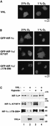
Fig. 5. Protection of HIF-1α against VHL-mediated degradation requires nuclear translocation of HIF-1α and a hypoxia-dependent intranuclear signal. (A) Subcellular localization of VHL. COS7 cells were transfected with pCMX/VHL and after 24 h of expression, the cells were incubated for 6 h at normoxia or hypoxia. The subcellular localization was determined by indirect immunofluorescence using anti-VHL antibodies. (B) Subcellular distribution of GFP–HIF-1α chimeric proteins. GFP fusion proteins spanning wild-type or mutant forms of HIF-1α were transiently expressed in COS7 cells and incubated as above. Photographs were taken using a Zeiss fluorescent microscope. (C) Effect of VHL on degradation of wild-type and mutant forms of HIF-1α showing constitutively cytoplasmic or nuclear localization. FLAG-tagged wild-type or mutant forms of HIF-1α were transiently expressed in the absence or presence of VHL and incubated for 12 h at normoxia or hypoxia. Whole-cell extracts were prepared and assayed as in Figure 1.
None
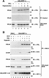
Fig. 6. VHL, Arnt and HIF-1α form a ternary nuclear complex. (A) VHL is not released from HIF-1α in hypoxic cells. GAL4, GAL4/HIF-1α and/or VHL were transiently coexpressed in COS7 cells in the presence of MG132 at normoxia or hypoxia for the indicated periods of time. Following immunoprecipitation of whole cell extracts by anti-GAL4 (upper panel) or control (middle panel) antibodies, VHL was detected by immunoblotting using anti-VHL antibodies. Ten percent of input whole-cell extracts is shown in the lower panel. (B) Association with Arnt. GAL4 or GAL4/HIF-1α were transiently expressed in COS7 cells in the absence or presence of Arnt or VHL at normoxia or hypoxia for 6 h, and analyzed as above.
None
Fig. 7. Mutant N-TAD maintains an hypoxia-inducible transactivation response. (A) Constitutive stabilization of PYI mutant N-TAD. GAL4-fused wild-type and mutant N-TAD were transiently expressed in COS7 cells at normoxia or hypoxia for 12 h. Whole-cell extracts were prepared and assayed as in Figure 1. (B) Hypoxia-inducible transactivation function of mutant N-TAD. The transcriptional activation function by GAL4-fused wild-type or mutant N-TAD proteins was monitored using a GAL4-responsive luciferase reporter gene as described in Materials and methods. Six hours after transfection, cells were incubated for 36 h at either normoxia or hypoxia. Reporter gene activities are expressed relative to the activity of the GAL4 DNA binding domain alone at normoxia, and normalized against internal control β-galactosidase activities. Values represent the mean ± SD of three independent experiments. (C) PYI mutant stabilizes full-length HIF-1α in normoxic cells. FLAG-tagged wild-type and mutant HIF-1α were transiently expressed in absence or presence of VHL and analyzed as above. (D) Transcription activation functions of wild-type and mutant HIF-1α proteins were assayed as in (B).
None
Fig. 8. Model of conditional regulation of HIF-1α function. Under normoxic conditions (left panel) HIF-1α is targeted for ubiquitin-proteasomal degradation by VHL. The shaded areas in VHL indicate mutational hotspots in tumors that coincide with the HIF-1α or the elongin C binding domain (CBD) of VHL, respectively. Mutations in either of these two domains stabilize HIF-1α protein. Hypoxia (right panel) leads to inhibition of degradation of HIF-1α by induction of nuclear translocation and an as yet unidentified nuclear regulatory signal that may be linked to recruitment of a partner DNA binding factor, Arnt, transcriptional coactivators and/or the redox regulator Ref-1. See text for details.

Similar articles

Cited by

References

    1. Arany Z., Huang,L.E., Eckner,R., Bhattacharya,S., Jiang,C., Goldberg,M.A., Bunn,H.F. and Livingston,D.M. (1996) An essential role for p300/CBP in the cellular response to hypoxia. Proc. Natl Acad. Sci. USA, 93, 12969–12973. - PMC - PubMed
    1. Carrero P., Okamoto,K., Coumailleau,P., O’Brien,S., Tanaka,H. and Poellinger,L. (2000) Redox-regulated recruitment of the transcriptional coactivators CBP and SRC-1 to the hypoxia-inducible factor-1α. Mol. Cell. Biol., 20, 402–415. - PMC - PubMed
    1. Ciechanover A. (1998) The ubiquitin-proteasome pathway: on protein death and cell life. EMBO J., 17, 7151–7160. - PMC - PubMed
    1. Ema M., Hirota,K., Mimura,J., Abe,H., Yodoi,J., Sogawa,K., Poellinger,L. and Fujii-Kuriyama,Y. (1999) Molecular mechanisms of transcription activation by HLF and HIF1α in response to hypoxia: their stabilization and redox signal-induced interaction with CBP/p300. EMBO J., 18, 1905–1914. - PMC - PubMed
    1. Gnarra J.R., Zhou,S., Merrill,M.J., Wagner,J.R., Krumm,A., Papavassiliou,E., Oldfield,E.H., Klausner,R.D. and Linehan,W.M. (1996) Post-transcriptional regulation of vascular endothelial growth factor mRNA by the product of the VHL tumor suppressor gene. Proc. Natl Acad. Sci. USA, 93, 10589–10594. - PMC - PubMed

Publication types

MeSH terms

Substances