

Insulin-like growth factor-1 inhibits mature oligodendrocyte apoptosis during primary demyelination
- PMID: 10908609
- PMCID: PMC6772563
- DOI: 10.1523/JNEUROSCI.20-15-05703.2000
Insulin-like growth factor-1 inhibits mature oligodendrocyte apoptosis during primary demyelination
Abstract
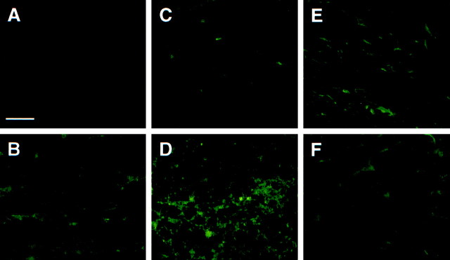
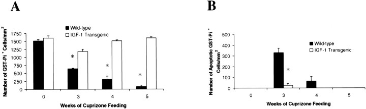
Metabolic insult results in apoptosis and depletion of mature oligodendrocytes during demyelination. To examine the role of insulin-like growth factor-1 (IGF-1) during acute demyelination and remyelination in the adult CNS, we exposed transgenic mice that continuously express IGF-1 (IGF-1 tg) to cuprizone intoxication. Demyelination was observed within the corpus callosum in both wild-type and IGF-1 tg mice 3 weeks after exposure to cuprizone. Wild-type mice showed significant apoptotic mature oligodendrocytes and a dramatic loss of these cells within the lesion that resulted in near complete depletion and demyelination by week 5. In contrast, the demyelinated corpus callosum of the IGF-1 tg mice was near full recovery by week 5. This rapid recovery was apparently caused by survival of the mature oligodendrocyte population because apoptosis was negligible, and by week 4, the mature oligodendrocyte population was completely restored. Furthermore, despite demyelination in both wild-type and IGF-1 tg mice, oligodendrocyte progenitors accumulated only in the absence of mature oligodendrocytes and failed to accumulate if the mature oligodendrocytes remained as demonstrated in the IGF-1 tg mice. These results suggest that IGF-1 may be important in preventing the depletion of mature oligodendrocytes in vivo and thus facilitates an early recovery from demyelination.
Figures


Similar articles
-
Mature oligodendrocyte apoptosis precedes IGF-1 production and oligodendrocyte progenitor accumulation and differentiation during demyelination/remyelination.J Neurosci Res. 2000 Aug 1;61(3):251-62. doi: 10.1002/1097-4547(20000801)61:3<251::AID-JNR3>3.0.CO;2-W. J Neurosci Res. 2000. PMID: 10900072
-
Platelet-derived growth factor promotes repair of chronically demyelinated white matter.J Neuropathol Exp Neurol. 2007 Nov;66(11):975-88. doi: 10.1097/NEN.0b013e3181587d46. J Neuropathol Exp Neurol. 2007. PMID: 17984680 Free PMC article.
-
Absence of fibroblast growth factor 2 promotes oligodendroglial repopulation of demyelinated white matter.J Neurosci. 2002 Oct 1;22(19):8574-85. doi: 10.1523/JNEUROSCI.22-19-08574.2002. J Neurosci. 2002. PMID: 12351731 Free PMC article.
-
The role of oligodendrocytes and oligodendrocyte progenitors in CNS remyelination.Adv Exp Med Biol. 1999;468:183-97. doi: 10.1007/978-1-4615-4685-6_15. Adv Exp Med Biol. 1999. PMID: 10635029 Review.
-
The neurotoxicant, cuprizone, as a model to study demyelination and remyelination in the central nervous system.Brain Pathol. 2001 Jan;11(1):107-16. doi: 10.1111/j.1750-3639.2001.tb00385.x. Brain Pathol. 2001. PMID: 11145196 Free PMC article. Review.
Cited by
-
Insulin-like growth factor-I ameliorates demyelination induced by tumor necrosis factor-alpha in transgenic mice.J Neurosci Res. 2007 Mar;85(4):712-22. doi: 10.1002/jnr.21181. J Neurosci Res. 2007. PMID: 17279553 Free PMC article.
-
Myelin repair: the role of stem and precursor cells in multiple sclerosis.Philos Trans R Soc Lond B Biol Sci. 2008 Jan 12;363(1489):171-83. doi: 10.1098/rstb.2006.2019. Philos Trans R Soc Lond B Biol Sci. 2008. PMID: 17282989 Free PMC article. Review.
-
CXCR2-positive neutrophils are essential for cuprizone-induced demyelination: relevance to multiple sclerosis.Nat Neurosci. 2010 Mar;13(3):319-26. doi: 10.1038/nn.2491. Epub 2010 Feb 14. Nat Neurosci. 2010. PMID: 20154684 Free PMC article.
-
Activation of proteasome by insulin-like growth factor-I may enhance clearance of oxidized proteins in the brain.Mech Ageing Dev. 2009 Nov-Dec;130(11-12):793-800. doi: 10.1016/j.mad.2009.10.005. Mech Ageing Dev. 2009. PMID: 19896963 Free PMC article.
-
The Role of Insulin-Like Growth Factors and Insulin-Like Growth Factor-Binding Proteins in the Nervous System.Biochem Insights. 2019 Apr 17;12:1178626419842176. doi: 10.1177/1178626419842176. eCollection 2019. Biochem Insights. 2019. PMID: 31024217 Free PMC article. Review.
References
-
- Barres BA, Schmid R, Sendnter M, Raff MC. Multiple extracellular signals are required for long-term oligodendrocyte survival. Development. 1993;118:283–295. - PubMed
-
- Cho KH, Kim MW, Kim SU. Tissue culture model of Krabbe's disease: psychosine cytotoxicity in rat oligodendrocyte cultures. Dev Neurosci. 1997;19:321–327. - PubMed
-
- Compston A. Remyelination of the central nervous system. Mult Scler. 1996;1:388–392. - PubMed
-
- Coetzee T, Fujita N, Dupree J, Shi R, Blight A, Suzuki K, Suzuki K, Popko B. Myelination in the absence of galactocerebroside and sulfatide: normal structure with abnormal function and regional stability. Cell. 1996;86:209–219. - PubMed
-
- D'Ercole AJ, Ye P, Calikoglu AS, Gutierrez-Ospina G. The role of the insulin-like growth factors in the central nervous system. Mol Neurobiol. 1996;13:227–255. - PubMed
Publication types
MeSH terms
Substances
Grants and funding
LinkOut - more resources
Full Text Sources
Miscellaneous