Kaposi's sarcoma-associated herpesvirus encodes two proteins that block cell surface display of MHC class I chains by enhancing their endocytosis
- PMID: 10859362
- PMCID: PMC16668
- DOI: 10.1073/pnas.140129797
Kaposi's sarcoma-associated herpesvirus encodes two proteins that block cell surface display of MHC class I chains by enhancing their endocytosis
Erratum in
- Proc Natl Acad Sci U S A 2001 Feb 13;98(4):2111
Abstract
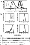
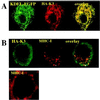
Down-regulation of the cell surface display of class I MHC proteins is an important mechanism of immune evasion by human and animal viruses. Herpesviruses in particular encode a variety of proteins that function to lower MHC I display by several mechanisms. These include binding and retention of MHC I chains in the endoplasmic reticulum, dislocation of class I chains from the ER, inhibition of the peptide transporter (TAP) involved in antigen presentation, and shunting of newly assembled chains to lysosomes. Kaposi's sarcoma (KS)-associated herpesvirus (KSHV) is a human herpesvirus strongly linked to the development of KS and to certain AIDS-associated lymphoproliferative disorders. Here we show that KSHV encodes two distinctive gene products that function to dramatically reduce cell surface MHC I expression. These viral proteins are localized predominantly to the ER. However, unlike previously described MHC I inhibitors, they do not interfere with the synthesis, translocation, or assembly of class I chains, nor do they retain them in the ER. Rather, they act to enhance endocytosis of MHC I from the cell surface; internalized class I chains are delivered to endolysosomal vesicles, where they undergo degradation. These KSHV proteins define a mechanism of class I down-regulation distinct from the mechanisms of other herpesviruses and are likely to contribute importantly to immune evasion during viral infection.
Figures


Similar articles
-
Seroreactivity to Kaposi's sarcoma-associated herpesvirus (human herpesvirus 8) latent nuclear antigen in AIDS-associated Kaposi's sarcoma patients depends on CD4+ T-cell count.J Med Virol. 2007 Oct;79(10):1562-8. doi: 10.1002/jmv.20949. J Med Virol. 2007. PMID: 17705173
-
Modulation of cell signaling pathways by Kaposi's sarcoma-associated herpesvirus (KSHVHHV-8).Cell Biochem Biophys. 2004;40(3):305-22. doi: 10.1385/CBB:40:3:305. Cell Biochem Biophys. 2004. PMID: 15211030 Review.
-
Notch signal transduction induces a novel profile of Kaposi's sarcoma-associated herpesvirus gene expression.J Microbiol. 2006 Apr;44(2):217-25. J Microbiol. 2006. PMID: 16728959
-
Immune evasion by Kaposi's sarcoma-associated herpesvirus.Nat Rev Immunol. 2007 May;7(5):391-401. doi: 10.1038/nri2076. Nat Rev Immunol. 2007. PMID: 17457345 Review.
-
Kaposi's sarcoma-associated herpesvirus (KSHV)/human herpesvirus 8 (HHV-8) as a tumour virus.Herpes. 2003 Dec;10(3):72-7. Herpes. 2003. PMID: 14759339 Review.
Cited by
-
Intracellular Transport Routes for MHC I and Their Relevance for Antigen Cross-Presentation.Front Immunol. 2015 Jul 2;6:335. doi: 10.3389/fimmu.2015.00335. eCollection 2015. Front Immunol. 2015. PMID: 26191062 Free PMC article. Review.
-
The Role of F-Box Proteins during Viral Infection.Int J Mol Sci. 2013 Feb 18;14(2):4030-49. doi: 10.3390/ijms14024030. Int J Mol Sci. 2013. PMID: 23429191 Free PMC article.
-
Equine herpesvirus type 4 UL56 and UL49.5 proteins downregulate cell surface major histocompatibility complex class I expression independently of each other.J Virol. 2012 Aug;86(15):8059-71. doi: 10.1128/JVI.00891-12. Epub 2012 May 23. J Virol. 2012. PMID: 22623773 Free PMC article.
-
Kaposi's sarcoma-associated herpesvirus K3 and K5 proteins down regulate both DC-SIGN and DC-SIGNR.PLoS One. 2013;8(2):e58056. doi: 10.1371/journal.pone.0058056. Epub 2013 Feb 27. PLoS One. 2013. PMID: 23460925 Free PMC article.
-
Regulation of the Macroautophagic Machinery, Cellular Differentiation, and Immune Responses by Human Oncogenic γ-Herpesviruses.Viruses. 2021 May 8;13(5):859. doi: 10.3390/v13050859. Viruses. 2021. PMID: 34066671 Free PMC article. Review.
References
MeSH terms
Substances
LinkOut - more resources
Full Text Sources
Other Literature Sources
Research Materials
Miscellaneous

