. 2000 Jun;20(11):4106-14.
doi: 10.1128/MCB.20.11.4106-4114.2000.

Analysis of fractalkine receptor CX(3)CR1 function by targeted deletion and green fluorescent protein reporter gene insertion

Affiliations

Analysis of fractalkine receptor CX(3)CR1 function by targeted deletion and green fluorescent protein reporter gene insertion

S Jung et al. Mol Cell Biol. 2000 Jun.

Abstract

The seven-transmembrane receptor CX(3)CR1 is a specific receptor for the novel CX(3)C chemokine fractalkine (FKN) (neurotactin). In vitro data suggest that membrane anchoring of FKN, and the existence of a shed, soluble FKN isoform allow for both adhesive and chemoattractive properties. Expression on activated endothelium and neurons defines FKN as a potential target for therapeutic intervention in inflammatory conditions, particularly central nervous system diseases. To investigate the physiological function of CX(3)CR1-FKN interactions, we generated a mouse strain in which the CX(3)CR1 gene was replaced by a green fluorescent protein (GFP) reporter gene. In addition to the creation of a mutant CX(3)CR1 locus, this approach enabled us to assign murine CX(3)CR1 expression to monocytes, subsets of NK and dendritic cells, and the brain microglia. Analysis of CX(3)CR1-deficient mice indicates that CX(3)CR1 is the only murine FKN receptor. Yet, defying anticipated FKN functions, absence of CX(3)CR1 interferes neither with monocyte extravasation in a peritonitis model nor with DC migration and differentiation in response to microbial antigens or contact sensitizers. Furthermore, a prominent response of CX(3)CR1-deficient microglia to peripheral nerve injury indicates unimpaired neuronal-glial cross talk in the absence of CX(3)CR1.


Figures

FIG. 1
FIG. 1
Targeted disruption of the murine CX3CR1 gene. (A) CX3CR1 targeting strategy. (B) Southern blot analysis of genomic DNA of E14 wt ES cells, the CX3CR1+/GFP ES clone 382, before and after Cre recombinase mediated neo gene deletion, and homozygous mutant CX3CR1GFP/GFP mice. DNA was digested with BglII and analyzed by Southern blotting using the indicated probe. (C) RT-PCR analysis of lymphoid tissues of wt, CX3CR1+/GFP, and CX3CR1GFP/GFP mice. Primer A, hybridizing to the upstream untranslated exon of CX3CR1, in combination with primer B results in amplification of a 390-bp fragment indicative of the wt CX3CR1 locus. Its combination with primer C yields a 760-bp fragment specific for the mutant CX3CR1GFP locus. (D) Flow cytometric analysis of peripheral blood of wt, CX3CR1+/GFP, and CX3CR1GFP/GFP mice. PBMNCs were enriched by Ficoll gradient. Cells were stained with NTN-Fc and Cy5-coupled goat anti-human IgG1. Cells are gated on live cells and according to scatter.
FIG. 2
FIG. 2
Flow cytometric analysis of peripheral blood and resting and activated T cells of wt (light grey) and CX3CR1+/GFP (dark grey) mice. (A) Heparinized peripheral blood was subjected to erythrocyte lysis. Cells were stained for the indicated cell surface markers, (CD11b, Gr1, NK1.1, CD3ɛ, and B220). Granulocyte and monocyte analysis was performed on cells gated for viability; lymphocyte analysis was performed after gating for viability and scatter. (B) Splenocytes were stained for the indicated cell surface markers (CD11c, NK1.1, and CD3ɛ). Histogram data are gated to exclude NK cells and DC. Dashed lines, wt; solid lines, CX3CR1+/GFP cells. (C) Concanavalin A-activated wt (dashed lines) and CX3CR1+/GFP (solid lines) T-cell blasts were harvested on day 2 of culture; wt cells were stained for CD69 and surface CX3CR1 with NTN-Fc/Cy5-labeled goat anti-human IgG1. CX3CR1+/GFP T-cell blasts were stained for CD4 and CD8.
FIG. 3
FIG. 3
.Thioglycolate-elicited monocyte extravasation in wt and CX3CR1GFP/GFP mice. (A) Flow cytometric analysis of peritoneal lavage of CX3CR1GFP/GFP mice. The lower panel shows GFP expression profiles of gated populations, i.e., resident macrophages (control) and elicited monocytes/macrophages (day 1 and day 3). Dashed lines, wt; solid lines, CX3CR1+/GFP cells. Note the absence of GFP expression in resident macrophages (control) and the transient appearance of neutrophils (CD11b+ Gr1high) day 1 postinjection. (B) Quantitative analysis of peritoneal lavages of wt and CX3CR1-deficient mice. B cells were defined as being CD19+, neutrophils were defined as being Gr1high CD11b+, and monocytes/macrophages were defined as being CD11b+ Gr1low-negative. Data represent mean (± standard deviation) of age-matched wt BALB/c mice (n = 3 per time point) and CX3CR1GFP/GFP BALB/c mice (N6) (n = 2 per time point).
FIG. 4
FIG. 4
Analysis of CX3CR1 function in DC. (A) Cryosection of paraformaldehyde-fixed spleens of CX3CR1GFP/GFP mice 6 h after PBS or STAg injection indicating STAg-induced recruitment of DC to central periarteriolar lymphoid sheaths. Note that there is no depletion of GFP-positive cells from the marginal zone in the STAg-injected spleen due to recruitment of CD11b+ CD11c blood monocytes. (B) Flow cytometric analysis of overnight-cultured DC isolated from STAg-injected spleens of wt, CX3CR1+/GFP, and CX3CR1GFP/GFP mice. Cells are gated according to scatter and CD11c expression as indicated. The fractions of IL-12-producing CD8+ DC were 50%, 50% (± 2.4%), and 49% (± 12%) for wt, heterozygous, and mutant mice, respectively. Note the absence of IL-12 (p40)-positive cells among the GFPbright DC of CX3CR1+/GFP and CX3CR1GFP/GFP mice, indicating that the FKN-positive CD8+ DC do not participate in IL-12 production. (C) Contact hypersensitivity assay. Data represent mean (± standard deviation) of results obtained from age-matched wt BALB/c mice and CX3CR1+/GFP and CX3CR1GFP/GFP BALB/c mice (N6) (n = 5 per time point). Open bars, ear thickness before challenge (day 6); black bars, ear thickness 24 h after oxazolone challenge; grey bars, control ear thickness 24 h after challenge with vehicle only.
FIG. 4
FIG. 4
Analysis of CX3CR1 function in DC. (A) Cryosection of paraformaldehyde-fixed spleens of CX3CR1GFP/GFP mice 6 h after PBS or STAg injection indicating STAg-induced recruitment of DC to central periarteriolar lymphoid sheaths. Note that there is no depletion of GFP-positive cells from the marginal zone in the STAg-injected spleen due to recruitment of CD11b+ CD11c blood monocytes. (B) Flow cytometric analysis of overnight-cultured DC isolated from STAg-injected spleens of wt, CX3CR1+/GFP, and CX3CR1GFP/GFP mice. Cells are gated according to scatter and CD11c expression as indicated. The fractions of IL-12-producing CD8+ DC were 50%, 50% (± 2.4%), and 49% (± 12%) for wt, heterozygous, and mutant mice, respectively. Note the absence of IL-12 (p40)-positive cells among the GFPbright DC of CX3CR1+/GFP and CX3CR1GFP/GFP mice, indicating that the FKN-positive CD8+ DC do not participate in IL-12 production. (C) Contact hypersensitivity assay. Data represent mean (± standard deviation) of results obtained from age-matched wt BALB/c mice and CX3CR1+/GFP and CX3CR1GFP/GFP BALB/c mice (N6) (n = 5 per time point). Open bars, ear thickness before challenge (day 6); black bars, ear thickness 24 h after oxazolone challenge; grey bars, control ear thickness 24 h after challenge with vehicle only.
FIG. 5
FIG. 5
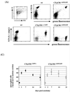
Analysis of CX3CR1 function in microglia. (A) Surface CX3CR1 staining of microglial cells isolated via Percoll density gradient from collagenase-digested brains of wt, CX3CR1+/GFP, and CX3CR1GFP/GFP mice. Cells were stained for the indicated surface markers (CD11b and CX3CR1) and gated according to scatter and viability as indicated. (B) Peripheral nerve transection experiment. Coronal section through operated and contralateral control facial nerve nuclei of axotomized CX3CR1+/GFP mouse day 7 after axotomy. Section were stained with an anti-neuronal nucleus-specific antibody (NeuN) and Cy5-coupled sheep anti-mouse IgG serum. (C) Quantitative evaluation of microglial reaction in response to facial nerve transection in operated CX3CR1+/GFP and CX3CR1GFP/GFP mice. The volume analyzed in the facial nerve nucleus cross sections represents about 0.25 mm2 by 12 μm. Results are presented as means (± standard deviations) of 16 sections obtained from four mice of each genotype per time point.
FIG. 5
FIG. 5
Analysis of CX3CR1 function in microglia. (A) Surface CX3CR1 staining of microglial cells isolated via Percoll density gradient from collagenase-digested brains of wt, CX3CR1+/GFP, and CX3CR1GFP/GFP mice. Cells were stained for the indicated surface markers (CD11b and CX3CR1) and gated according to scatter and viability as indicated. (B) Peripheral nerve transection experiment. Coronal section through operated and contralateral control facial nerve nuclei of axotomized CX3CR1+/GFP mouse day 7 after axotomy. Section were stained with an anti-neuronal nucleus-specific antibody (NeuN) and Cy5-coupled sheep anti-mouse IgG serum. (C) Quantitative evaluation of microglial reaction in response to facial nerve transection in operated CX3CR1+/GFP and CX3CR1GFP/GFP mice. The volume analyzed in the facial nerve nucleus cross sections represents about 0.25 mm2 by 12 μm. Results are presented as means (± standard deviations) of 16 sections obtained from four mice of each genotype per time point.

Similar articles

Cited by

References

    1. Bazan J F, Bacon K B, Hardiman G, Wang W, Soo K, Rossi D, Greaves D R, Zlotnik A, Schall T J. A new class of membrane-bound chemokine with a CX3C motif. Nature. 1997;385:640–644. - PubMed
    1. Butcher E C, Picker L J. Lymphocyte homing and homeostasis. Science. 1996;272:60–66. - PubMed
    1. Campbell J J, Hedrick J, Zlotnik A, Siani M A, Thompson D A, Butcher E C. Chemokines and the arrest of lymphocytes rolling under flow conditions. Science. 1998;279:381–384. - PubMed
    1. Fong A M, Robinson L A, Steeber D A, Tedder T F, Yoshie O, Imai T, Patel D D. Fractalkine and CX3CR1 mediate a novel mechanism of leukocyte capture, firm adhesion, and activation under physiologic flow. J Exp Med. 1998;188:1413–1419. - PMC - PubMed
    1. Foussat A, Coulomb-L'Hermine A, Gosling J, Krzysiek R, Durand-Gasselin I, Schall T, Balian A, Richard Y, Galanaud P, Emilie D. Fractalkine receptor expression by T lymphocyte subpopulations and in vivo production of fractalkine in human. Eur J Immunol. 2000;30:87–97. - PubMed

Publication types