

Glatiramer acetate (Copaxone) induces degenerate, Th2-polarized immune responses in patients with multiple sclerosis
- PMID: 10749576
- PMCID: PMC377485
- DOI: 10.1172/JCI8970
Glatiramer acetate (Copaxone) induces degenerate, Th2-polarized immune responses in patients with multiple sclerosis
Abstract
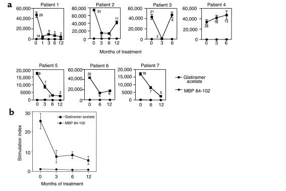
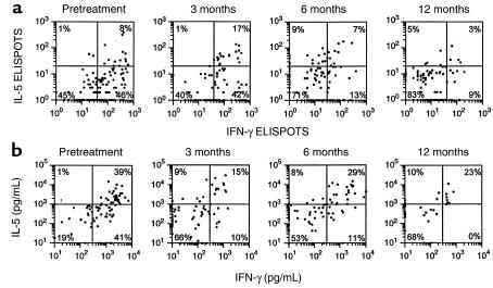
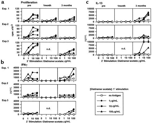
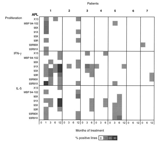
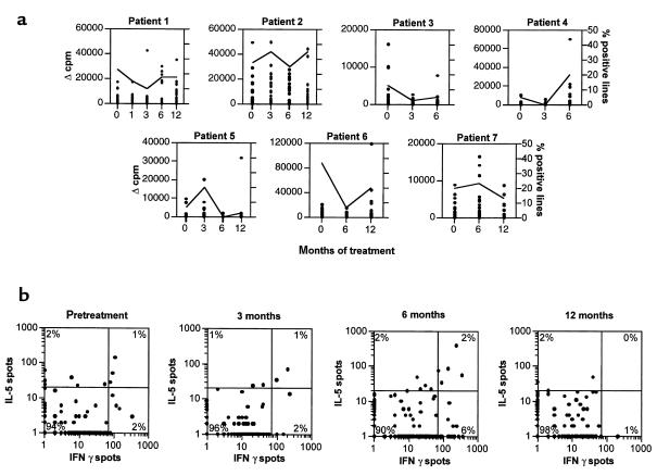
We examined the effect of glatiramer acetate, a random copolymer of alanine, lysine, glutamic acid, and tyrosine, on antigen-specific T-cell responses in patients with multiple sclerosis (MS). Glatiramer acetate (Copaxone) functioned as a universal antigen, inducing proliferation, independent of any prior exposure to the polymer, in T-cell lines prepared from MS or healthy subjects. However, for most patients, daily injections of glatiramer acetate abolished this T-cell response and promoted the secretion of IL-5 and IL-13, which are characteristic of Th2 cells. The surviving glatiramer acetate-reactive T cells exhibited a greater degree of degeneracy as measured by cross-reactive responses to combinatorial peptide libraries. Thus, it appears that, in some individuals, in vivo administration of glatiramer acetate induces highly cross-reactive T cells that secrete Th2 cytokines. To our knowledge, glatiramer acetate is the first agent that suppresses human autoimmune disease and alters immune function by engaging the T-cell receptor. This compound may be useful in a variety of autoimmune disorders in which immune deviation to a Th2 type of response is desirable.
Figures
Similar articles
-
Glatiramer acetate-reactive peripheral blood mononuclear cells respond to multiple myelin antigens with a Th2-biased phenotype.J Neuroimmunol. 2003 Jul;140(1-2):163-71. doi: 10.1016/s0165-5728(03)00170-x. J Neuroimmunol. 2003. PMID: 12864985
-
Glatiramer acetate induces a Th2-biased response and crossreactivity with myelin basic protein in patients with MS.Mult Scler. 2001 Aug;7(4):209-19. doi: 10.1177/135245850100700401. Mult Scler. 2001. PMID: 11548979 Clinical Trial.
-
Characterization of T cell lines derived from glatiramer-acetate-treated multiple sclerosis patients.J Neuroimmunol. 2000 Aug 1;108(1-2):201-6. doi: 10.1016/s0165-5728(00)00263-0. J Neuroimmunol. 2000. PMID: 10900354
-
Glatiramer acetate (Copaxone) therapy for multiple sclerosis.Pharmacol Ther. 2003 May;98(2):245-55. doi: 10.1016/s0163-7258(03)00036-6. Pharmacol Ther. 2003. PMID: 12725872 Review.
-
Immunomodulatory vaccines against autoimmune diseases.Rejuvenation Res. 2006 Spring;9(1):126-33. doi: 10.1089/rej.2006.9.126. Rejuvenation Res. 2006. PMID: 16608409 Review.
Cited by
-
Specific Th2 cells accumulate in the central nervous system of mice protected against experimental autoimmune encephalomyelitis by copolymer 1.Proc Natl Acad Sci U S A. 2000 Oct 10;97(21):11472-7. doi: 10.1073/pnas.97.21.11472. Proc Natl Acad Sci U S A. 2000. PMID: 11027347 Free PMC article.
-
Role of apoptosis in autoimmunity.J Clin Immunol. 2004 Jan;24(1):1-11. doi: 10.1023/B:JOCI.0000018057.89066.c6. J Clin Immunol. 2004. PMID: 14997028 Review.
-
Immunomodulatory synergy by combination of atorvastatin and glatiramer acetate in treatment of CNS autoimmunity.J Clin Invest. 2006 Apr;116(4):1037-44. doi: 10.1172/JCI25805. Epub 2006 Mar 16. J Clin Invest. 2006. PMID: 16543951 Free PMC article.
-
Glatiramer acetate: a review of its use in relapsing-remitting multiple sclerosis.CNS Drugs. 2002;16(12):825-50. doi: 10.2165/00023210-200216120-00004. CNS Drugs. 2002. PMID: 12421116 Review.
-
Glatiramer acetate (Copaxone) therapy induces CD8(+) T cell responses in patients with multiple sclerosis.J Clin Invest. 2002 Mar;109(5):641-9. doi: 10.1172/JCI14380. J Clin Invest. 2002. PMID: 11877472 Free PMC article.
References
-
- Grewal IS, et al. Requirement for CD40 ligand in costimulation induction, T cell activation, and experimental allergic encephalomyelitis. Science. 1996;273:1864–1867. - PubMed
-
- Lehmann PV, Forsthuber T, Miller A, Sercarz EE. Spreading of T-cell autoimmunity to cryptic determinants of an autoantigen. Nature. 1992;358:155–157. - PubMed
-
- Windhagen A, et al. Cytokine secretion of myelin basic protein reactive T cells in patients with multiple sclerosis. J Neuroimmunol. 1998;91:1–9. - PubMed
Publication types
MeSH terms
Substances
Grants and funding
LinkOut - more resources
Full Text Sources
Other Literature Sources
Medical
Molecular Biology Databases
