. 2000 Apr;105(7):887-95.
doi: 10.1172/JCI8688.

Pharmacological chaperones rescue cell-surface expression and function of misfolded V2 vasopressin receptor mutants

Affiliations

Pharmacological chaperones rescue cell-surface expression and function of misfolded V2 vasopressin receptor mutants

J P Morello et al. J Clin Invest. 2000 Apr.

Abstract

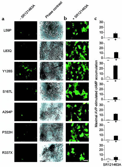
Over 150 mutations within the coding sequence of the V2 vasopressin receptor (V2R) gene are known to cause nephrogenic diabetes insipidus (NDI). A large number of these mutant receptors fail to fold properly and therefore are not routed to the cell surface. Here we show that selective, nonpeptidic V2R antagonists dramatically increase cell-surface expression and rescue the function of 8 mutant NDI-V2Rs by promoting their proper folding and maturation. A cell-impermeant V2R antagonist could not mimic these effects and was unable to block the rescue mediated by a permeant agent, indicating that the nonpeptidic antagonists act intracellularly, presumably by binding to and stabilizing partially folded mutants. In addition to opening new therapeutic avenues for NDI patients, these data demonstrate that by binding to newly synthesized mutant receptors, small ligands can act as pharmacological chaperones, promoting the proper folding and maturation of receptors and their targeting to the cell surface.


Figures

Figure 1
Figure 1
The del 62-64 V2R mutant is not expressed at the cell surface and is retained in the ER. (a and b) Nonpermeabilized cell immunofluorescence microscopy of COS-1 cells expressing the myc-tagged wild-type (WT) or myc-tagged del 62-64 V2R, respectively. (c) The same field of cells as in b stained with rhodamine-conjugated phalloidin to label actin filaments. (d and e) Permeabilized cell immunofluorescence microscopy of COS-1 cells expressing the myc-tagged WT or myc-tagged del 62-64 V2R, respectively. Bar = 50 μm. (fh) Confocal immunofluorescence microscopy on permeabilized COS-1 cells expressing the myc-tagged del 62-64 V2R using the anti-myc antibody (f), an antibody directed against the ER-resident protein calreticulin (g). (h) Illustration of the superimposed localization of both proteins. Bar = 20 μm. Representative of 5 separate experiments.
Figure 2
Figure 2
Cells expressing the del 62-64 V2R mutant do not display AVP-binding or signaling on whole cells. (a) Saturation-binding isotherm of [3H]AVP to whole cells expressing the WT (open circles) and the del 62-64 V2R (filled circles). COS-1 cells were transfected with 15 μg of plasmid encoding either WT or del 62-64 V2R. Specific binding is expressed as picomoles per well of [3H]AVP bound per well. Representative of 3 separate experiments. (b) Accumulation of cAMP in cells expressing WT or del 62-64 V2R. COS-1 cells were transfected with the indicated amounts of plasmid DNA. The data are expressed as an increase in cAMP levels above basal levels obtained at a 10–5 M AVP concentration and expressed as the mean ± SEM (n = 4).
Figure 3
Figure 3
SR121463A treatment increases the presence of the del 62-64 V2R at the cell surface. Nonpermeabilized cell immunofluorescence microscopy of HEK-293 cells stably expressing the del 62-64 V2R that were incubated for 16 hours in the absence (a) or presence (b) of 10–5 M SR121463A. (c and d) Identical treatment as a and b performed on HEK-293 cells expressing the WT V2R. Bar = 50 μm. (eh) Same field of cells as ad, viewed by phase-contrast microscopy. Representative of 3 separate experiments.
Figure 4
Figure 4
Concentration-dependent increase of cell-surface del 62-64 V2R after SR121463A treatment. Cell-surface receptor expression was measured by FACS of HEK-293 cells stably expressing either del 62-64 (a) or WT (b) V2R. Cells were treated with the indicated concentrations of SR121463A. Representative of 3 separate experiments. (c) Dose-dependent effect of SR121463A treatment on cell-surface del 62-64 (filled triangles) and WT (filled squares) V2R expression as quantified by the mean cell-surface fluorescence intensity obtained by FACS. The 100% point was taken from cells exposed to a 10–5 M concentration of SR121463A, and the 0% point was taken from cells incubated without anti-myc antibody. There was no difference in cell-surface fluorescence intensity between untransfected HEK-293 cells incubated with both antibodies and HEK-293 cells expressing the del 62-64 V2R that were incubated without anti-myc antibody. There was no difference in cell-surface fluorescence intensity between untransfected HEK-293 cells incubated with both antibodies and HEK-293 cells expressing the del 62-64 V2R that were incubated without anti-myc antibody. The data are expressed as means ± SEM (n = 3).
Figure 5
Figure 5
Rescued signaling activity of cells expressing the del 62-64 V2R after pretreatment with SR121463A. (a) AVP-stimulated cAMP accumulation was measured in whole HEK-293 cells stably expressing the del 62-64 V2R. Cells were treated (or not) with 10–5 M SR121463A for 16 hours, washed extensively, and incubated (or not) with 10–5 M AVP for 20 minutes. The data are expressed as means ± SEM (n = 3). (b) Concentration-dependent AVP-stimulated cAMP accumulation was measured in whole COS-1 cells transiently expressing the del 62-64 V2R. Cells were treated with (filled triangles) or without (filled squares) 10–5 M SR121463A for 16 hours, washed extensively, and incubated with the indicated AVP concentrations. Representative of 3 separate experiments. (c) Dose-dependent effect of SR121463A on AVP-stimulated cAMP accumulation. HEK-293 cells stably expressing the del 62-64 V2R were treated for 16 hours with the indicated concentrations of SR121463A before the determination of cAMP accumulation. The data are expressed as means ± SEM (n = 4). (d) Onset of the effect of SR121463A pretreatment on AVP-dependent cAMP accumulation. HEK-293 cells stably expressing the del 62-64 V2R were treated with 10–6 M SR121463A for the indicated times before assessing AVP-stimulated cAMP accumulation. Representative of 3 separate experiments.
Figure 6
Figure 6
SR121463A treatment promotes del 62-64 V2R protein maturation. Metabolic labeling of the WT (a, b) and del 62-64 (d, e) V2R in HEK-293 cells incubated in the absence or presence of 10–5 M SR121463A for 16 hours. Labeling was carried out with 150 μCi/mL–1 [35S]methionine/cysteine for 30 minutes followed by the indicated times of chase in complete medium. The migration of the precursor (open arrowhead) and mature (filled arrowhead) species is indicated. Representative of 3 separate experiments. (c and f) Mature/precursor receptor ratio used as an index of the maturation efficiency was calculated at the 60-minute chase time by densitometric analysis of the autoradiograms.
Figure 7
Figure 7
Sensitivity of the mature and precursor V2R species to Endo H and PNGase F. The nature of the del 62-64 V2R species was assessed in pulse-chase metabolic-labeling experiments carried out in cells treated (or not) with SR121463A. The glycosylation state was determined at the 60-minute chase time by treating the purified receptor with the indicated enzymes. In the absence of SR121463A treatment (a), only the precursor (open arrowhead) form that is sensitive to both enzymes is observed, whereas following a 10–5-M SR121463A treatment for 16 hours (b), the Endo H–resistant mature species is observed (closed arrowhead). (c) Concentration dependence of the SR121463A-promoted maturation of the V2R. The effect of the indicated concentrations of SR121463A (for 16 hours) on the maturation of the del 62-64 V2R was assessed at the 60–minute chase time. (d) The mature/precursor receptor ratio of the autoradiogram shown in c was calculated by densitometric analysis of the autoradiogram. Representative of 3 separate experiments.
Figure 8
Figure 8
Intracellular mode of action of SR121463A on the rescue of cell-surface del 62-64 V2R. HEK-293 cells stably expressing the del 62-64 V2R were treated with or without 10–5 M of the peptidic antagonist d(CH2)51,D-Tyr(Et)2,Val4,Arg8,des-Gly9-vasopressin for 1 hour before the addition of SR121463A. A concentration of 10–7 M of SR121463A was then added (or not) to the cells for 6 hours. To ensure that the peptidic antagonist did not become degraded during the incubation period it was added every hour. Cell-surface receptor expression was measured by FACS® as described above. Cells were scored as positive based on a 0% point taken without anti-myc antibody and without any drug treatments. The data are expressed as means ± SEM (n = 3).
Figure 9
Figure 9
SR121463A increases the cell-surface expression and signaling activity of several distinct NDI-V2R mutants. (a) cos-1 cells transiently transfected with plasmids encoding the indicated myc-tagged mutant V2 receptors detected by whole-cell immunofluorescence and phase-contrast microscopy. All mutants were found to be poorly expressed at the cell surface. (b) Treatment with 10–5 M SR121463A for 16 hours leads to cell-surface appearance of these receptor mutants as assessed by fluorescence microscopy. Bar = 50 μm. (c) Effects of a pretreatment with 10–5 M SR121463A for 16 hours on vasopressin-stimulated cAMP accumulation in cos-1 cells expressing the various mutants as indicated in a. The position and nature of the amino acid replacement in each NDI-causing mutation is indicated by the single letter amino acid code. X indicates a stop codon. Representative of 3 separate experiments.

Comment in

  • Antagonists to the rescue.
    Welch WJ, Howard M. Welch WJ, et al. J Clin Invest. 2000 Apr;105(7):853-4. doi: 10.1172/JCI9158. J Clin Invest. 2000. PMID: 10749562 Free PMC article. Review. No abstract available.

Similar articles

Cited by

References

    1. Knoers NV, Monnens LL. Nephrogenic diabetes insipidus. Semin Nephrol. 1999;19:344–352. - PubMed
    1. Bichet, D.G., and Fujiwara, T.M. 2000. Nephrogenic diabetes insipidus. In The metabolic and molecular basis of inherited disease. 8th edition. C.R. Scriver et al., editors. McGraw-Hill. New York, NY. In press.
    1. Schülein R, Rutz C, Rosenthal W. Membrane targeting and determination of transmembrane topology of the human vasopressin V2 receptor. J Biol Chem. 1996;271:28844–28852. - PubMed
    1. Wenkert D, et al. Functional characterization of five V2 vasopressin receptor gene mutations. Mol Cell Endocrinol. 1996;124:43–50. - PubMed
    1. Sadeghi HM, Innamorati G, Birnbaumer M. An X-linked NDI mutation reveals a requirement for cell surface V2R expression. Mol Endocrinol. 1997;11:706–713. - PubMed

Publication types

MeSH terms