

Mucin secretion is modulated by luminal factors in the isolated vascularly perfused rat colon
- PMID: 10644316
- PMCID: PMC1727811
- DOI: 10.1136/gut.46.2.218
Mucin secretion is modulated by luminal factors in the isolated vascularly perfused rat colon
Abstract
Background: Mucins play an important protective role in the colonic mucosa. Luminal factors modulating colonic mucus release have been not fully identified.
Aim: To determine the effect of some dietary compounds on mucus discharge in rat colon.
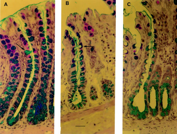
Methods: An isolated vascularly perfused rat colon model was used. Mucus secretion was induced by a variety of luminal factors administered as a bolus of 1 ml for 30 minutes in the colonic loop. Mucin release was evaluated using a sandwich enzyme linked immunosorbent assay supported by histological analysis.
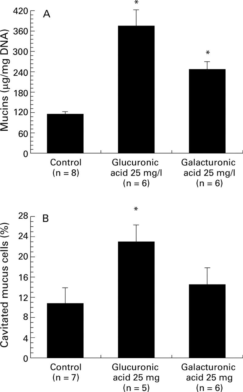
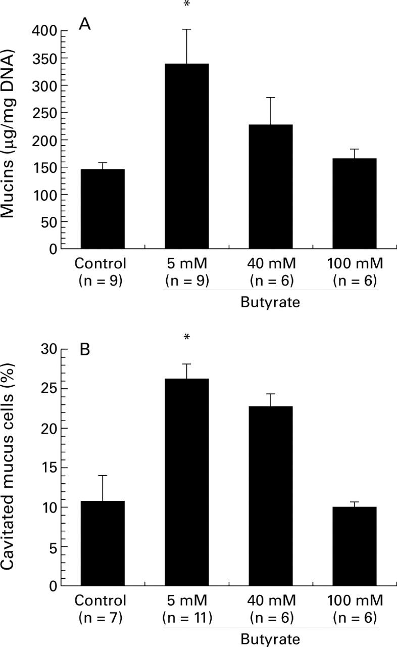
Results: The three dietary fibres tested in this study (pectin, gum arabic, and cellulose) did not provoke mucus secretion. Luminal administration of sodium alginate (an algal polysaccharide used as a food additive) or ulvan (a sulphated algal polymer) induced a dose dependent increase in mucin discharge over the concentration range 1-25 mg/l (p<0.05 for 25 mg/l alginate and p<0.05 for 10 and 25 mg/l ulvan). Glucuronic acid and galacturonic acid, which are major constituents of a variety of fibres, produced significant mucin secretion (p<0.05). Hydrogen sulphide and mercaptoacetate, two sulphides produced in the colonic lumen by microbial fermentation of sulphated polysaccharides, did not modify mucin secretion. Among the short chain fatty acids, acetate (5-100 mM) induced a dose dependent release of mucus (p<0.05 for 100 mM acetate). Interestingly, butyrate at a concentration of 5 mM produced colonic mucin secretion (p<0.05), but increasing its concentration to 100 mM provoked a gradual decrease in mucus discharge. Propionate (5-100 mM) did not induce mucin release. Several dietary phenolic compounds (quercetin, epicatechin, resveratrol) did not provoke mucus discharge.
Conclusions: Two algal polysaccharides (alginate and ulvan), two uronic acids (glucuronic acid and galacturonic acid), and the short chain fatty acids acetate and butyrate induce mucin secretion in rat colon. Taken together, these data suggest that some food constituents and their fermentation products may regulate the secretory function of colonic goblet cells.
Figures

Similar articles
-
Luminal peptide YY-releasing factors in the isolated vascularly perfused rat colon.J Endocrinol. 1996 Dec;151(3):421-9. doi: 10.1677/joe.0.1510421. J Endocrinol. 1996. PMID: 8994387
-
Colonic mucin discharge by a cholinergic agonist, prostaglandins, and peptide YY in the isolated vascularly perfused rat colon.Digestion. 1997;58(2):168-75. doi: 10.1159/000201440. Digestion. 1997. PMID: 9144307
-
Sulfated polysaccharides, but not cellulose, increase colonic mucus in rats with loperamide-induced constipation.Dig Dis Sci. 2001 Jul;46(7):1482-9. doi: 10.1023/a:1010644021888. Dig Dis Sci. 2001. PMID: 11478500
-
Use of viscous fibres in beverages for appetite control: a review of studies.Int J Food Sci Nutr. 2015;66(5):479-90. doi: 10.3109/09637486.2015.1034252. Epub 2015 May 22. Int J Food Sci Nutr. 2015. PMID: 26001088 Review.
-
Famine, fiber, fatty acids, and failed colonic absorption: does fiber fermentation ameliorate diarrhea?JPEN J Parenter Enteral Nutr. 1994 Jan-Feb;18(1):4-8. doi: 10.1177/014860719401800104. JPEN J Parenter Enteral Nutr. 1994. PMID: 8164301 Review.
Cited by
-
Implication of fructans in health: immunomodulatory and antioxidant mechanisms.ScientificWorldJournal. 2015;2015:289267. doi: 10.1155/2015/289267. Epub 2015 Mar 16. ScientificWorldJournal. 2015. PMID: 25961072 Free PMC article. Review.
-
Polymers in the gut compress the colonic mucus hydrogel.Proc Natl Acad Sci U S A. 2016 Jun 28;113(26):7041-6. doi: 10.1073/pnas.1602789113. Epub 2016 Jun 14. Proc Natl Acad Sci U S A. 2016. PMID: 27303035 Free PMC article.
-
Dietary fructo-oligosaccharides and inulin decrease resistance of rats to salmonella: protective role of calcium.Gut. 2004 Apr;53(4):530-5. doi: 10.1136/gut.2003.023499. Gut. 2004. PMID: 15016747 Free PMC article.
-
Respiratory and Intestinal Microbiota in Pediatric Lung Diseases-Current Evidence of the Gut-Lung Axis.Int J Mol Sci. 2022 Jun 18;23(12):6791. doi: 10.3390/ijms23126791. Int J Mol Sci. 2022. PMID: 35743234 Free PMC article. Review.
-
Development of a self-limiting model of methotrexate-induced mucositis reinforces butyrate as a potential therapy.Sci Rep. 2021 Nov 25;11(1):22911. doi: 10.1038/s41598-021-02308-w. Sci Rep. 2021. PMID: 34824316 Free PMC article.
References
Publication types
MeSH terms
Substances
LinkOut - more resources
Full Text Sources
Other Literature Sources