. 1999 Nov 1;147(3):631-44.
doi: 10.1083/jcb.147.3.631.

N-cadherin promotes motility in human breast cancer cells regardless of their E-cadherin expression

Affiliations

N-cadherin promotes motility in human breast cancer cells regardless of their E-cadherin expression

M T Nieman et al. J Cell Biol. .

Abstract

E-cadherin is a transmembrane glycoprotein that mediates calcium-dependent, homotypic cell-cell adhesion and plays a role in maintaining the normal phenotype of epithelial cells. Decreased expression of E-cadherin has been correlated with increased invasiveness of breast cancer. In other systems, inappropriate expression of a nonepithelial cadherin, such as N-cadherin, by an epithelial cell has been shown to downregulate E-cadherin expression and to contribute to a scattered phenotype. In this study, we explored the possibility that expression of nonepithelial cadherins may be correlated with increased motility and invasion in breast cancer cells. We show that N-cadherin promotes motility and invasion; that decreased expression of E-cadherin does not necessarily correlate with motility or invasion; that N-cadherin expression correlates both with invasion and motility, and likely plays a direct role in promoting motility; that forced expression of E-cadherin in invasive, N-cadherin-positive cells does not reduce their motility or invasive capacity; that forced expression of N-cadherin in noninvasive, E-cadherin-positive cells produces an invasive cell, even though these cells continue to express high levels of E-cadherin; that N-cadherin-dependent motility may be mediated by FGF receptor signaling; and that cadherin-11 promotes epithelial cell motility in a manner similar to N-cadherin.


Figures

Figure 1
Figure 1
Cadherin and β-catenin expression in breast carcinoma cell lines. Confluent monolayers of MCF-7, BT-20, SUM 149, SKBr3, MDA-MB-453, SUM 1315, MDA-MB-435, MDA-MB-436, BT-549, Hs578t, SUM 159PT, or MDA-MB-231 were extracted with NP-40. 20 μg total protein from each cell extract was resolved by SDS-PAGE, transferred to nitrocellulose, and blotted with antibodies against E-cadherin (HECD1), N-cadherin, P-cadherin, cadherin-11, or β-catenin.
Figure 2
Figure 2
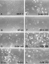
Morphological analysis of breast cancer cell lines. Living monolayers of MCF-7 (A), BT-20 (B), SUM 149 (C), SKBr3 (D), SUM 1315 (E), MDA-MB-435 (F), MDA-MB-436 (G), or SUM 159PT (H) cells were photographed using an inverted Zeiss microscope at 200×. Bar, 10 μm.
Figure 3
Figure 3
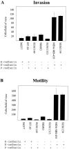
N-cadherin expression correlates with increased invasiveness and motility in breast carcinoma cell lines. Cells were plated on Matrigel-coated or noncoated membranes for invasion assays or motility assays, respectively. The cells were incubated for 24 h, and those that did not migrate through the pores in the membrane were removed by scraping the membrane with a cotton swab. The remaining cells were stained, and the number transversing the membrane was determined by averaging ten random fields of view at 100×. The data are expressed as the number of cells/field of view and is the average of three independent experiments. Error bars indicate SD of the average.
Figure 4
Figure 4
Expression of N-cadherin by BT-20 cells. BT-20 cells were transfected with N-cadherin (BT-20N) and expression induced with dexamethasone. A, Phase-microscopy of living BT-20N cells. Bar, 10 μm. B and C, Cells were grown on glass coverslips and processed for coimmunofluorescence localization with antibodies against E-cadherin (Jelly; B) and N-cadherin (C). D, BT-20 and BT-20N cells were extracted with NP-40 and 20 μg protein from each extract was resolved by SDS-PAGE, transferred to nitrocellulose, and immunoblotted for E-cadherin (HECD1), N-cadherin, or P-cadherin. E, Extracts of BT-20N cells were immunoprecipitated with antibodies against N-cadherin or E-cadherin (HECD1). The immunoprecipitation reactions, as well as cell extracts, were resolved by SDS-PAGE, transferred to nitrocellulose, and immunoblotted for N-cadherin and β-catenin (lanes 1 and 2) or E-cadherin (HECD1) and β-catenin (lanes 3 and 4).
Figure 5
Figure 5
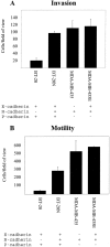
Exogenous expression of N-cadherin by BT-20 cells (BT-20N) increases their invasiveness, whereas exogenous expression of E-cadherin by MDA-MB-435 cells (MDA-MB-435E) does not effect their behavior. Cells were plated on Matrigel-coated or noncoated membranes for invasion assays or motility assays, respectively. The cells were incubated for 24 h, and those that did not migrate through the pores in the membrane were removed by scraping the membrane with a cotton swab. The remaining cells were stained, and the number transversing the membrane was determined by averaging ten random fields of view at 100×. The data are expressed as the number of cells/field of view and is the average of three independent experiments. Error bars indicate SD of the average.
Figure 6
Figure 6
Expression of E-cadherin by MDA-MB-435 cells. MDA-MB-435 cells were transfected with E-cadherin (MDA-MB-435E) and expression was induced with dexamethasone. A, Phase-microscopy of MDA-MB-435E cells. Bar, 10 μm. B and C, Cells were grown on glass coverslips and processed for coimmunofluorescence localization with antibodies against E- (Jelly; B) and N-cadherin (C). D, MDA-MB-435 and several clones of MDA-MB-435E cells were extracted with NP-40 and 20 μg protein from each extract was resolved by SDS-PAGE, transferred to nitrocellulose, and immunoblotted for E- (HECD1) and N-cadherin. Clone 2 (cl2) expressed the highest level of E-cadherin and was chosen for subsequent studies. E, Extracts of MDA-MB-435 and MDA-MB-435E cells were immunoprecipitated with antibodies against N- or E-cadherin (HECD1). The immunoprecipitation reactions, along with cell extracts, were resolved by SDS-PAGE, transferred to nitrocellulose, and immunoblotted for N-cadherin and β-catenin (lanes 1 and 2), or E-cadherin (HECD1) and β-catenin (lanes 3 and 4).
Figure 7
Figure 7
BT-20N cells do not mix with HT1080 cells. 5 × 10⁴ BT-20 or BT-20N cells were mixed with an equal number of HT1080 cells, allowed to settle on coverslips, and processed for immunofluorescence with an mAb against N- (13A9) or E-cadherin (Jelly). A and B are a mix of BT-20 and HT1080 cells stained for E- and N-cadherin, respectively. The encircled cells are a group of E-cadherin–negative, N-cadherin–positive HT1080 cells. C and D are a mix of BT-20N and HT1080 cells stained for E- and N-cadherin, respectively. The encircled cells are a group of E-cadherin–negative, N-cadherin–positive HT1080 cells.
Figure 8
Figure 8
Exogenous expression of cadherin-11 by BT-20 cells (BT-20cad11) increases their invasiveness. BT-20 cells were transfected with cadherin-11 (BT-20cad11) and expression induced with dexamethasone. A, Phase-microscopy of living BT-20cad11 cells. Bars, 10 μm. B and C, Cells were grown on glass coverslips and processed for coimmunofluorescence localization with antibodies against E-cadherin (Jelly; B) and cadherin-11 (C). D and E, Cells were plated on Matrigel-coated or noncoated membranes for invasion assays or motility assays, respectively. The cells were incubated for 24 h, and those that did not migrate through the pores in the membrane were removed by scraping the membrane with a cotton swab. The remaining cells were stained, and the number transversing the membrane was determined by averaging ten random fields of view at 100 ×. The data are expressed as the number of cells/field of view and is the average of three independent experiments. Error bars indicate SD of the average.
Figure 8
Figure 8
Exogenous expression of cadherin-11 by BT-20 cells (BT-20cad11) increases their invasiveness. BT-20 cells were transfected with cadherin-11 (BT-20cad11) and expression induced with dexamethasone. A, Phase-microscopy of living BT-20cad11 cells. Bars, 10 μm. B and C, Cells were grown on glass coverslips and processed for coimmunofluorescence localization with antibodies against E-cadherin (Jelly; B) and cadherin-11 (C). D and E, Cells were plated on Matrigel-coated or noncoated membranes for invasion assays or motility assays, respectively. The cells were incubated for 24 h, and those that did not migrate through the pores in the membrane were removed by scraping the membrane with a cotton swab. The remaining cells were stained, and the number transversing the membrane was determined by averaging ten random fields of view at 100 ×. The data are expressed as the number of cells/field of view and is the average of three independent experiments. Error bars indicate SD of the average.
Figure 9
Figure 9
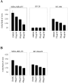
The diacylglycerol lipase inhibitor RHC80267 decreases motility of N-cadherin– and cadherin-11–expressing cells. Cells were plated on noncoated membranes for motility assays. The cells were incubated for 24 h in the presence of RHC80267 at varying concentrations, and those that did not migrate through the pores in the membrane were removed by scraping the membrane with a cotton swab. The remaining cells were stained, and the number transversing the membrane was determined by averaging ten random fields of view at 100×. The data are expressed as the number of cells/field of view and is the average of three (A) or two (B) independent experiments. Error bars indicate SD of the average.

Similar articles

Cited by

References

    1. Aberle H., Butz S., Stappert J., Weissig H., Kemler R., Hoschuetzky H. Assembly of the cadherin–catenin complex in vitro with recombinant proteins. J. Cell Sci. 1994;107:3655–3663. - PubMed
    1. Aono S., Nakagawa S., Reynolds A.B., Takeichi M. p120(ctn) acts as an inhibitory regulator of cadherin function in colon carcinoma cells. J. Cell Biol. 1999;145:551–562. - PMC - PubMed
    1. Behrens J., Mareel M., van Roy F.M., Birchmeier W. Dissecting tumor cell invasionepithelial cells acquire invasive properties after the loss of uvomorulin-mediated cell–cell adhesion. J. Cell Biol. 1989;108:2435–2447. - PMC - PubMed
    1. Behrens J., Weidner K.M., Frixen U.H., Schipper J.H., Sachs M., Arakaki N., Daikuhara Y., Birchmeier W. The role of E-cadherin and scatter factor in tumor invasion and cell motility. Exper. Suppl. 1991;59:109–126. - PubMed
    1. Berx G., Cleton-Jansen A.M., Nollet F., de Leeuw W.J.F., van de Vijver M.J., Cornelisse M.J., van Roy F. E-cadherin is a tumor/invasion suppressor gene mutated in human lobular breast cancers. EMBO (Eur. Mol. Biol. Organ.) J. 1995;14:6107–6115. - PMC - PubMed

Publication types

MeSH terms