

Polarized vascular endothelial growth factor secretion by human retinal pigment epithelium and localization of vascular endothelial growth factor receptors on the inner choriocapillaris. Evidence for a trophic paracrine relation
- PMID: 10433935
- PMCID: PMC1866848
- DOI: 10.1016/S0002-9440(10)65138-3
Polarized vascular endothelial growth factor secretion by human retinal pigment epithelium and localization of vascular endothelial growth factor receptors on the inner choriocapillaris. Evidence for a trophic paracrine relation
Abstract
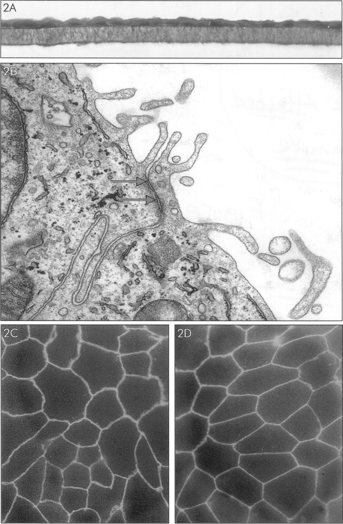
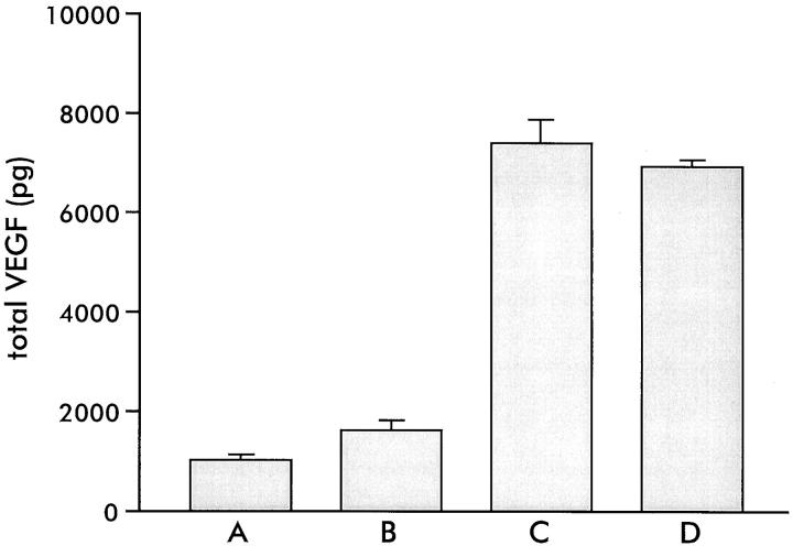
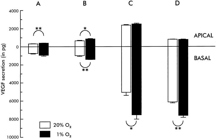
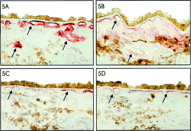
The retinal pigment epithelium (RPE) maintains the choriocapillaris (CC) in the normal eye and is involved in the pathogenesis of choroidal neovascularization in age-related macular degeneration. Vascular endothelial growth factor-A (VEGF) is produced by differentiated human RPE cells in vitro and in vivo and may be involved in paracrine signaling between the RPE and the CC. We investigated whether there is a polarized secretion of VEGF by RPE cells in vitro. Also, the localization of VEGF receptors in the human retina was investigated. We observed that highly differentiated human RPE cells, cultured on transwell filters in normoxic conditions, produced two- to sevenfold more VEGF toward their basolateral side as compared to the apical side. In hypoxic conditions, VEGF-A secretion increased to the basal side only, resulting in a three- to 10-fold higher basolateral secretion. By immunohistochemistry in 30 human eyes and in two cynomolgus monkey eyes, KDR (VEGFR-2) and flt-4 (VEGFR-3) were preferentially localized at the side of the CC endothelium facing the RPE cell layer, whereas flt-1 (VEGFR-1) was found on the inner CC and on other choroidal vessels. Our results indicate that RPE secretes VEGF toward its basal side where its receptor KDR is located on the adjacent CC endothelium, suggesting a role of VEGF in a paracrine relation, possibly in cooperation with flt-4 and its ligand. This can explain the known trophic function of the RPE in the maintenance of the CC and its fenestrated permeable phenotype and points to a role for VEGF in normal eye functioning. Up-regulated basolateral VEGF secretion by RPE in hypoxia or loss of polarity of VEGF production may play a role in the pathogenesis of choroidal neovascularization.
Figures

Similar articles
-
Immunohistochemical localization of vascular endothelial growth factor (VEGF) and its two specific receptors, Flt-1 and KDR, in the porcine placenta and non-pregnant uterus.Placenta. 1999 Jan;20(1):35-43. doi: 10.1053/plac.1998.0350. Placenta. 1999. PMID: 9950143
-
Expression of vascular endothelial growth factor (VEGF) and its receptors is regulated in eyes with intra-ocular tumours.J Pathol. 1998 Nov;186(3):306-12. doi: 10.1002/(SICI)1096-9896(1998110)186:3<306::AID-PATH183>3.0.CO;2-B. J Pathol. 1998. PMID: 10211121
-
Role of hypoxia and extracellular matrix-integrin binding in the modulation of angiogenic growth factors secretion by retinal pigmented epithelial cells.J Cell Biochem. 1999 Jul 1;74(1):135-43. J Cell Biochem. 1999. PMID: 10381270
-
Possible involvement of VEGF-FLT tyrosine kinase receptor system in normal and tumor angiogenesis.Princess Takamatsu Symp. 1994;24:162-70. Princess Takamatsu Symp. 1994. PMID: 8983073 Review.
-
Vascular endothelial growth factors and angiogenesis in eye disease.Prog Retin Eye Res. 2003 Jan;22(1):1-29. doi: 10.1016/s1350-9462(02)00043-5. Prog Retin Eye Res. 2003. PMID: 12597922 Review.
Cited by
-
Fire in the ashes: can failed Alzheimer's disease drugs succeed with second chances?Alzheimers Dement. 2013 Jan;9(1):50-7. doi: 10.1016/j.jalz.2012.01.007. Epub 2012 Mar 30. Alzheimers Dement. 2013. PMID: 22465172 Free PMC article.
-
Activated Protein C (APC) and 3K3A-APC-Induced Regression of Choroidal Neovascularization (CNV) Is Accompanied by Vascular Endothelial Growth Factor (VEGF) Reduction.Biomolecules. 2021 Feb 26;11(3):358. doi: 10.3390/biom11030358. Biomolecules. 2021. PMID: 33652861 Free PMC article.
-
Photoreceptor avascular privilege is shielded by soluble VEGF receptor-1.Elife. 2013 Jun 18;2:e00324. doi: 10.7554/eLife.00324. Elife. 2013. PMID: 23795287 Free PMC article.
-
Choroidal Neovascularization: Mechanisms of Endothelial Dysfunction.Front Pharmacol. 2019 Nov 29;10:1363. doi: 10.3389/fphar.2019.01363. eCollection 2019. Front Pharmacol. 2019. PMID: 31849644 Free PMC article. Review.
-
Attainment of polarity promotes growth factor secretion by retinal pigment epithelial cells: relevance to age-related macular degeneration.Aging (Albany NY). 2009 Dec 27;2(1):28-42. doi: 10.18632/aging.100111. Aging (Albany NY). 2009. PMID: 20228934 Free PMC article.
References
-
- Yi X, Ogata N, Komada M, Yamamoto C, Takahashi K, Omori K, Uyama M: Vascular endothelial growth factor expression in choroidal neovascularization in rats. Graefe’s Arch Clin Exp Ophthalmol 1997, 235:313-319 - PubMed
-
- Korte GE, Gerszberg T, Pua F, Henkind P: Choriocapillaris atrophy after experimental destruction of the retinal pigment epithelium in the rat. A study in thin sections and vascular casts. Acta Anat 1986, 127:171-175 - PubMed
-
- Sakamoto T, Sakamoto H, Murphy TL, Spee C, Soriano D, Ishibashi T, Hinton DR: Vessel formation by choroidal endothelial cells in vitro is modulated by pigment epithelial cells. Arch Ophthalmol 1995, 113:512-520 - PubMed
-
- Miller H, Miller B, Ryan SJ: The role of retinal pigment epithelium in the involution of subretinal neovascularization. Invest Ophthalmol Vis Sci 1986, 27:1644-1652 - PubMed
-
- Glaser BM: Extracellular modulating factors and the control of intraocular neovascularization. An overview. Arch Ophthalmol 1988, 106:603-607 - PubMed
Publication types
MeSH terms
Substances
LinkOut - more resources
Full Text Sources
Other Literature Sources
Miscellaneous
