Review
. 1999 Jun;63(2):266-92.
doi: 10.1128/MMBR.63.2.266-292.1999.

Pseudomonas syringae phytotoxins: mode of action, regulation, and biosynthesis by peptide and polyketide synthetases

Affiliations
Review

Pseudomonas syringae phytotoxins: mode of action, regulation, and biosynthesis by peptide and polyketide synthetases

C L Bender et al. Microbiol Mol Biol Rev. 1999 Jun.

Abstract

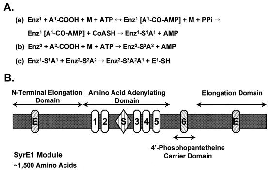
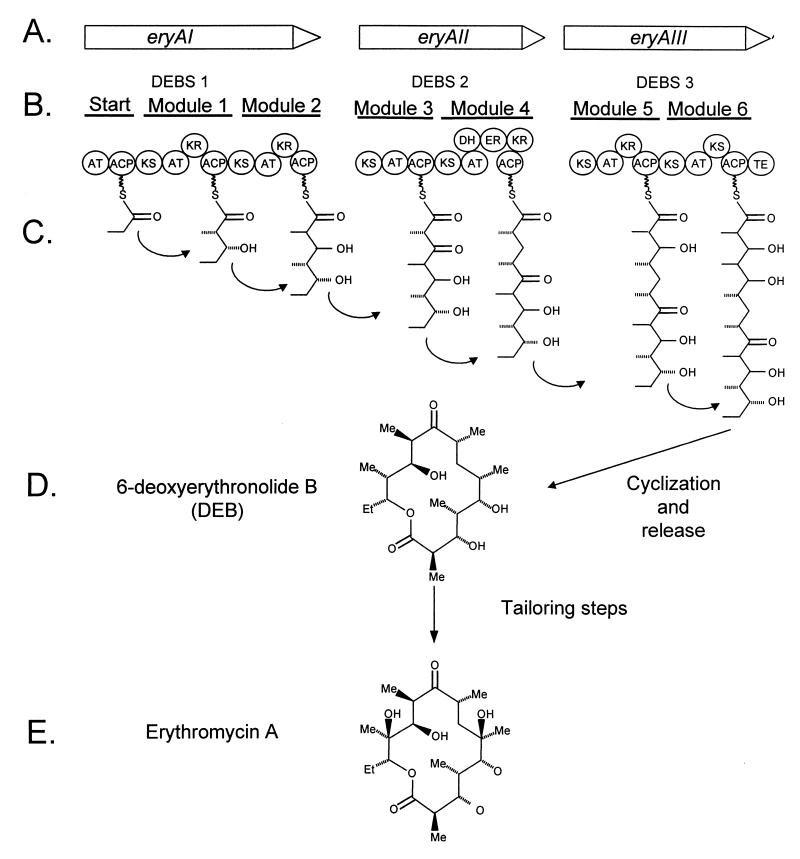
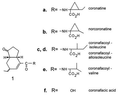
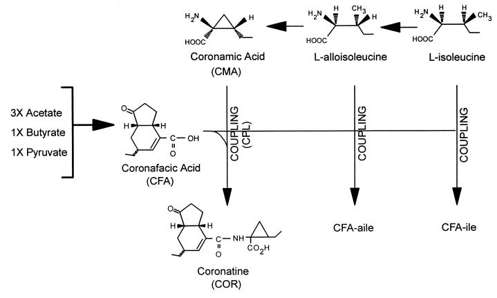
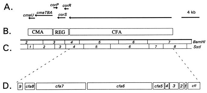
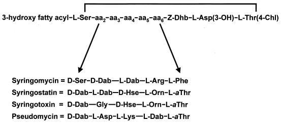
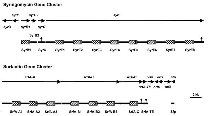
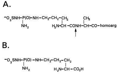
Coronatine, syringomycin, syringopeptin, tabtoxin, and phaseolotoxin are the most intensively studied phytotoxins of Pseudomonas syringae, and each contributes significantly to bacterial virulence in plants. Coronatine functions partly as a mimic of methyl jasmonate, a hormone synthesized by plants undergoing biological stress. Syringomycin and syringopeptin form pores in plasma membranes, a process that leads to electrolyte leakage. Tabtoxin and phaseolotoxin are strongly antimicrobial and function by inhibiting glutamine synthetase and ornithine carbamoyltransferase, respectively. Genetic analysis has revealed the mechanisms responsible for toxin biosynthesis. Coronatine biosynthesis requires the cooperation of polyketide and peptide synthetases for the assembly of the coronafacic and coronamic acid moieties, respectively. Tabtoxin is derived from the lysine biosynthetic pathway, whereas syringomycin, syringopeptin, and phaseolotoxin biosynthesis requires peptide synthetases. Activation of phytotoxin synthesis is controlled by diverse environmental factors including plant signal molecules and temperature. Genes involved in the regulation of phytotoxin synthesis have been located within the coronatine and syringomycin gene clusters; however, additional regulatory genes are required for the synthesis of these and other phytotoxins. Global regulatory genes such as gacS modulate phytotoxin production in certain pathovars, indicating the complexity of the regulatory circuits controlling phytotoxin synthesis. The coronatine and syringomycin gene clusters have been intensively characterized and show potential for constructing modified polyketides and peptides. Genetic reprogramming of peptide and polyketide synthetases has been successful, and portions of the coronatine and syringomycin gene clusters could be valuable resources in developing new antimicrobial agents.


Figures

FIG. 1
FIG. 1
(A) Reaction sequence catalyzed by multifunctional peptide synthetases. (a) Carboxyl activation of the first amino acid (A1) and formation of the aminoacyl adenylate; (b) activation of the second amino acid (A2) and formation of the aminoacyl adenylate; and (c) the condensation reaction. Abbreviations: Enz, enzyme; A, amino acid, M, divalent metal ion (Mg2+, Mn2+, Ca2+). Numbering indicates specific domains within an individual multienzyme; for example, Enz1 and Enz2 are two distinct domains within the same enzyme. Amino acids (A1 and A2) are two individual amino acids. For additional information, see reference . (B) Domain structure of the amino acid-activating module SyrE1. The SyrE1 module contains approximately 1,500 amino acids organized into four domains as defined by Stein and Vater (252). The relative positions of conserved core sequences are shown for each domain. The two elongation domains contain a characteristic HHxxxDG motif (E) (54). The five core sequences described by Stachelhaus and Marahiel (247) are located within the amino acid-adenylating domain. Core 2 has a sequence (SGTTGxPKGV) resembling the Walker type A motif involved in ATP binding, and the core 4 sequence (TGD) carries a motif associated with ATPase activity. A role in catalyzing aminoacyl adenylate formation is suggested for cores 3 and 5 (247). A region between cores 2 and 3 is associated with substrate recognition (S) (47), and SyrE1 exhibits substrate specificity for l-Ser (95). Core 6, with a characteristic LGGHSL motif, is located in the 4′-phosphopantetheine carrier domain. The motif contains a conserved serine to which the 4′-phosphopantetheine cofactor is covalently attached, which in turn is the site of thioester formation. The next module, SyrE2, carrying three domains (i.e., amino acid adenylating, 4′-phosphopantetheine carrier, and elongation domains), follows the SyrE1 module.
FIG. 2
FIG. 2
Reaction sequence in the synthesis of fatty acids. The starting units for the fatty acid synthase are acetyl-CoA and malonyl-CoA; these are converted into acetyl-ACP and malonyl-ACP by acetyl and malonyl transacylase, respectively. The fatty acid synthase proceeds with the condensation of these two precursors and then continues with a cycle of reduction, dehydration, and further reduction of the keto group (asterisk). Two key differences between polyketide synthases (PKS) and fatty acid synthase include the choice of starter units and the extent of reduction.
FIG. 3
FIG. 3
Biosynthesis of erythromycin A. (A) The nucleotide sequence of the eryA region contains three ORFs designated eryAI, eryAII, and eryAIII (29, 60). (B) These genes encode three proteins which constitute erythromycin B synthetase (DEBS); these are designated DEBS 1, DEBS 2, and DEBS 3 (39). (C) Each DEBS protein contains two modules, each with domains for acetyltransferase (AT), ACP, and β-keto synthase (KS) activity. Some modules contain additional domains for dehydratase (DH), enoyl reductase (ER), ketoreductase (KR), and TE activity. (D) Cyclization and release of the DEBS 3 product results in the formation of 6-deoxyerythronolide B (DEB), and additional tailoring steps result in the production of erythromycin A (E). Modified from reference .
FIG. 4
FIG. 4
Structures of COR and coronafacoyl compounds.
FIG. 5
FIG. 5
Biochemical pathways involved in the synthesis of COR and coronafacoyl compounds in P. syringae pv. glycinea PG4180. COR consists of a polyketide component, CFA, coupled (CPL) via amide-bond formation to an amino acid component, CMA. CFA is synthesized as a branched polyketide from three acetate units, one pyruvate unit, and one butyrate unit via an unknown sequence of events (196). CMA is derived from isoleucine via alloisoleucine and cyclized by an unknown mechanism (160, 195). CMA functions as an intermediate in the COR biosynthetic pathway, which indicates that cyclization of l-alloisoleucine to form CMA occurs before CFA and CMA are coupled (170). The coronafacoyl analogues, CFA-Ile and CFA-aIle result from amide bond formation between CFA and isoleucine and alloisoleucine, respectively, and are not utilized further in the synthesis of COR.
FIG. 6
FIG. 6
Functional and physical map of the COR biosynthetic gene cluster. (A) Horizontal lines with arrowheads indicate the transcriptional organization of the COR gene cluster. (B) Functional regions of the COR biosynthetic cluster: CMA, CMA biosynthetic gene cluster; REG, regulatory region; CFA, CFA biosynthetic gene cluster. (C) Physical map of the COR gene cluster; the enzymes used for restriction mapping were BamHI and SstI. (D) Expanded view of SstI fragments 4 to 8, which contain the CFA biosynthetic gene cluster. Abbreviations: 1, cfa1; 2, cfa2; 3, cfa3; 4, cfa4; 9, cfa9.
FIG. 7
FIG. 7
Structures of syringomycin, syringostatin, syringotoxin, and pseudomycin. The four lipodepsinonapeptides differ in the amino acid sequence between positions 2 and 6. The 3-hydroxy fatty acyl group is a derivative of either decanoic acid (syringomycin), dodecanoic acid (syringomycin and syringostatin), tetradecanoic acid (all four lipodepsinonapeptides), or hexadecanoic acid (pseudomycin); some forms of pseudomycin are acylated by 3,4-dihydroxytetradecanoate or 3,4-dihydroxyhexadecanoate. Abbreviations of nonstandard amino acids: Asp(3-OH), 3-hydroxyaspartic acid; Dab, 2,4-diaminobutyric acid; Dhb, 2,3-dehydroaminobutyric acid; Hse, homoserine; Orn, ornithine; Thr(4-Chl), 4-chlorothreonine; aThr, allothreonine.
FIG. 8
FIG. 8
Physical map of the syringomycin gene cluster of P. syringae pv. syringae compared to that of the surfactin gene cluster of B. subtilis. The amino acid-adenylating domains and 4′-phosphopantetheine carrier domains are indicated by cross-hatched and stippled regions, respectively, for each module. The small circles attached above the proteins indicate the regions carrying motifs characteristic of thioesterases. The 37-kb syr gene cluster encodes four proteins (SyrB1, SyrB2, SyrC, and SyrE) involved in biosynthesis. The syrD and syrP genes encode proteins predicted to be involved in secretion and regulation, respectively. The 31-kb srf gene cluster encodes five proteins (SrfA-A, SrfA-B, SrfA-C, SrfA-TE, and Sfp) involved in surfactin synthesis. The sfp gene product has been proposed to function as a 4′-phosphopantetheine transferase (147). The functions of the products of orf5 through orf8 are unknown.
FIG. 9
FIG. 9
Structures of phenolic plant signal molecules known to activate the syrB gene involved in syringomycin biosynthesis by P. syringae pv. syringae. Arbutin is found in several plant species including pear (Pyrus communis L.). The flavonol glycosides (quercetin 3-rutinosyl-4′-glucoside and kaempferol 3-rutinosyl-4′-glucoside) and the flavanone glucoside (dihydrowogonin 7-glucoside) are abundant in the leaves of sweet cherry (Prunus avium L.).
FIG. 10
FIG. 10
Structures of syringopeptin forms SP22 and SP25. The fatty acid can be either 3-hydroxydecanoic or 3-hydroxydodecanoic acid. Abbreviations of nonstandard amino acids: Dab, 2,4-diaminobutyric acid; Dhb, 2,3-dehydroaminobutyric acid; aThr, allothreonine. d-Amino acids are common in both SP22 (13 of 22 residues) and SP25 (15 of 25 residues). A P. syringae pv. syringae strain from laurel produces a form of SP25 that differs from the above structure by the replacement of Phe with Tyr at the C terminus (232). Strain SC1 from sugarcane produces a form of SP22 that differs from the above structure by the replacement of Leu at amino acid positions 4 and 7 and 2-aminodehydropropionic acid (dehydroalanine) at position 9 (110).
FIG. 11
FIG. 11
Structure of tabtoxin, which consists of the toxic moiety, TβL, linked via an amide bond to threonine. The arrow shows the site of aminopeptidase cleavage, which releases TβL.
FIG. 12
FIG. 12
Biochemical pathways involved in the synthesis of lysine and TβL. In lysine biosynthesis, the first committed step is the DapA-catalyzed condensation of aspartic acid semialdehyde with pyruvate to form DHDPA. DHDPA is then reduced to THDPA, a reaction catalyzed by DapB. Tabtoxin biosynthesis is thought to diverge from the lysine biosynthetic pathway after THDPA synthesis and before DAP formation (vertical arrow). TabB is related to DapD, a gene encoding THDPA succinyl-CoA succinyltransferase. TabB is thought to function as an acetyltransferase that converts an unknown compound (xTHDPA) to an acetyl derivative, which is further metabolized to TβL.
FIG. 13
FIG. 13
Structure of phaseolotoxin (A) and octicidine (B). Plant peptidases cleave phaseolotoxin (arrow) to release the alanine and homoarginine residues, a reaction which results in octicidine formation.
FIG. 14
FIG. 14
Mechanism of action of octicidine (PSorn), the toxic moiety of phaseolotoxin. PSorn is an irreversible inhibitor of OCTase, a critical enzyme in the urea cycle which converts ornithine and carbamoyl phosphate to citrulline. Inhibition of OCTase causes an accumulation of ornithine and a deficiency in intracellular pools of arginine, leading to chlorosis. Diagonal parallel lines indicate the block in the urea cycle elicited by PSorn.
FIG. 15
FIG. 15
Construct used for obtaining transgenic tobacco with resistance to phaseolotoxin. LB and RB indicate the left and right border regions, respectively, of the T-DNA region in the Ti plasmid of Agrobacterium tumefaciens. The argK gene encodes ROCTase, the toxin-resistant form of ornithine carbamoyltransferase. A transit peptide (tp) sequence was inserted at the 5′ end of argK to facilitate targeting of the gene product to the chloroplast. The argK gene was expressed under control of the 35S promoter of CaMV. The neomycin transferase gene (NPTII) was used as a selective marker and expressed by using the nos (nopaline synthetase) promoter. Arrowheads indicate the direction of transcription. See reference for additional details.

Similar articles

Cited by

References

    1. Agron P G, Monson E K, Ditta G S, Helinski D R. Oxygen regulation of expression of nitrogen fixation genes in Rhizobium meliloti. Res Microbiol. 1994;145:454–459. - PubMed
    1. Akers C P, Hoff J E. Simultaneous formation of chymopapain inhibitor activity and cubical crystals in tomato leaves. Can J Bot. 1980;58:1000–1003.
    1. Alfano J B, Collmer A. Bacterial pathogens in plants: life up against the wall. Plant Cell. 1996;8:1683–1698. - PMC - PubMed
    1. Ankenbauer R G, Nester E W. Sugar-mediated induction of Agrobacterium tumefaciens virulence genes: structural specificity and activities of monosaccharides. J Bacteriol. 1990;172:6442–6446. - PMC - PubMed
    1. Anthamatten D, Hennecke H. The regulatory status of the fixL- and fixJ-like genes in Bradyrhizobium japonicum may be different from that in Rhizobium meliloti. Mol Gen Genet. 1991;225:38–48. - PubMed

Publication types

MeSH terms

LinkOut - more resources