ABSTRACT
The mechanisms underlying bone development, repair and regeneration are reliant on the interplay and communication between osteoclasts and other surrounding cells. Osteoclasts are multinucleated monocyte lineage cells with resorptive abilities, forming the bone marrow cavity during development. This marrow cavity, essential to hematopoiesis and osteoclast-osteoblast interactions, provides a setting to investigate the origin of osteoclasts and their multi-faceted roles. This Review examines recent developments in the embryonic understanding of osteoclast origin, as well as interactions within the immune environment to regulate normal and pathological bone development, homeostasis and repair.
KEY WORDS: Osteoclast, Bone development, Hematopoietic stem cell, Yolk sac
Summary: This Review summarizes recent developments in our understanding of the embryonic origin of osteoclasts and their interactions with the immune environment, highlighting their role in the regulation of normal and pathological bone development, homeostasis and repair.
Introduction
Interaction between different cell types is fundamental for development, repair and regeneration. In bone, recent data has uncovered that interactions between immune-regulated monocyte/macrophage lineage cells (osteoclasts) and mesenchymal cells that form the structural components of bone (osteoblasts) are crucial for normal bone homeostasis and its successful repair (Ambrosi et al., 2021; Kim et al., 2020; Cawley et al., 2020). Osteoclasts are multinucleated monocyte-lineage cells uniquely present in the bone, where they are required to form the bone marrow cavity, a site necessary for adult hematopoiesis (Sugiyama and Nagasawa, 2012; Aguila and Rowe, 2005). In addition, osteoclasts are essential for the establishment and maintenance of bone homeostasis, as well as repair after injury (Novack and Teitelbaum, 2008).
The multinucleated morphology of osteoclasts has generated much discussion about their origin, function and regulation since their discovery by Kolliker in 1873 (Nijweide et al., 1986). As a resident bone cell population, there was initially an assumed commonality between osteoclasts and osteoblasts in the early 20th century (Tonna, 1960; Young, 1962; Bloom et al., 1941). However, mounting evidence began to support a ‘biphyletic origin’ theory between the two bone cell types, and it was observed that there were morphological similarities between mature osteoclast and macrophage-derived cells (Hancox, 1949). Early rodent parabiosis and avian egg transplantation experiments provided evidence that osteoclasts shared a common hematopoietic origin with macrophages, explaining how osteoclasts could be recruited via blood (Jotereau and Douarin, 1978; Nijweide et al., 1986; Walker, 1973). Additional reciprocal rescue and induction analyses of osteopetrosis from spleen and bone marrow cell suspensions continued to delineate osteoclasts' hematopoietic origins (Walker, 1975a,b).
In recent years, our understanding of osteoclast biology faced a paradigm shift owing to advances in mouse genomics, intravital imaging and the emergence of single-cell genomics. Prior dogmas have been revisited and replaced by new concepts. Indeed, newly discovered notions of osteoclast origin and cell recycling have changed our interpretations of osteoclast cell fate, determination and longevity, and provide new insights into osteoclast formation and maintenance.
Here, we review these recent developments in understanding the origin and roles of osteoclasts. We describe the embryonic and adult origins of osteoclasts and their role in the regulation of normal and pathological bone development, homeostasis and repair. Finally, we discuss the immune functions of osteoclasts during homeostasis and disease.
Origins of the osteoclasts
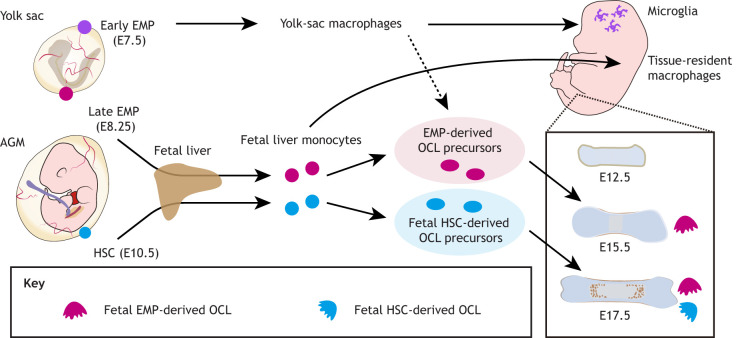
The hematopoietic system is initially established in mammals by several sequential and overlapping waves during embryonic development (Hoeffel et al., 2015; Schulz et al., 2012; Sheng et al., 2015; Munro and Hughes, 2017) (Figs 1 and 2). The first wave of hematopoiesis in mammals (also termed ‘primitive’) starts in the blood islands of the yolk sac and gives rise to nucleated erythroblasts, megakaryocytes and macrophages (Moore and Metcalf, 1970; Palis et al., 1999; Hoeffel and Ginhoux, 2018; Tober et al., 2006; Naito et al., 1989; Takahashi et al., 1989). The yolk-sac hemogenic endothelium produces early erythromyeloid progenitors (EMPs) around embryonic day (E) 7-E7.5 in the yolk sac (Ginhoux et al., 2010; Italiani and Boraschi, 2017), which can become colony-stimulating factor 1 receptor (CSF1R)+ yolk-sac macrophages by E8.5. These ‘early’ EMPs develop independently of the transcription factor MYB and give rise to primitive yolk-sac macrophages without passing through monocyte intermediates (Gomez Perdiguero et al., 2015; Hoeffel and Ginhoux, 2018). Around E8.25, the yolk-sac vascular system connects to the intra-embryonic circulation (McGrath et al., 2003) and yolk-sac macrophages can then colonize embryonic organs, such as the brain and liver (Kierdorf et al., 2013; Gomez Perdiguero et al., 2015). The second wave of hematopoiesis develops from E8.25, in which MYB-dependent ‘late’ EMPs emerge in the yolk sac and migrate into the fetal liver to produce fetal liver monocytes (Hoeffel et al., 2015; McGrath et al., 2015; Mass et al., 2016). Later in development, the final wave of hematopoiesis is initiated by hematopoietic stem cell (HSC) precursors, which emerge in the aorta-gonad-mesonephros region at E10.5. Subsequently, HSCs serve a central role in maintaining the hematopoietic system throughout the life of the organism (Medvinsky et al., 1993; Müller et al., 1994). HSCs mature and expand in the fetal liver, and later colonize the bone marrow for adult hematopoiesis. Bone marrow HSCs eventually establish circulating monocyte-derived macrophages. Initially, it was believed that the yolk-sac wave of hematopoiesis was a transient blood supply that was completely replaced by HSC-derived cells. However, accumulating evidence suggests that some adult macrophages are established during fetal development and maintained in postnatal tissues via proliferation (reviewed by Lee and Ginhoux, 2022; Epelman et al., 2014; Gomez Perdiguero et al., 2015). Both EMP-derived macrophages and HSC-derived monocytes can produce embryonic and postnatal osteoclasts (Jacome-Galarza et al., 2019; Yahara et al., 2020) (Fig. 1).
Fig. 1.
Schematic showing the developmental origin of osteoclasts. Early erythromyeloid progenitors (EMPs; purple lineage) appear around E7-E7.5 in the yolk sac and differentiate into yolk-sac macrophages that give rise to tissue-resident macrophage populations, such as brain microglia. Late EMPs (pink lineage) emerge in the yolk sac at around E8.25-E9 and migrate to the fetal liver to produce fetal liver monocytes. These EMP-derived monocytes/macrophages can give rise to embryonic osteoclasts (OCLs), forming the bone marrow cavity around E15.5, which is necessary for hematopoiesis during development. Fetal hematopoietic stem cells (HSCs; blue lineage) emerge at E10.5 in the aorta-gonad-mesonephros (AGM) region and also migrate to the fetal liver, where they give rise to OCL precursors.
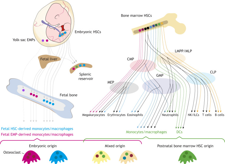
Fig. 2.
Complexity and variety of osteoclasts with multiple developmental origins. Schematic showing the diversity of osteoclasts. Early and late embryonic yolk-sac erythromyeloid progenitors (EMPs; purple and pink lineages, respectively) and fetal hematopoietic stem cells (HSCs; blue) can produce fetal monocyte-derived osteoclast progenitors. Postnatal bone marrow HSCs form monocytes/macrophages and dendritic cells (DCs) through continuous differentiation processes, which give rise to osteoclasts (green) (schematic based on Laurenti and Göttgens, 2018). Postnatal bone marrow HSC-derived, fetal HSC-derived and embryonic late EMP-derived osteoclast precursors fuse to form a diverse and complex osteoclast diversity (yellow multinucleated cells). CLP, common myeloid progenitor; CMP, common myeloid progenitor; GMP, granulocyte-monocyte progenitors; ILCs, innate lymphoid cells; LMPP, lymphoid-primed multipotent progenitor; MEP, megakaryocyte-erythrocyte progenitors; MLP, multi-lymphoid progenitor; NK, natural killer cells.
EMP-derived osteoclasts
A recent study using genetic lineage tracing demonstrated that EMP-derived osteoclasts are crucial for development of the normal fetal skeleton, and their absence impairs tooth eruption, skull formation and long bone formation (Jacome-Galarza et al., 2019). Genetic lineage-tracing experiments showed that EMP-derived osteoclasts are long-lived, surviving at least 6 months after birth, and are responsible for steady-state bone remodeling and fracture healing (Yahara et al., 2020). In addition, EMP-derived osteoclast precursors can migrate through the bloodstream to the site of bone injury and differentiate into mature osteoclasts (Yahara et al., 2020). EMP-derived osteoclast precursors are gradually replaced by HSC-derived, mononuclear, monocyte-progenitor cells (Jacome-Galarza et al., 2019). However, some monocyte-progenitor cells can fuse with long-lived EMP-derived osteoclasts, thus maintaining the population throughout adulthood (Jacome-Galarza et al., 2019).
HSC-derived osteoclasts
Many studies demonstrated that HSCs, and their myeloid progeny, provide osteoclast progenitor cells (Box 1; Fig. 2). In the classical model of hematopoiesis, bone marrow HSCs differentiate into multipotent progenitors (MPPs), resulting in lineage-restricted precursors (Kawamoto et al., 2010; Seita and Weissman, 2010). The precursors then give rise to oligopotent progenitors: common myeloid progenitors (CMPs) and common lymphoid progenitors (CLPs). CMPs produce megakaryocyte/erythrocyte progenitors (MEPs) and granulocyte/macrophage progenitors (GMPs) (Fig. 2). Furthermore, GMPs differentiate into a common macrophage/osteoclast/dendritic cell (DC) progenitor (MODP), with DCs functioning as an antigen-presenting cell (APC) population that assists in the adaptive immune system (Jacome-Galarza et al., 2013; Geissmann et al., 2008). These MODPs later give rise to mature osteoclasts through interaction with the receptor activator of the nuclear factor-kappa B (NF-κB) ligand (RANKL; also known as TNFSF11) and colony stimulating factor 1 [CSF1; also known as macrophage colony-stimulating factor (M-CSF)], which are essential for the continued differentiation of mature osteoclasts (Miyamoto et al., 2001; Cecchini et al., 1997).
Box 1. New insights into hematopoiesis.
The classical model of hematopoiesis is based on a long-held dogma that HSCs are at the top of the hierarchy, capable of self-renewal and generation of all blood lineage cells. However, with the advancement of next-generation techniques, such as scRNA-seq combined with genetic lineage tracing, in the last few years this classical view of hematopoiesis has been questioned (Notta et al., 2016; Velten et al., 2017; Paul et al., 2015; Grün et al., 2016). For example, recent studies identified several hematopoietic cell populations and unexpected branching points of hematopoiesis, suggesting that hematopoiesis is better described as a continuous process rather than a discrete hierarchy (Velten et al., 2017; Laurenti and Göttgens, 2018; Cheng et al., 2020) (Fig. 2). HSCs and early progenitor cells are heterogeneous and contain distinct subpopulations with different differentiation potentials, suggesting that the lineages are determined at an early stage of hematopoiesis. Although most hematopoietic progenitors can develop into one mature cell type (i.e. uni-lineage potential), bi- and multi-lineage progenitors are rare, but present (Karamitros et al., 2018). GMPs, defined as a LIN-KIT+, CD34high and CD16/32high lineage-primed progeny derived from CMPs, already contain early committed neutrophil progenitors, which increase extensively in the early stages of inflammation at the expense of monocyte differentiation (Kwok et al., 2020). Lymphoid-primed multipotent progenitors (LMPPs) can give rise to lymphoid and myeloid cells, including DCs (Fig. 2). A recent study showed that DC-lineage specification starts close to the HSC stage (Lee et al., 2017) and the transcription factor interferon regulatory factor 8 (IRF8) regulates chromatin at the LMPP stage to induce early commitment toward DCs (Kurotaki et al., 2019). Whether osteoclasts can be derived through alternative routes, such as LMPP-derived DCs, remains to be shown. Further study is necessary to elucidate when osteoclast-lineage specification starts and how their fate is determined.
Although bone marrow-derived HSCs and their descendants provide precursor cells for osteoclasts, there is controversy regarding whether differentiated monocytes/macrophages and DCs can give rise to osteoclasts. Udagawa and colleagues confirmed that osteoclasts originate from monocytes/macrophages (Udagawa et al., 1990). Arai and colleagues found that a KIT proto-oncogene receptor tyrosine kinase (KIT)+ and CSF1R+ population of mouse bone marrow mononuclear cells can be a source of early-stage osteoclast precursors (Arai et al., 1999). In human pediatric bone marrow, interleukin 3 receptor subunit alpha (IL3Rα)-expressing cells are the common osteoclast precursor (Xiao et al., 2015). Although monocyte/macrophage cells were thought to be the precursors of osteoclasts, some reports proposed that osteoclasts also arise through DCs (Rivollier et al., 2004; Speziani et al., 2007). Furthermore, a recent study using single-cell RNA sequencing (scRNA-seq) provided more detailed insights into the stepwise cell fate transition from the bone marrow-derived precursors to mature osteoclasts (Tsukasaki et al., 2020b). The differentiation trajectory of osteoclasts clearly shows that monocyte-like progenitor cells give rise to mature osteoclasts via CD11c (ITGAX)+ DC populations (Fig. 2). In addition, ablation of RANK (TNFRSF11a) in CD11c+ cells suppressed osteoclast differentiation, suggesting that this CD11c+ DC-like precursor population is primed for osteoclast differentiation and maturation (Tsukasaki et al., 2020b).
Osteoclast multinucleation, differentiation and maturation
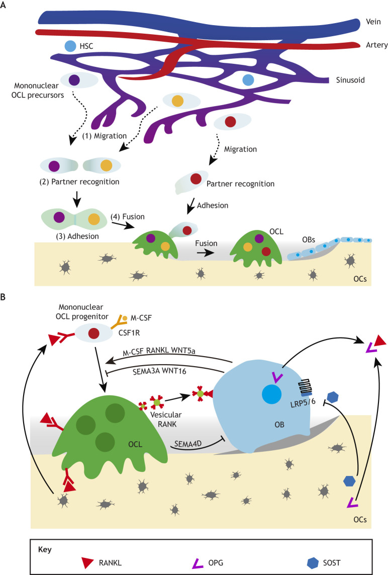
Cell-cell fusion of mononuclear osteoclast precursors and multinucleation are essential processes for osteoclast maturation and its bone resorption capacity (Fig. 3A). Osteoclast fusion is classically thought to consist of sequential steps: migration, recognition, intercellular adhesion and membrane fusion (Søe, 2020). Osteoclast precursor cells reside in the bone marrow cavity, adjacent to collagen fibers and vascular networks. Following cell-cell fusion with mature osteoclasts, single-nucleated precursors migrate from the bone marrow cavity to sites of resorption on the bone surface. Hobolt-Pedersen and colleagues proposed that osteoclast precursors fuse selectively, not randomly, to recognize their fusion partners (Hobolt-Pedersen et al., 2014). This selectivity is based on the intercellular heterogeneity, such as maturity, mobility and nuclearity (Hobolt-Pedersen et al., 2014; Søe, 2020; Søe et al., 2015). Additionally, osteoclast fusion factor, CD47, DC-specific transmembrane protein (DC-STAMP) and syncytin-1 are involved in the development of this heterogeneity (Søe, 2020). Mononuclear osteoclast precursors and their fusion partners need to move towards and adhere to each other for multinucleation to proceed. αVβ3 integrin, which localizes to podosomes at the leading edge of osteoclasts, plays an important role in migration and formation of the sealing zone (Nakamura et al., 1999; McHugh et al., 2000). In mice deficient for the DC-STAMP, d2 isoform of vacuolar (H+) ATPase (v-ATPase) V0 domain (ATP6v0d2) and osteoclast stimulatory transmembrane protein (OC-STAMP), only osteoclasts with a single nucleus were found, indicating that these proteins are essential for the multinucleation of osteoclasts (Yagi et al., 2005; Miyamoto et al., 2012; Lee et al., 2006). Impairment of osteoclast multinucleation reduced bone resorption activity resulting in osteopetrosis. CD44, CD47, syncytin-1, PIN1 (peptidyl-prolyl cis-trans isomerase NIMA-interacting 1) and tetraspanins (CD9, CD81) have also been reported to be crucial for osteoclast fusion and multinucleation (Sterling et al., 1998; Takeda et al., 2003; Cui et al., 2006; Søe et al., 2011; Islam et al., 2014; Møller et al., 2017). Thus, osteoclast fusion and multinucleation are not random processes, but are controlled through a strict mechanism by which fusion partners are selected based on the heterogeneity of osteoclast precursors.
Fig. 3.
Osteoclast specification. (A) Osteoclast (OCL) fusion and maturation. The osteoclast fusion consists of sequential steps: (1) migration, (2) partner recognition, (3) adhesion and (4) fusion. Osteoclast progenitor cells reside in the bone marrow cavity proximal to vascular networks. Single-nucleated osteoclast precursors (shown with purple, orange and red nuclei) migrate from the bone marrow cavity to sites of resorption on the bone surface. Osteoclast progenitors selectively recognize their fusion partners, followed by cell-cell fusion and maturation. (B) Osteoclast-osteoblast-osteocyte interactions. Osteoblasts (OBs), osteocytes (OCs) and osteoclasts directly communicate each other through either cell-to-cell interaction or paracrine signaling molecules. Osteoblasts secrete M-CSF, RANKL and WNT5A to promote osteoclast differentiation. OPG, SEMA3A and WNT16 secreted by osteoblasts inhibit osteoclast differentiation. SEMA4D suppresses osteoblast differentiation. Osteocytes regulate the balance of bone formation and resorption by secreting RANKL/OPG. SOST from osteocytes interacts with LRP5/6 and suppresses preosteoblast differentiation via inhibitory effects on the WNT/β-catenin pathway. RANK-enriched vesicles secreted from osteoclasts can increase bone formation by triggering RANKL reverse signaling. HSC, hematopoietic stem cell.
The earliest step in osteoclast formation begins with a mononuclear, monocytic progenitor cell expressing the E26 transformation-specific (ETS) domain transcription factor PU.1 (SPI1), which is indispensable for hematopoietic cell fate (Burda et al., 2010). PU.1 regulates the expression of CSF1R and RANK gene expression in myeloid progenitor cells, resulting in the establishment of osteoclast-specific transcriptional pattern driven by RANKL-RANK signaling (Tondravi et al., 1997; Kwon et al., 2005). In the bone microenvironment, osteoblasts, osteocytes and osteoclasts directly communicate with each other through either cell-to-cell interaction or paracrine signaling molecules (Furuya et al., 2018; Kim et al., 2020) (Fig. 3B). Osteoblasts secrete M-CSF (MacDonald et al., 1986; Fuller et al., 1993; Tanaka et al., 1993), RANKL (Kong et al., 1999; Lacey et al., 1998; Yasuda et al., 1998b) and Wnt gene family 5A (WNT5A) (Maeda et al., 2012) to promote osteoclast differentiation. In contrast, osteoprotegerin (OPG; TNFRSF11b), a decoy receptor of RANKL (Simonet et al., 1997; Tsuda et al., 1997; Yasuda et al., 1998a), semaphorin 3A (SEMA3A) (Hayashi et al., 2012) and WNT16 (Kobayashi et al., 2015) secreted by osteoblasts inhibit osteoclast differentiation. Osteoclast-derived factors that affect osteoblast differentiation and function include bone morphogenetic protein 6 (BMP6) (Pederson et al., 2008), collagen triple helix repeat containing 1 (CTHRC1) (Takeshita et al., 2013), sphingosine 1-phosphate (S1P) (Ryu et al., 2006; Pederson et al., 2008), SEMA4D (Negishi-Koga et al., 2011) and cardiotrophin-1 (CT-1; CTF1) (Walker et al., 2008). Osteocytes are multifunctional and dynamic cells that can integrate both mechanical cues (Qin et al., 2020) and hormonal signals (Wein, 2018) to modulate the development and function of osteoclasts and osteoblasts. Osteocytes are a prime source of RANKL, a major osteoclast differentiation facilitator (Nakashima et al., 2011; Xiong et al., 2011), and sclerostin (SOST), which inhibits preosteoblast differentiation via inhibitory effects on the WNT/β-catenin pathway (Winkler et al., 2003; Bezooijen et al., 2005). During osteoclast differentiation and maturation, osteoblasts and osteoclasts communicate with each other through direct cell-to-cell interaction allowing bidirectional signal transduction. Ephrin B2, which is expressed on the cell surface of osteoclasts, binds to the osteoblast surface molecule ephrin type-B receptor 4 (EPHB4). Reverse signaling through ephrin B2 into osteoclast precursors suppresses osteoclast differentiation by inhibiting the FOS/NFATc1 (nuclear factor of activated T cells c1) axis (Zhao et al., 2006). In contrast, forward signaling through EPHB4 into osteoblasts promotes osteoblast differentiation (Zhang et al., 2010). SEMA3A is produced by osteoblast lineage cells and works as a potent osteoprotective effector by simultaneously inhibiting bone resorption and promoting bone formation (Hayashi et al., 2012, 2019). By binding to neuropilin 1 (NRP1), SEMA3A inhibits RANKL-induced osteoclast function and promotes osteoblast differentiation via the WNT/β-catenin pathway (Hayashi et al., 2012). RANK-enriched vesicles secreted from osteoclasts can increase bone formation by promoting RANKL reverse signaling through activating runt-related transcription factor 2 (RUNX2) (Ikebuchi et al., 2018). This cell-cell interaction and the bone microenvironment promote osteoclast differentiation, eventually giving rise to mature osteoclasts. Mature osteoclasts drive bone resorption, a crucial component in the maintenance of bone homeostasis.
The role of osteoclasts in bone development
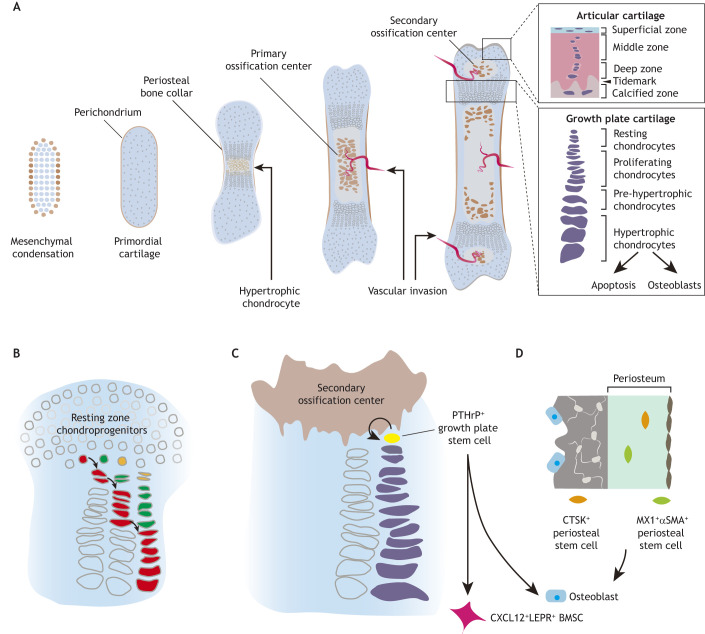
The embryonic skeleton begins its formation via intramembranous and endochondral ossification. Intramembranous ossification is characterized by mesenchymal cells directly differentiating into osteoblasts, as is the case for the skull, mandibles and clavicles (Berendsen and Olsen, 2015). Endochondral ossification begins with the condensation of mesenchymal cells to form a primordial bone template (Berendsen and Olsen, 2015) (Fig. 4A). The mesenchymal cells differentiate into chondrocytes, which then proliferate, increasing the size of the condensation (Berendsen and Olsen, 2015). The perichondrium covers the periphery of the primordial cartilage (Kronenberg, 2003). Chondrocytes differentiate into hypertrophic chondrocytes forming a hypertrophic zone that then mineralizes (Kronenberg, 2003). The surrounding perichondrium also differentiates and forms the osteogenic periosteum (Colnot et al., 2004). The epiphyses are at the ends of the bone and are composed entirely of growth plate cartilage and are separated by the primary ossification center with vascular invasion (Berendsen and Olsen, 2015). Subsequently, the hypertrophic chondrocytes undergo apoptosis or are directly converted to osteoblasts (Yang et al., 2014; Zhou et al., 2014b; Ono et al., 2014). Shortly after birth, a secondary ossification center appears in the epiphysis. The secondary ossification center is then vascularized and forms the articular cartilage and epiphyseal growth plate (Fig. 4A).
Fig. 4.
Endochondral ossification and the skeletal stem cell niche. (A) Schematic of endochondral ossification and long bone development. Mesenchymal cells condensate and form primordial cartilage. Chondrocytes start to proliferate and then differentiate into hypertrophic chondrocytes with the mineralization. The diaphysis is separated by the primary ossification center with vascular invasion. Hypertrophic chondrocytes undergo apoptosis or are directly converted to osteoblasts. The secondary ossification center appears in the epiphysis, which is vascularized and forms the articular cartilage and epiphyseal growth plate. (B) The round-shaped resting-zone chondroprogenitors in the epiphysis are recruited into the proliferative columns during fetal bone development, leading to their gradual consumption. (C) After forming the secondary ossification center, PTHrP+ chondroprogenitors (yellow) are present in the resting zone; these cells start renewing and generate long columns from single clones. PTHrP+ skeletal stem cells can give rise to CXCL12+ LEPR+ bone marrow stromal cells (BMSCs; pink) and osteoblasts (blue). (D) CTSK+ (orange) and MX1+αSMA+ (green) skeletal stem cells in the periosteum, which can give rise to osteogenic cells.
Osteoblast origins and differentiation
The origin of the cells that produce bone is an area of continued investigation; these cells can come from several sources, including skeletal stem cells (SSCs), vascular cells and growth plate chondrocytes that differentiate into osteoblasts. SSCs with multi-lineage differentiation potential were thought to be present in the bone marrow niche, identified based on combinations of cell markers, such as platelet-derived growth factor receptor alpha (PDGFRα)+/stem cell antigen-1 (SCA-1; LY6A)+/CD45 (PTPRC)−TER119 (LY76)− cells (Morikawa et al., 2009), CD73 (NT5E)+/CD31 (PECAM)− cells (Breitbach et al., 2018), CD271 (NGFR)+/CD45− cells (Álvarez-Viejo et al., 2015; Coutu et al., 2017), and leptin receptor (LEPR)+ cells (Zhou et al., 2014a; Yue et al., 2016). However, recent in vivo lineage-tracing studies using mouse genetic models demonstrated that the growth plate and skeleton surrounding the periosteum containing SSCs plays an essential role in bone development and formation (Newton et al., 2019). During fetal and neonatal bone growth, chondroprogenitors in the resting zone are gradually consumed and recruited into the proliferative columns (Newton et al., 2019). Therefore, individual rows of proliferating and hypertrophic chondrocytes are not derived from a single resting chondrocyte but have multiple origins (Fig. 4B). However, after forming the secondary ossification center, chondroprogenitors start renewing and generate long columns from single clones (Newton et al., 2019). Parathyroid hormone-related peptide (PTHrP; PTHLH)+ chondroprogenitors are present in the resting zone of the mouse growth plate, and give rise to columnar chondrocytes, osteoblasts and a subset of long-lived stem cells in the bone marrow niche (Mizuhashi et al., 2018) (Fig. 4C). Furthermore, recent studies also identified cathepsin K (CTSK)+/CD200+ cells (Debnath et al., 2018; Han et al., 2019) and MX dynamin-like GTPase 1 (MX1)+/α-smooth muscle actin (αSMA; ACTA2)+ cells (Ortinau et al., 2019) in the periosteum, which can give rise to bone-forming osteoblasts and participate in the healing of fractures (Fig. 4D). Thus, bone contains multiple stem cell niches with different physiological functions, which can change with development, growth and disease.
The role of osteoclasts in establishing the bone cavity
HSCs home and migrate from the fetal liver to vascularized regions of bone marrow to initiate definitive hematopoiesis as early as E16.5 (Coşkun et al., 2014). However, osteoclasts are a crucial cell population that create a space for hematopoiesis before bone marrow is established. Osteoclast precursors initially appear at the mesenchyme surrounding the bone rudiments (Taniguchi et al., 2007; Blavier and Delaisse, 1995) and differentiate into TRAP (ACP5)+ cells at E15.5 (Jacome-Galarza et al., 2019). Fate-mapping experiments demonstrated the presence of EMP-derived TRAP+ multinucleated osteoclasts in the bone marrow cavity at E15.5 (Jacome-Galarza et al., 2019). Furthermore, TRAP+ multinucleated osteoclasts were also observed at E16.5 in mouse embryos in which HSC-derived osteoclastic progenitors had been eliminated. These findings suggest that osteoclasts arising in the primary ossification center during early osteogenesis are derived from EMPs, rather than HSCs (Jacome-Galarza et al., 2019). During this short period, EMP-derived embryonic osteoclasts play an important role in generation of the bone marrow cavity, facilitating the migration of hematopoietic stem cells and myeloid cells (Fig. 1).
Osteoclasts are thought to undergo apoptosis when bone resorption is finished (Jansen et al., 2012; Yagi et al., 2005; Miyamoto et al., 2012). The survival time of mouse osteoclasts is thought to be less than 6 weeks (Marks and Seifert, 1985) and the lifespan of activated osteoclasts is estimated to be 2-3 weeks (Manolagas, 2000; Weinstein and Manolagas, 2000). However, a recent study suggested that individual osteoclasts can be long-lived and acquire a new nucleus every 4-8 weeks from circulating HSC-derived blood cells (Jacome-Galarza et al., 2019) (Fig. 5A). Thus, it would take 6 months to renew five nuclei in an individual osteoclast. Furthermore, data from intravital imaging revealed an unexpected cell fate of mature osteoclasts that divide into smaller, motile cells termed ‘osteomorphs’ (McDonald et al., 2021). Osteomorphs can fuse with neighboring osteoclasts, or in some cases with each other, and are ‘recycled’ into mature bone-resorbing osteoclasts under the control of RANKL signaling (Fig. 5B). An accumulation of osteomorphs caused by RANKL inhibition was observed in some patients who discontinue osteoclast inhibitory therapy (denosumab treatment). This recycling system of osteoclasts and osteomorphs may contribute to energy efficiency and rapid osteoclast regeneration.
Fig. 5.
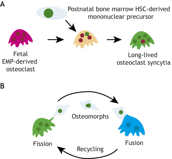
Osteoclast maintenance and recycling. (A) Postnatal maintenance of osteoclasts in the long-lived syncytia occurs through the sequential acquisition of new nuclei from hematopoietic stem cell (HSC)-derived precursors in the blood. (B) Osteoclasts divide into smaller, more motile daughter cells called osteomorphs, which are recycled by fusing to form functional osteoclasts.
The role of osteoclasts in bone homeostasis and repair
Bone is a dynamic organ that is continuously resorbed by osteoclasts and subsequently rebuilt with new bone by osteoblasts throughout life. This remodeling activity is a tightly controlled process to maintain serum elements, such as calcium, and the mechanical strength of the skeleton. It is regulated by various hormones, cytokines, chemokines, extracellular vesicles and biomechanical stimuli (Hattner et al., 1965; Takayanagi, 2007; Li et al., 2016; Deng et al., 2015; Ikebuchi et al., 2018). These sites of remodeling activity occur asynchronously at basic multicellular units (BMUs), and the BMUs in cortical and trabecular bone differ because of their structure (Hattner et al., 1965).
Bone remodeling
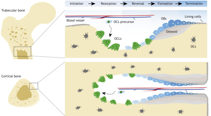
The bone remodeling cycle consists of overlapping phases: initiation, resorption, reversal, formation and termination (Fig. 6). The entire process is achieved by the coordinated actions of osteoclasts, osteoblasts and other osteoblast-lineage cells, such as bone-lining cells and osteocytes. Biological and functional differences between EMP-derived osteoclasts and HSC-derived osteoclasts in bone homeostasis need to be further explored.
Fig. 6.
Schematic of trabecular (top) and cortical (bottom) bone remodeling. The bone remodeling cycle consists of overlapping phases: initiation, resorption, reversal, formation and termination. The lining cells and osteocytes (OCs) release local factors that attract osteoclast precursors from the perivascular and bone marrow niches to the remodeling compartment. Osteoclasts (OCLs) initiate bone resorption, followed by the breakdown and removal of old bone. Osteoclasts then begin to interact directly or indirectly with osteoblasts (OBs), which deposit osteoid and new lamellar bone. Osteoblasts trapped in the bone matrix differentiate into osteocytes, whereas others die or turn into quiescent lining cells on the bone surface. The resting bone environment is maintained until the next wave of remodeling cycle is initiated.
Initiation phase
Initiation of bone remodeling occurs when necessary, such as in the event of injury or old age. In the initiation phase, mechanical loading and microdamage are sensed by osteocytes through their extensive network of dendritic processes, which leads to the release of paracrine factors that increase local angiogenesis and recruit osteoclast precursors (Dallas et al., 2013; Nakashima et al., 2011; Xiong et al., 2011; Cabahug-Zuckerman et al., 2016).
Resorption phase
In the resorption phase, osteoclastogenesis is stimulated by RANKL, M-CSF and ligands for immunoglobulin-like receptors, which are produced by osteoblast-lineage cells, including osteocytes. The cytoskeleton of the osteoclast is realigned and a sealing zone is formed, enhancing the secretory surface. After completing bone resorption, osteoclasts undergo apoptosis and the bone resorption phase is terminated, ensuring that excess resorption does not occur.
Reversal phase
In the early reversal phase, scattered osteoclasts on the bone surface release secreted factors, matrix-released factors and extra vesicles (Lassen et al., 2017; Sims and Martin, 2020). These osteoclasts also make direct cell-cell contacts, allowing a signal to recruit the osteoblast lineage on the bone surface. Mature osteoclasts initiate bone resorption when they are not in contact with osteoblasts, whereas mature osteoclasts in contact with mature osteoblasts do not resorb the bone in the reversal phase (Furuya et al., 2018). Osteoblasts, especially for the decorin (DCN)high subset, directly suppress osteoclast production by producing OPG, which plays a role in terminating the resorption phase (Cawley et al., 2020; Tsukasaki et al., 2020a). The number of osteoblast-lineage cells increases, reaching sufficient mass to promote their bone formation activity (Lassen et al., 2017).
Formation and termination phase
The formation phase is distinct by the complete replacement of osteoclast with osteoblastic cells. In the formation phase, the resorption lacuna is synthesized to a new bone matrix and then mineralized to fill the resorption lacuna. During this process, osteoblasts deposit a new bone matrix called the osteoid, which gradually mineralizes and terminally differentiates into osteocytes (Dallas and Bonewald, 2010). Osteocytes play a key role in signaling the termination of the remodeling cycle through the secretion of antagonists to osteogenesis, such as SOST (van Bezooijen et al., 2004). The resting bone environment is maintained until the next wave of the remodeling cycle is initiated.
Bone fracture repair
Bone fractures are common during the lifetime of an organism, and effective repair is crucial for survival. During fracture repair, the commonly occurring secondary bone healing begins with an inflammatory response, followed by the recruitment of various immune and mesenchymal cells at the fracture site. In the initial phase, bleeding from the fracture site causes hematomas, which further develop into vascularized and innervated granulation tissue (Loi et al., 2016; Hu et al., 2017). A temporary scaffold created by the hematoma is characterized by hypoxia and low pH, enabling the invasion of hematopoietic cells, such as neutrophils, lymphocytes and macrophages (Claes et al., 2012). The influx of various cytokine-secreting immune cells evokes acute inflammation (Salhotra et al., 2020). Subsequently, mesenchymal cells are recruited at the fracture site by factors, such as PDGF, transforming growth factor beta (TGFβ) and CXCL12 (Dimitriou et al., 2005; Kitaori et al., 2009; Granero-Moltó et al., 2009). Secreted factors, including BMP-4, vascular endothelial growth factor (VEGF), interleukin (IL) 17A (IL17A), IL6, tumor necrosis factor alpha (TNFα) and CCL2, are also released at the fracture site, promoting osteogenic differentiation of the mesenchymal cells (Peng et al., 2002; Ono et al., 2016; Ono and Takayanagi, 2017; Yang et al., 2007; Wallace et al., 2011; Ishikawa et al., 2014; Xing et al., 2010). At the callus periphery and inside cortical area where bone is repaired by intramembranous ossification, mesenchymal progenitor cells differentiate directly into osteoblasts, whereas mesenchymal progenitor cells that accumulate around damaged bone differentiate into fibroblasts, osteoblasts and mainly chondroblasts through endochondral ossification (Granero-Moltó et al., 2009; Debnath et al., 2018; Duchamp de Lageneste et al., 2018). In endochondral ossification, these cells synthesize the cartilage matrix to form a soft callus, which is then replaced by woven bone. Subsequently, woven bone is transformed into a hard callus, which is further remodeled by osteoclasts and osteoblasts to restore its original shape and function. This remodeling process is crucial for effective and adequate bone repair, and perturbations in osteoclast-mediated bone and cartilage resorption may negatively impact fracture repair (Lin and O'Connor, 2017; Flick et al., 2003; Vi et al., 2015; Yahara et al., 2021).
Osteoclasts can be divided into two subtypes based on their activation phase during fracture repair. Early-induced osteoclasts, which are present before callus formation, have high mobility and a low resorption profile. Late-induced osteoclasts have strong adhesion ability with a high bone resorption profile (Takeyama et al., 2014; Schell et al., 2006). Furthermore, increasing evidence suggests that, although bone marrow cells are a major source of osteoclast precursors in homeostasis, circulating monocytes could be a source of osteoclasts in pathogenic conditions, including during fracture repair (Novak et al., 2020; Kotani et al., 2013). EMP-derived osteoclast precursors could migrate through the bloodstream from the spleen to the fracture site (Yahara et al., 2020), although the mechanism of splenic cell mobilization to the fracture site remains to be identified. However, several studies have shown that reservoirs of macrophages and monocytes play an essential role in tissue inflammation and repair (Hoyer et al., 2019; Kotani et al., 2013).
Immune functions of osteoclasts
A principal function of skeletal bone marrow is to provide a specialized HSC niche to maintain postnatal hematopoiesis, which is crucial for regulating the development of immune cells and, thus, immune responses. Bone cells and the immune system are closely related through cellular and molecular interactions in the bone marrow microenvironment (Tsukasaki and Takayanagi, 2019; Takayanagi, 2007). It follows that osteoclasts are associated with many pathological conditions, including osteoporosis, rheumatoid arthritis (RA), chronic inflammation and cancer. Furthermore, similar to other members of the monocytic lineage, osteoclasts exhibit a wide range of phenotypic and functional heterogeneity involved in anti/pro-inflammatory effects and antigen presentation, depending on their environment.
Immune disease
Osteoclasts are activated in immune diseases. The interaction between osteoclasts and the immune system is observed in the autoimmune disease RA, a form of inflammatory polyarthritis that can lead to joint destruction, deformity and loss of function. LY6Chigh (Charles et al., 2012) and fragment crystallizable (Fc) γ receptor IV+ inflammatory monocyte cells are the primary sources of osteoclasts in RA (Seeling et al., 2013). The arthritis-committed osteoclast precursors express CX3CR1highLY6CintF4/80 (ADGRE1)+I-A+/I-E+ in mouse RA synovium (Hasegawa et al., 2019). Osteoclast differentiation is accelerated by T cell activation and their differentiation into type 17 helper T (Th17) cells. Th17 cells then stimulate synovial inflammation and produce proinflammatory mediators, such as IL1 and TNFα from synovial fibroblasts, macrophages and chondrocytes. Furthermore, IL17A induces RANKL expression in synovial fibroblasts and osteoblasts, which induces osteoclast differentiation and bone destruction associated with RA. Thus, Th17 cells have been proposed as an osteoclastogenesis-inducer in pathogenic arthritis.
APCs
Osteoclasts also play a role as APCs (Rivollier et al., 2004; Li et al., 2010; Ibáñez et al., 2016; Grassi et al., 2011). Human osteoclasts derived from monocytes express major histocompatibility complex (MHC) class I and II, CD80, CD86 and CD40, and have the potential to present allogeneic cells, resulting in the activation of both CD4+ and CD8+ (cytotoxic) T cells (Li et al., 2010). Furthermore, T cell receptors (TCRs) on CD8+ and CD4+ T cells can initiate T-cell activation and trigger TCR signaling by binding to MHC class I and II complexes, respectively (Li et al., 2010). Thus, osteoclasts can enfold soluble antigens and present them on their cell surfaces, demonstrating that osteoclasts can function as APCs.
Immunosuppressive roles
T regulatory (Treg) lymphocytes are a subpopulation of CD4+ T lymphocytes, which can cause immunosuppression, maintain peripheral tolerance and prevent chronic inflammation. The suppressive effects of Tregs, which express the forkhead box protein P3 (FOXP3) and CD25 (IL2RA) (Fontenot et al., 2003; Hori et al., 2003), are due to modulation of the effector functions of inflammatory cells (such as T cells, B cells, neutrophils and macrophages) (Alvarez et al., 2020). Tregs inhibit osteoclastogenesis through direct interaction with osteoclast precursors via cytotoxic T-lymphocyte-associated protein 4 (CTLA4) (Zaiss et al., 2007). Enhancing the activity of Treg cells improved the clinical signs of RA and suppressed local and systemic bone destruction in the TNF-mediated arthritis model (Zaiss et al., 2010). In contrast, CX3CR1− inflammatory osteoclasts have significantly higher bone resorption capacity in vitro than the CX3CR1+ fraction (Madel et al., 2020). Both subsets can function as APCs, but the T-cell activation capacity is higher in the CX3CR1− inflammatory osteoclast subset. Both can induce TNFα-producing CD4+ T cells, resulting in accelerated inflammation; however, the CX3CR1+ osteoclast subset expresses high levels of co-suppressor molecules, such as the programmed death-ligand 1 (PD-L1; CD274), Galectin-9 and herpes virus entry mediator (HVEM; TNFRSF14). These factors are the major immune checkpoint molecules involved in immunosuppression by autoimmune diseases and tumors (Madel et al., 2020). Thus, a subset of osteoclasts are thought to act as immunosuppressive cells that emerge in response to inflammatory signals and regulate inflammation. However, much remains to be clarified to understand the role of osteoclasts as immune cells in all aspects of biology.
Conclusions
EMP-derived macrophage and osteoclast precursor populations persist during adult life and produce long-lived cells that can self-renew locally. Cell populations established during embryonic development behave differently from the HSC lineage, having distinct roles in tissue homeostasis and repair. However, the principal mechanisms causing the differences between the HSC- and EMP-derived macrophages and osteoclasts remain to be elucidated. Further delineation of the role of this embryonic cell population will determine its function in bone homeostasis. As there are insufficient therapeutic agents for the treatment of disorders caused by osteoclasts, differences in the regulation of osteoclast precursors from different origins could be exploited to develop optimal methods to target osteoclasts therapeutically. Developing a new framework for osteoclast biology along with technological advancements could thus not only allow us to improve our understanding of osteoclast biology, but also help in the development of novel treatments for bone disease.
Footnotes
Competing interests
The authors declare no competing or financial interests.
Funding
This work was funded by grants from the National Institute on Aging (R01 AG072058 and R01 AG049745) and supported in part by KAKENHI grants from the Japan Society for the Promotion of Science (19K18544 and 20K18023) and the Japan Science and Technology Agency Precursory Research for Embryonic Science and Technology (PRESTO) (JPMJPR214C). Deposited in PMC for release after 12 months.
References
- Aguila, H. L. and Rowe, D. W. (2005). Skeletal development, bone remodeling, and hematopoiesis. Immunol. Rev. 208, 7-18. 10.1111/j.0105-2896.2005.00333.x [DOI] [PubMed] [Google Scholar]
- Álvarez-Viejo, M., Menéndez-Menéndez, Y. and OTERO-Hernández, J. (2015). CD271 as a marker to identify mesenchymal stem cells from diverse sources before culture. World J. Stem Cells 7, 470-476. 10.4252/wjsc.v7.i2.470 [DOI] [PMC free article] [PubMed] [Google Scholar]
- Alvarez, C., Suliman, S., Almarhoumi, R., Vega, M. E., Rojas, C., Monasterio, G., Galindo, M., Vernal, R. and Kantarci, A. (2020). Regulatory T cell phenotype and anti-osteoclastogenic function in experimental periodontitis. Sci. Rep. 10, 19018. 10.1038/s41598-020-76038-w [DOI] [PMC free article] [PubMed] [Google Scholar]
- Ambrosi, T. H., Marecic, O., Mcardle, A., Sinha, R., Gulati, G. S., Tong, X., Wang, Y., Steininger, H. M., Hoover, M. Y., Koepke, L. S.et al. (2021). Aged skeletal stem cells generate an inflammatory degenerative niche. Nature 597, 256-262. 10.1038/s41586-021-03795-7 [DOI] [PMC free article] [PubMed] [Google Scholar]
- Arai, F., Miyamoto, T., Ohneda, O., Inada, T., Sudo, T., Brasel, K., Miyata, T., Anderson, D. M. and Suda, T. (1999). Commitment and differentiation of osteoclast precursor cells by the sequential expression of c-Fms and receptor activator of nuclear factor kappaB (RANK) receptors. J. Exp. Med. 190, 1741-1754. 10.1084/jem.190.12.1741 [DOI] [PMC free article] [PubMed] [Google Scholar]
- Berendsen, A. D. and Olsen, B. R. (2015). Bone development. Bone 80, 14-18. 10.1016/j.bone.2015.04.035 [DOI] [PMC free article] [PubMed] [Google Scholar]
- Bezooijen, R. L. V., Dijke, P. T., Papapoulos, S. E. and Löwik, C. W. G. M. (2005). SOST/sclerostin, an osteocyte-derived negative regulator of bone formation. Cytokine Growth Factor. Rev. 16, 319-327. 10.1016/j.cytogfr.2005.02.005 [DOI] [PubMed] [Google Scholar]
- Blavier, L. and Delaisse, J. M. (1995). Matrix metalloproteinases are obligatory for the migration of preosteoclasts to the developing marrow cavity of primitive long bones. J. Cell Sci. 108, 3649-3659. 10.1242/jcs.108.12.3649 [DOI] [PubMed] [Google Scholar]
- Bloom, W., Bloom, M. A. and McLean, F. C. (1941). Calcification and ossification. Medullary bone changes in the reproductive cycle of female pigeons. Anat. Record. 81, 443-475. [Google Scholar]
- Breitbach, M., Kimura, K., Luis, T. C., Fuegemann, C. J., Woll, P. S., Hesse, M., Facchini, R., Rieck, S., Jobin, K., Reinhardt, J.et al. (2018). In Vivo labeling by CD73 marks multipotent stromal cells and highlights endothelial heterogeneity in the bone marrow niche. Cell Stem Cell 22, 262-276. 10.1016/j.stem.2018.01.008 [DOI] [PubMed] [Google Scholar]
- Burda, P., Laslo, P. and Stopka, T. (2010). The role of PU.1 and GATA-1 transcription factors during normal and leukemogenic hematopoiesis. Leukemia 24, 1249-1257. 10.1038/leu.2010.104 [DOI] [PubMed] [Google Scholar]
- Cabahug-Zuckerman, P., Frikha-Benayed, D., Majeska, R. J., Tuthill, A., Yakar, S., Judex, S. and Schaffler, M. B. (2016). Osteocyte apoptosis caused by hindlimb unloading is required to trigger osteocyte RANKL production and subsequent resorption of cortical and trabecular bone in mice femurs. J. Bone Miner. Res. 31, 1356-1365. 10.1002/jbmr.2807 [DOI] [PMC free article] [PubMed] [Google Scholar]
- Cawley, K. M., Bustamante-Gomez, N. C., Guha, A. G., Macleod, R. S., Xiong, J., Gubrij, I., Liu, Y., Mulkey, R., Palmieri, M., Thostenson, J. D.et al. (2020). Local production of osteoprotegerin by osteoblasts suppresses bone resorption. Cell Rep 32, 108052. 10.1016/j.celrep.2020.108052 [DOI] [PMC free article] [PubMed] [Google Scholar]
- Cecchini, M. G., Hofstetter, W., Halasy, J., Wetterwald, A. and Felix, R. (1997). Role of CSF-1 in bone and bone marrow development. Mol. Reprod. Dev. 46, 75-83. [DOI] [PubMed] [Google Scholar]
- Charles, J. F., Hsu, L. Y., Niemi, E. C., Weiss, A., Aliprantis, A. O. and Nakamura, M. C. (2012). Inflammatory arthritis increases mouse osteoclast precursors with myeloid suppressor function. J. Clin. Invest. 122, 4592-4605. 10.1172/JCI60920 [DOI] [PMC free article] [PubMed] [Google Scholar]
- Cheng, H., Zheng, Z. and Cheng, T. (2020). New paradigms on hematopoietic stem cell differentiation. Protein Cell 11, 34-44. 10.1007/s13238-019-0633-0 [DOI] [PMC free article] [PubMed] [Google Scholar]
- Claes, L., Recknagel, S. and Ignatius, A. (2012). Fracture healing under healthy and inflammatory conditions. Nat. Rev. Rheumatol. 8, 133-143. 10.1038/nrrheum.2012.1 [DOI] [PubMed] [Google Scholar]
- Colnot, C., Lu, C., Hu, D. and Helms, J. A. (2004). Distinguishing the contributions of the perichondrium, cartilage, and vascular endothelium to skeletal development. Dev. Biol. 269, 55-69. 10.1016/j.ydbio.2004.01.011 [DOI] [PubMed] [Google Scholar]
- Coşkun, S., Chao, H., Vasavada, H., Heydari, K., Gonzales, N., Zhou, X., De Crombrugghe, B. and Hirschi, K. K. (2014). Development of the fetal bone marrow niche and regulation of HSC quiescence and homing ability by emerging osteolineage cells. Cell Rep 9, 581-590. 10.1016/j.celrep.2014.09.013 [DOI] [PMC free article] [PubMed] [Google Scholar]
- Coutu, D. L., Kokkaliaris, K. D., Kunz, L. and Schroeder, T. (2017). Three-dimensional map of nonhematopoietic bone and bone-marrow cells and molecules. Nat. Biotechnol. 35, 1202-1210. 10.1038/nbt.4006 [DOI] [PubMed] [Google Scholar]
- Cui, W., Ke, J. Z., Zhang, Q., Ke, H.-Z., Chalouni, C. C. and Vignery, A. S. (2006). The intracellular domain of CD44 promotes the fusion of macrophages. Blood 107, 796-805. 10.1182/blood-2005-05-1902 [DOI] [PMC free article] [PubMed] [Google Scholar]
- Dallas, S. L. and Bonewald, L. F. (2010). Dynamics of the transition from osteoblast to osteocyte. Ann. N. Y. Acad. Sci. 1192, 437-443. 10.1111/j.1749-6632.2009.05246.x [DOI] [PMC free article] [PubMed] [Google Scholar]
- Dallas, S. L., Prideaux, M. and Bonewald, L. F. (2013). The osteocyte: an endocrine cell ... and more. Endocr. Rev. 34, 658-690. 10.1210/er.2012-1026 [DOI] [PMC free article] [PubMed] [Google Scholar]
- Debnath, S., Yallowitz, A. R., Mccormick, J., Lalani, S., Zhang, T., Xu, R., Li, N., Liu, Y., Yang, Y. S., Eiseman, M.et al. (2018). Discovery of a periosteal stem cell mediating intramembranous bone formation. Nature 562, 133-139. 10.1038/s41586-018-0554-8 [DOI] [PMC free article] [PubMed] [Google Scholar]
- Deng, L., Wang, Y., Peng, Y., Wu, Y., Ding, Y., Jiang, Y., Shen, Z. and Fu, Q. (2015). Osteoblast-derived microvesicles: A novel mechanism for communication between osteoblasts and osteoclasts. Bone 79, 37-42. 10.1016/j.bone.2015.05.022 [DOI] [PubMed] [Google Scholar]
- Dimitriou, R., Tsiridis, E. and Giannoudis, P. V. (2005). Current concepts of molecular aspects of bone healing. Injury 36, 1392-1404. 10.1016/j.injury.2005.07.019 [DOI] [PubMed] [Google Scholar]
- Duchamp De Lageneste, O., Julien, A., Abou-Khalil, R., Frangi, G., Carvalho, C., Cagnard, N., Cordier, C., Conway, S. J. and Colnot, C. (2018). Periosteum contains skeletal stem cells with high bone regenerative potential controlled by Periostin. Nat. Commun. 9, 773. 10.1038/s41467-018-03124-z [DOI] [PMC free article] [PubMed] [Google Scholar]
- Epelman, S., Lavine, K. J., Beaudin, A. E., Sojka, D. K., Carrero, J. A., Calderon, B., Brija, T., Gautier, E. L., Ivanov, S., Satpathy, A. T.et al. (2014). Embryonic and adult-derived resident cardiac macrophages are maintained through distinct mechanisms at steady state and during inflammation. Immunity 40, 91-104. 10.1016/j.immuni.2013.11.019 [DOI] [PMC free article] [PubMed] [Google Scholar]
- Flick, L. M., Weaver, J. M., Ulrich-Vinther, M., Abuzzahab, F., Zhang, X., Dougall, W. C., Anderson, D., O'keefe, R. J. and Schwarz, E. M. (2003). Effects of receptor activator of NFκB (RANK) signaling blockade on fracture healing. J. Orthop. Res. 21, 676-684. 10.1016/S0736-0266(03)00011-1 [DOI] [PubMed] [Google Scholar]
- Fontenot, J. D., Gavin, M. A. and Rudensky, A. Y. (2003). Foxp3 programs the development and function of CD4+CD25+ regulatory T cells. Nat. Immunol. 4, 330-336. 10.1038/ni904 [DOI] [PubMed] [Google Scholar]
- Fuller, K., Owens, J. M., Jagger, C. J., Wilson, A., Moss, R. and Chambers, T. J. (1993). Macrophage colony-stimulating factor stimulates survival and chemotactic behavior in isolated osteoclasts. J. Exp. Med. 178, 1733-1744. 10.1084/jem.178.5.1733 [DOI] [PMC free article] [PubMed] [Google Scholar]
- Furuya, M., Kikuta, J., Fujimori, S., Seno, S., Maeda, H., Shirazaki, M., Uenaka, M., Mizuno, H., Iwamoto, Y., Morimoto, A.et al. (2018). Direct cell–cell contact between mature osteoblasts and osteoclasts dynamically controls their functions in vivo. Nat. Commun. 9, 300. 10.1038/s41467-017-02541-w [DOI] [PMC free article] [PubMed] [Google Scholar]
- Geissmann, F., Auffray, C., Palframan, R., Wirrig, C., Ciocca, A., Campisi, L., Narni-Mancinelli, E. and Lauvau, G. (2008). Blood monocytes: distinct subsets, how they relate to dendritic cells, and their possible roles in the regulation of T-cell responses. Immunol. Cell Biol. 86, 398-408. 10.1038/icb.2008.19 [DOI] [PubMed] [Google Scholar]
- Ginhoux, F., Greter, M., Leboeuf, M., Nandi, S., See, P., Gokhan, S., Mehler, M. F., Conway, S. J., Ng, L. G., Stanley, E. R.et al. (2010). Fate mapping analysis reveals that adult microglia derive from primitive macrophages. Science 330, 841-845. 10.1126/science.1194637 [DOI] [PMC free article] [PubMed] [Google Scholar]
- Gomez Perdiguero, E., Klapproth, K., Schulz, C., Busch, K., Azzoni, E., Crozet, L., Garner, H., Trouillet, C., De Bruijn, M. F., Geissmann, F.et al. (2015). Tissue-resident macrophages originate from yolk-sac-derived erythro-myeloid progenitors. Nature 518, 547-551. 10.1038/nature13989 [DOI] [PMC free article] [PubMed] [Google Scholar]
- Grün, D., Muraro, M. J., Boisset, J.-C., Wiebrands, K., Lyubimova, A., Dharmadhikari, G., Van Den Born, M., Van Es, J., Jansen, E., Clevers, H.et al. (2016). De novo prediction of stem cell identity using single-cell transcriptome data. Cell Stem Cell 19, 266-277. 10.1016/j.stem.2016.05.010 [DOI] [PMC free article] [PubMed] [Google Scholar]
- Granero-Moltó, F., Weis, J. A., Miga, M. I., Landis, B., Myers, T. J., O'rear, L., Longobardi, L., Jansen, E. D., Mortlock, D. P. and Spagnoli, A. (2009). Regenerative effects of transplanted mesenchymal stem cells in fracture healing. Stem Cells 27, 1887-1898. 10.1002/stem.103 [DOI] [PMC free article] [PubMed] [Google Scholar]
- Grassi, F., Manferdini, C., Cattini, L., Piacentini, A., Gabusi, E., Facchini, A. and Lisignoli, G. (2011). T cell suppression by osteoclasts in vitro. J. Cell. Physiol. 226, 982-990. 10.1002/jcp.22411 [DOI] [PubMed] [Google Scholar]
- Han, Y., Feng, H., Sun, J., Liang, X., Wang, Z., Xing, W., Dai, Q., Yang, Y., Han, A., Wei, Z.et al. (2019). Lkb1 deletion in periosteal mesenchymal progenitors induces osteogenic tumors through mTORC1 activation. J. Clin. Invest. 129, 1895-1909. 10.1172/JCI124590 [DOI] [PMC free article] [PubMed] [Google Scholar]
- Hancox, N. M. (1949). The osteoclast. Biol. Rev. Camb. Philos. Soc. 24, 448-471. 10.1111/j.1469-185X.1949.tb00583.x [DOI] [PubMed] [Google Scholar]
- Hasegawa, T., Kikuta, J., Sudo, T., Matsuura, Y., Matsui, T., Simmons, S., Ebina, K., Hirao, M., Okuzaki, D., Yoshida, Y.et al. (2019). Identification of a novel arthritis-associated osteoclast precursor macrophage regulated by FoxM1. Nat. Immunol. 20, 1631-1643. 10.1038/s41590-019-0526-7 [DOI] [PubMed] [Google Scholar]
- Hattner, R., Epker, B. N. and Frost, H. M. (1965). Suggested sequential mode of control of changes in cell behaviour in adult bone remodelling. Nature 206, 489-490. 10.1038/206489a0 [DOI] [PubMed] [Google Scholar]
- Hayashi, M., Nakashima, T., Yoshimura, N., Okamoto, K., Tanaka, S. and Takayanagi, H. (2019). Autoregulation of osteocyte sema3a orchestrates estrogen action and counteracts bone aging. Cell Metab. 29, 627-637. 10.1016/j.cmet.2018.12.021 [DOI] [PubMed] [Google Scholar]
- Hayashi, M., Nakashima, T., Taniguchi, M., Kodama, T., Kumanogoh, A. and Takayanagi, H. (2012). Osteoprotection by semaphorin 3A. Nature 485, 69-74. 10.1038/nature11000 [DOI] [PubMed] [Google Scholar]
- Hobolt-Pedersen, A. S., Delaissé, J. M. and Søe, K. (2014). Osteoclast fusion is based on heterogeneity between fusion partners. Calcif. Tissue Int. 95, 73-82. 10.1007/s00223-014-9864-5 [DOI] [PMC free article] [PubMed] [Google Scholar]
- Hoeffel, G. and Ginhoux, F. (2018). Fetal monocytes and the origins of tissue-resident macrophages. Cell. Immunol. 330, 5-15. 10.1016/j.cellimm.2018.01.001 [DOI] [PubMed] [Google Scholar]
- Hoeffel, G., Chen, J., Lavin, Y., Low, D., Almeida, F. F., See, P., Beaudin, A. E., Lum, J., Low, I., Forsberg, E. C.et al. (2015). C-Myb+ erythro-myeloid progenitor-derived fetal monocytes give rise to adult tissue-resident macrophages. Immunity 42, 665-678. 10.1016/j.immuni.2015.03.011 [DOI] [PMC free article] [PubMed] [Google Scholar]
- Hori, S., Nomura, T. and Sakaguchi, S. (2003). Control of regulatory T cell development by the transcription factor Foxp3. Science 299, 1057-1061. 10.1126/science.1079490 [DOI] [PubMed] [Google Scholar]
- Hoyer, F. F., Naxerova, K., Schloss, M. J., Hulsmans, M., Nair, A. V., Dutta, P., Calcagno, D. M., Herisson, F., Anzai, A., Sun, Y.et al. (2019). Tissue-specific macrophage responses to remote injury impact the outcome of subsequent local immune challenge. Immunity 51, 899-914. 10.1016/j.immuni.2019.10.010 [DOI] [PMC free article] [PubMed] [Google Scholar]
- Hu, D. P., Ferro, F., Yang, F., Taylor, A. J., Chang, W., Miclau, T., Marcucio, R. S. and Bahney, C. S. (2017). Cartilage to bone transformation during fracture healing is coordinated by the invading vasculature and induction of the core pluripotency genes. Development 144, 221-234. 10.1242/dev.130807 [DOI] [PMC free article] [PubMed] [Google Scholar]
- Ibáñez, L., Abou-Ezzi, G., Ciucci, T., Amiot, V., Belatd, N., Obino, D., Mansour, A., Rouleau, M., Wakkach, A. and Blin-Wakkach, C. (2016). Inflammatory osteoclasts prime TNFα-producing CD4(+) T cells and express CX(3) CR1. J. Bone Miner. Res. 31, 1899-1908. 10.1002/jbmr.2868 [DOI] [PubMed] [Google Scholar]
- Ikebuchi, Y., Aoki, S., Honma, M., Hayashi, M., Sugamori, Y., Khan, M., Kariya, Y., Kato, G., Tabata, Y., Penninger, J. M.et al. (2018). Coupling of bone resorption and formation by RANKL reverse signalling. Nature 561, 195-200. 10.1038/s41586-018-0482-7 [DOI] [PubMed] [Google Scholar]
- Ishikawa, M., Ito, H., Kitaori, T., Murata, K., Shibuya, H., Furu, M., Yoshitomi, H., Fujii, T., Yamamoto, K. and Matsuda, S. (2014). MCP/CCR2 signaling is essential for recruitment of mesenchymal progenitor cells during the early phase of fracture healing. PLoS ONE 9, e104954. 10.1371/journal.pone.0104954 [DOI] [PMC free article] [PubMed] [Google Scholar]
- Islam, R., Bae, H.-S., Yoon, W.-J., Woo, K.-M., Baek, J.-H., Kim, H.-H., Uchida, T. and Ryoo, H.-M. (2014). Pin1 regulates osteoclast fusion through suppression of the master regulator of cell fusion DC-STAMP. J. Cell. Physiol. 229, 2166-2174. 10.1002/jcp.24679 [DOI] [PubMed] [Google Scholar]
- Italiani, P. and Boraschi, D. (2017). Development and functional differentiation of tissue-resident versus monocyte-derived macrophages in inflammatory reactions. Results Probl. Cell Differ. 62, 23-43. 10.1007/978-3-319-54090-0_2 [DOI] [PubMed] [Google Scholar]
- Jacome-Galarza, C. E., Lee, S.-K., Lorenzo, J. A. and Aguila, H. L. (2013). Identification, characterization, and isolation of a common progenitor for osteoclasts, macrophages, and dendritic cells from murine bone marrow and periphery. J. Bone Miner. Res. 28, 1203-1213. 10.1002/jbmr.1822 [DOI] [PMC free article] [PubMed] [Google Scholar]
- Jacome-Galarza, C. E., Percin, G. I., Muller, J. T., Mass, E., Lazarov, T., Eitler, J., Rauner, M., Yadav, V. K., Crozet, L., Bohm, M.et al. (2019). Developmental origin, functional maintenance and genetic rescue of osteoclasts. Nature 568, 541-545. 10.1038/s41586-019-1105-7 [DOI] [PMC free article] [PubMed] [Google Scholar]
- Jansen, I. D. C., Vermeer, J. A. F., Bloemen, V., Stap, J. and Everts, V. (2012). Osteoclast fusion and fission. Calcif. Tissue Int. 90, 515-522. 10.1007/s00223-012-9600-y [DOI] [PMC free article] [PubMed] [Google Scholar]
- Jotereau, F. V. and Douarin, N. M. L. (1978). The development relationship between osteocytes and osteoclasts: a study using the quail-chick nuclear marker in endochondral ossification. Dev. Biol. 63, 253-265. 10.1016/0012-1606(78)90132-X [DOI] [PubMed] [Google Scholar]
- Karamitros, D., Stoilova, B., Aboukhalil, Z., Hamey, F., Reinisch, A., Samitsch, M., Quek, L., Otto, G., Repapi, E., Doondeea, J.et al. (2018). Single-cell analysis reveals the continuum of human lympho-myeloid progenitor cells. Nat. Immunol. 19, 85-97. 10.1038/s41590-017-0001-2 [DOI] [PMC free article] [PubMed] [Google Scholar]
- Kawamoto, H., Ikawa, T., Masuda, K., Wada, H. and Katsura, Y. (2010). A map for lineage restriction of progenitors during hematopoiesis: the essence of the myeloid-based model. Immunol. Rev. 238, 23-36. 10.1111/j.1600-065X.2010.00959.x [DOI] [PubMed] [Google Scholar]
- Kierdorf, K., Erny, D., Goldmann, T., Sander, V., Schulz, C., Perdiguero, E. G., Wieghofer, P., Heinrich, A., Riemke, P., Hölscher, C.et al. (2013). Microglia emerge from erythromyeloid precursors via Pu.1- and Irf8-dependent pathways. Nat. Neurosci. 16, 273-280. 10.1038/nn.3318 [DOI] [PubMed] [Google Scholar]
- Kim, J.-M., Lin, C., Stavre, Z., Greenblatt, M. B. and Shim, J.-H. (2020). Osteoblast-osteoclast communication and bone homeostasis. Cells 9, 2073. 10.3390/cells9092073 [DOI] [PMC free article] [PubMed] [Google Scholar]
- Kitaori, T., Ito, H., Schwarz, E. M., Tsutsumi, R., Yoshitomi, H., Oishi, S., Nakano, M., Fujii, N., Nagasawa, T. and Nakamura, T. (2009). Stromal cell-derived factor 1/CXCR4 signaling is critical for the recruitment of mesenchymal stem cells to the fracture site during skeletal repair in a mouse model. Arthritis. Rheum. 60, 813-823. 10.1002/art.24330 [DOI] [PubMed] [Google Scholar]
- Kobayashi, Y., Thirukonda, G. J., Nakamura, Y., Koide, M., Yamashita, T., Uehara, S., Kato, H., Udagawa, N. and Takahashi, N. (2015). Wnt16 regulates osteoclast differentiation in conjunction with Wnt5a. Biochem. Biophys. Res. Commun. 463, 1278-1283. 10.1016/j.bbrc.2015.06.102 [DOI] [PubMed] [Google Scholar]
- Kong, Y. Y., Yoshida, H., Sarosi, I., Tan, H. L., Timms, E., Capparelli, C., Morony, S., Oliveira-Dos-Santos, A. J., Van, G., Itie, A.et al. (1999). OPGL is a key regulator of osteoclastogenesis, lymphocyte development and lymph-node organogenesis. Nature 397, 315-323. 10.1038/16852 [DOI] [PubMed] [Google Scholar]
- Kotani, M., Kikuta, J., Klauschen, F., Chino, T., Kobayashi, Y., Yasuda, H., Tamai, K., Miyawaki, A., Kanagawa, O., Tomura, M.et al. (2013). Systemic circulation and bone recruitment of osteoclast precursors tracked by using fluorescent imaging techniques. J. Immunol. 190, 605-612. 10.4049/jimmunol.1201345 [DOI] [PubMed] [Google Scholar]
- Kronenberg, H. M. (2003). Developmental regulation of the growth plate. Nature 423, 332-336. 10.1038/nature01657 [DOI] [PubMed] [Google Scholar]
- Kurotaki, D., Kawase, W., Sasaki, H., Nakabayashi, J., Nishiyama, A., Morse, H. C., III, Ozato, K., Suzuki, Y. and Tamura, T. (2019). Epigenetic control of early dendritic cell lineage specification by the transcription factor IRF8 in mice. Blood 133, 1803-1813. 10.1182/blood-2018-06-857789 [DOI] [PMC free article] [PubMed] [Google Scholar]
- Kwon, O. H., Lee, C. K., Lee, Y. I., Paik, S. G. and Lee, H. J. (2005). The hematopoietic transcription factor PU.1 regulates RANK gene expression in myeloid progenitors. Biochem. Biophys. Res. Commun. 335, 437-446. 10.1016/j.bbrc.2005.07.092 [DOI] [PubMed] [Google Scholar]
- Kwok, I., Becht, E., Xia, Y., Ng, M., Teh, Y. C., Tan, L., Evrard, M., Li, J. L. Y., Tran, H. T. N., Tan, Y.et al. (2020). Combinatorial single-cell analyses of granulocyte-monocyte progenitor heterogeneity reveals an early uni-potent neutrophil progenitor. Immunity 53, 303-318. 10.1016/j.immuni.2020.06.005 [DOI] [PubMed] [Google Scholar]
- Lacey, D. L., Timms, E., Tan, H. L., Kelley, M. J., Dunstan, C. R., Burgess, T., Elliott, R., Colombero, A., Elliott, G., Scully, S.et al. (1998). Osteoprotegerin ligand is a cytokine that regulates osteoclast differentiation and activation. Cell 93, 165-176. 10.1016/S0092-8674(00)81569-X [DOI] [PubMed] [Google Scholar]
- Lassen, N. E., Andersen, T. L., Pløen, G. G., Søe, K., Hauge, E. M., Harving, S., Eschen, G. E. T. and Delaisse, J.-M. (2017). Coupling of bone resorption and formation in real time: new knowledge gained from human haversian BMUs. J. Bone Miner. Res. 32, 1395-1405. 10.1002/jbmr.3091 [DOI] [PubMed] [Google Scholar]
- Laurenti, E. and Göttgens, B. (2018). From haematopoietic stem cells to complex differentiation landscapes. Nature 553, 418-426. 10.1038/nature25022 [DOI] [PMC free article] [PubMed] [Google Scholar]
- Lee, C. Z. W. and Ginhoux, F. (2022). Biology of resident tissue macrophages. Development 149, dev200270. 10.1242/dev.200270 [DOI] [PubMed] [Google Scholar]
- Lee, S.-H., Rho, J., Jeong, D., Sul, J.-Y., Kim, T., Kim, N., Kang, J.-S., Miyamoto, T., Suda, T., Lee, S.-K.et al. (2006). v-ATPase V0 subunit d2–deficient mice exhibit impaired osteoclast fusion and increased bone formation. Nat. Med. 12, 1403-1409. 10.1038/nm1514 [DOI] [PubMed] [Google Scholar]
- Lee, J., Zhou, Y. J., Ma, W., Zhang, W., Aljoufi, A., Luh, T., Lucero, K., Liang, D., Thomsen, M., Bhagat, G.et al. (2017). Lineage specification of human dendritic cells is marked by IRF8 expression in hematopoietic stem cells and multipotent progenitors. Nat. Immunol. 18, 877-888. 10.1038/ni.3789 [DOI] [PMC free article] [PubMed] [Google Scholar]
- Li, D., Liu, J., Guo, B., Liang, C., Dang, L., Lu, C., He, X., Cheung, H. Y., Xu, L., Lu, C.et al. (2016). Osteoclast-derived exosomal miR-214-3p inhibits osteoblastic bone formation. Nat. Commun. 7, 10872. 10.1038/ncomms10872 [DOI] [PMC free article] [PubMed] [Google Scholar]
- Li, H., Hong, S., Qian, J., Zheng, Y., Yang, J. and Yi, Q. (2010). Cross talk between the bone and immune systems: osteoclasts function as antigen-presenting cells and activate CD4+ and CD8+ T cells. Blood 116, 210-217. 10.1182/blood-2009-11-255026 [DOI] [PMC free article] [PubMed] [Google Scholar]
- Lin, H. N. and O'Connor, J. P. (2017). Osteoclast depletion with clodronate liposomes delays fracture healing in mice. J. Orthop. Res. 35, 1699-1706. 10.1002/jor.23440 [DOI] [PMC free article] [PubMed] [Google Scholar]
- Loi, F., Córdova, L. A., Pajarinen, J., Lin, T. H., Yao, Z. and Goodman, S. B. (2016). Inflammation, fracture and bone repair. Bone 86, 119-130. 10.1016/j.bone.2016.02.020 [DOI] [PMC free article] [PubMed] [Google Scholar]
- Müller, A. M., Medvinsky, A., Strouboulis, J., Grosveld, F. and Dzierzakt, E. (1994). Development of hematopoietic stem cell activity in the mouse embryo. Immunity 1, 291-301. 10.1016/1074-7613(94)90081-7 [DOI] [PubMed] [Google Scholar]
- Macdonald, B. R., Mundy, G. R., Clark, S., Wang, E. A., Kuehl, T. J., Stanley, E. R. and Roodman, G. D. (1986). Effects of human recombinant CSF-GM and highly purified CSF-1 on the formation of multinucleated cells with osteoclast characteristics in long-term bone marrow cultures. J. Bone Miner. Res. 1, 227-233. 10.1002/jbmr.5650010210 [DOI] [PubMed] [Google Scholar]
- Madel, M. B., Ibáñez, L., Ciucci, T., Halper, J., Rouleau, M., Boutin, A., Hue, C., Duroux-Richard, I., Apparailly, F., Garchon, H. J.et al. (2020). Dissecting the phenotypic and functional heterogeneity of mouse inflammatory osteoclasts by the expression of Cx3cr1. Elife 9, e54493. 10.7554/eLife.54493 [DOI] [PMC free article] [PubMed] [Google Scholar]
- Maeda, K., Kobayashi, Y., Udagawa, N., Uehara, S., Ishihara, A., Mizoguchi, T., Kikuchi, Y., Takada, I., Kato, S., Kani, S.et al. (2012). Wnt5a-Ror2 signaling between osteoblast-lineage cells and osteoclast precursors enhances osteoclastogenesis. Nat. Med. 18, 405-412. 10.1038/nm.2653 [DOI] [PubMed] [Google Scholar]
- Manolagas, S. C. (2000). Birth and death of bone cells: basic regulatory mechanisms and implications for the pathogenesis and treatment of osteoporosis*. Endocr. Rev. 21, 115-137. [DOI] [PubMed] [Google Scholar]
- Marks, S. C., Jr and Seifert, M. F. (1985). The lifespan of osteoclasts: experimental studies using the giant granule cytoplasmic marker characteristic of beige mice. Bone 6, 451-455. 10.1016/8756-3282(85)90223-6 [DOI] [PubMed] [Google Scholar]
- Mass, E., Ballesteros, I., Farlik, M., Halbritter, F., Günther, P., Crozet, L., Jacome-Galarza, C. E., Händler, K., Klughammer, J., Kobayashi, Y.et al. (2016). Specification of tissue-resident macrophages during organogenesis. Science 353, aaf4238. 10.1126/science.aaf4238 [DOI] [PMC free article] [PubMed] [Google Scholar]
- Mcdonald, M. M., Khoo, W. H., Ng, P. Y., Xiao, Y., Zamerli, J., Thatcher, P., Kyaw, W., Pathmanandavel, K., Grootveld, A. K., Moran, I.et al. (2021). Osteoclasts recycle via osteomorphs during RANKL-stimulated bone resorption. Cell 184, 1330-1347. 10.1016/j.cell.2021.02.002 [DOI] [PMC free article] [PubMed] [Google Scholar]
- Mcgrath, K. E., Koniski, A. D., Malik, J. and Palis, J. (2003). Circulation is established in a stepwise pattern in the mammalian embryo. Blood 101, 1669-1676. 10.1182/blood-2002-08-2531 [DOI] [PubMed] [Google Scholar]
- Mcgrath, K. E., Frame, J. M., Fegan, K. H., Bowen, J. R., Conway, S. J., Catherman, S. C., Kingsley, P. D., Koniski, A. D. and Palis, J. (2015). Distinct Sources of Hematopoietic Progenitors Emerge before HSCs and Provide Functional Blood Cells in the Mammalian Embryo. Cell Reports 11, 1892-1904. 10.1016/j.celrep.2015.05.036 [DOI] [PMC free article] [PubMed] [Google Scholar]
- Mchugh, K. P., Hodivala-Dilke, K., Zheng, M. H., Namba, N., Lam, J., Novack, D., Feng, X., Ross, F. P., Hynes, R. O. and Teitelbaum, S. L. (2000). Mice lacking beta3 integrins are osteosclerotic because of dysfunctional osteoclasts. J. Clin. Invest. 105, 433-440. 10.1172/JCI8905 [DOI] [PMC free article] [PubMed] [Google Scholar]
- Medvinsky, A. L., Samoylina, N. L., Müller, A. M. and Dzierzak, E. A. (1993). An early pre-liver intraembryonic source of CFU-S in the developing mouse. Nature 364, 64-67. 10.1038/364064a0 [DOI] [PubMed] [Google Scholar]
- Miyamoto, T., Ohneda, O., Arai, F., Iwamoto, K., Okada, S., Takagi, K., Anderson, D. M. and Suda, T. (2001). Bifurcation of osteoclasts and dendritic cells from common progenitors. Blood 98, 2544-2554. 10.1182/blood.V98.8.2544 [DOI] [PubMed] [Google Scholar]
- Miyamoto, H., Suzuki, T., Miyauchi, Y., Iwasaki, R., Kobayashi, T., Sato, Y., Miyamoto, K., Hoshi, H., Hashimoto, K., Yoshida, S.et al. (2012). Osteoclast stimulatory transmembrane protein and dendritic cell–specific transmembrane protein cooperatively modulate cell–cell fusion to form osteoclasts and foreign body giant cells. J. Bone Miner. Res. 27, 1289-1297. 10.1002/jbmr.1575 [DOI] [PubMed] [Google Scholar]
- Mizuhashi, K., Ono, W., Matsushita, Y., Sakagami, N., Takahashi, A., Saunders, T. L., Nagasawa, T., Kronenberg, H. M. and Ono, N. (2018). Resting zone of the growth plate houses a unique class of skeletal stem cells. Nature 563, 254-258. 10.1038/s41586-018-0662-5 [DOI] [PMC free article] [PubMed] [Google Scholar]
- Møller, A. M. J., Delaissé, J.-M. and Søe, K. (2017). Osteoclast fusion: time-lapse reveals involvement of CD47 and syncytin-1 at different stages of nuclearity. J. Cell. Physiol. 232, 1396-1403. 10.1002/jcp.25633 [DOI] [PMC free article] [PubMed] [Google Scholar]
- Moore, M. A. and Metcalf, D. (1970). Ontogeny of the haemopoietic system: yolk sac origin of in vivo and in vitro colony forming cells in the developing mouse embryo. Br. J. Haematol. 18, 279-296. 10.1111/j.1365-2141.1970.tb01443.x [DOI] [PubMed] [Google Scholar]
- Morikawa, S., Mabuchi, Y., Kubota, Y., Nagai, Y., Niibe, K., Hiratsu, E., Suzuki, S., Miyauchi-Hara, C., Nagoshi, N., Sunabori, T.et al. (2009). Prospective identification, isolation, and systemic transplantation of multipotent mesenchymal stem cells in murine bone marrow. J. Exp. Med. 206, 2483-2496. 10.1084/jem.20091046 [DOI] [PMC free article] [PubMed] [Google Scholar]
- Munro, D. A. D. and Hughes, J. (2017). The origins and functions of tissue-resident macrophages in kidney development. Front. Physiol. 8, 837. 10.3389/fphys.2017.00837 [DOI] [PMC free article] [PubMed] [Google Scholar]
- Naito, M., Yamamura, F., Nishikawa, S.-L. and Takahashi, K. (1989). Development, differentiation, and maturation of fetal mouse yolk sac macrophages in cultures. J. Leukoc. Biol. 46, 1-10. 10.1002/jlb.46.1.1 [DOI] [PubMed] [Google Scholar]
- Nakamura, I., Pilkington, M. F., Lakkakorpi, P. T., Lipfert, L., Sims, S. M., Dixon, S. J., Rodan, G. A. and Duong, L. T. (1999). Role of alpha(v)beta(3) integrin in osteoclast migration and formation of the sealing zone. J. Cell Sci. 112, 3985-3993. 10.1242/jcs.112.22.3985 [DOI] [PubMed] [Google Scholar]
- Nakashima, T., Hayashi, M., Fukunaga, T., Kurata, K., Oh-Hora, M., Feng, J. Q., Bonewald, L. F., Kodama, T., Wutz, A., Wagner, E. F.et al. (2011). Evidence for osteocyte regulation of bone homeostasis through RANKL expression. Nat. Med. 17, 1231-1234. 10.1038/nm.2452 [DOI] [PubMed] [Google Scholar]
- Negishi-Koga, T., Shinohara, M., Komatsu, N., Bito, H., Kodama, T., Friedel, R. H. and Takayanagi, H. (2011). Suppression of bone formation by osteoclastic expression of semaphorin 4D. Nat. Med. 17, 1473-1480. 10.1038/nm.2489 [DOI] [PubMed] [Google Scholar]
- Newton, P. T., Li, L., Zhou, B., Schweingruber, C., Hovorakova, M., Xie, M., Sun, X., Sandhow, L., Artemov, A. V., Ivashkin, E.et al. (2019). A radical switch in clonality reveals a stem cell niche in the epiphyseal growth plate. Nature 567, 234-238. 10.1038/s41586-019-0989-6 [DOI] [PubMed] [Google Scholar]
- Nijweide, P. J., Burger, E. H. and Feyen, J. H. (1986). Cells of bone: proliferation, differentiation, and hormonal regulation. Physiol. Rev. 66, 855-886. 10.1152/physrev.1986.66.4.855 [DOI] [PubMed] [Google Scholar]
- Notta, F., Zandi, S., Takayama, N., Dobson, S., Gan, O. I., Wilson, G., Kaufmann, K. B., Mcleod, J., Laurenti, E., Dunant, C. F.et al. (2016). Distinct routes of lineage development reshape the human blood hierarchy across ontogeny. Science 351, aab2116. 10.1126/science.aab2116 [DOI] [PMC free article] [PubMed] [Google Scholar]
- Novack, D. V. and Teitelbaum, S. L. (2008). The osteoclast: friend or foe? Annu. Rev. Pathol. 3, 457-484. 10.1146/annurev.pathmechdis.3.121806.151431 [DOI] [PubMed] [Google Scholar]
- Novak, S., Roeder, E., Kalinowski, J., Jastrzebski, S., Aguila, H. L., Lee, S. K., Kalajzic, I. and Lorenzo, J. A. (2020). Osteoclasts derive predominantly from bone marrow-resident CX(3)CR1(+) precursor cells in homeostasis, whereas circulating CX(3)CR1(+) cells contribute to osteoclast development during fracture repair. J. Immunol. 204, 868-878. 10.4049/jimmunol.1900665 [DOI] [PMC free article] [PubMed] [Google Scholar]
- Ono, N., Ono, W., Nagasawa, T. and Kronenberg, H. M. (2014). A subset of chondrogenic cells provides early mesenchymal progenitors in growing bones. Nat. Cell Biol. 16, 1157-1167. 10.1038/ncb3067 [DOI] [PMC free article] [PubMed] [Google Scholar]
- Ono, T., Okamoto, K., Nakashima, T., Nitta, T., Hori, S., Iwakura, Y. and Takayanagi, H. (2016). IL-17-producing γδ T cells enhance bone regeneration. Nat. Commun. 7, 10928. 10.1038/ncomms10928 [DOI] [PMC free article] [PubMed] [Google Scholar]
- Ono, T. and Takayanagi, H. (2017). Osteoimmunology in bone fracture healing. Curr. Osteoporos Rep. 15, 367-375. 10.1007/s11914-017-0381-0 [DOI] [PubMed] [Google Scholar]
- Ortinau, L. C., Wang, H., Lei, K., Deveza, L., Jeong, Y., Hara, Y., Grafe, I., Rosenfeld, S. B., Lee, D., Lee, B.et al. (2019). Identification of functionally distinct Mx1+αSMA+ periosteal skeletal stem cells. Cell Stem Cell 25, 784-796. 10.1016/j.stem.2019.11.003 [DOI] [PMC free article] [PubMed] [Google Scholar]
- Palis, J., Robertson, S., Kennedy, M., Wall, C. and Keller, G. (1999). Development of erythroid and myeloid progenitors in the yolk sac and embryo proper of the mouse. Development 126, 5073-5084. 10.1242/dev.126.22.5073 [DOI] [PubMed] [Google Scholar]
- Paul, F., Arkin, Y. A., Giladi, A., Jaitin, D. A., Kenigsberg, E., Keren-Shaul, H., Winter, D., Lara-Astiaso, D., Gury, M., Weiner, A.et al. (2015). Transcriptional heterogeneity and lineage commitment in myeloid progenitors. Cell 163, 1663-1677. 10.1016/j.cell.2015.11.013 [DOI] [PubMed] [Google Scholar]
- Pederson, L., Ruan, M., Westendorf, J. J., Khosla, S. and Oursler, M. J. (2008). Regulation of bone formation by osteoclasts involves Wnt/BMP signaling and the chemokine sphingosine-1-phosphate. Proc. Natl. Acad. Sci. USA 105, 20764-20769. 10.1073/pnas.0805133106 [DOI] [PMC free article] [PubMed] [Google Scholar]
- Peng, H., Wright, V., Usas, A., Gearhart, B., Shen, H. C., Cummins, J. and Huard, J. (2002). Synergistic enhancement of bone formation and healing by stem cell-expressed VEGF and bone morphogenetic protein-4. J. Clin. Invest. 110, 751-759. 10.1172/JCI15153 [DOI] [PMC free article] [PubMed] [Google Scholar]
- Qin, L., Liu, W., Cao, H. and Xiao, G. (2020). Molecular mechanosensors in osteocytes. Bone Res. 8, 23. 10.1038/s41413-020-0099-y [DOI] [PMC free article] [PubMed] [Google Scholar]
- Rivollier, A., Mazzorana, M. N., Tebib, J., Piperno, M., Aitsiselmi, T., Rabourdin-Combe, C., Jurdic, P. and Servet-Delprat, C. (2004). Immature dendritic cell transdifferentiation into osteoclasts: a novel pathway sustained by the rheumatoid arthritis microenvironment. Blood 104, 4029-4037. 10.1182/blood-2004-01-0041 [DOI] [PubMed] [Google Scholar]
- Ryu, J., Kim, H. J., Chang, E. J., Huang, H., Banno, Y. and Kim, H. H. (2006). Sphingosine 1-phosphate as a regulator of osteoclast differentiation and osteoclast-osteoblast coupling. EMBO J. 25, 5840-5851. 10.1038/sj.emboj.7601430 [DOI] [PMC free article] [PubMed] [Google Scholar]
- Salhotra, A., Shah, H. N., Levi, B. and Longaker, M. T. (2020). Mechanisms of bone development and repair. Nat. Rev. Mol. Cell Biol. 21, 696-711. 10.1038/s41580-020-00279-w [DOI] [PMC free article] [PubMed] [Google Scholar]
- Schell, H., Lienau, J., Epari, D. R., Seebeck, P., Exner, C., Muchow, S., Bragulla, H., Haas, N. P. and Duda, G. N. (2006). Osteoclastic activity begins early and increases over the course of bone healing. Bone 38, 547-554. 10.1016/j.bone.2005.09.018 [DOI] [PubMed] [Google Scholar]
- Schulz, C., Perdiguero, E. G., Chorro, L., Szabo-Rogers, H., Cagnard, N., Kierdorf, K., Prinz, M., Wu, B., Jacobsen, S. E. W., Pollard, J. W.et al. (2012). A Lineage of Myeloid Cells Independent of Myb and Hematopoietic Stem Cells. Science 336, 86-90. 10.1126/science.1219179 [DOI] [PubMed] [Google Scholar]
- Seeling, M., Hillenhoff, U., David, J. P., Schett, G., Tuckermann, J., Lux, A. and Nimmerjahn, F. (2013). Inflammatory monocytes and Fcγ receptor IV on osteoclasts are critical for bone destruction during inflammatory arthritis in mice. Proc. Natl. Acad. Sci. USA 110, 10729-10734. 10.1073/pnas.1301001110 [DOI] [PMC free article] [PubMed] [Google Scholar]
- Seita, J. and Weissman, I. L. (2010). Hematopoietic stem cell: self-renewal versus differentiation. Wiley Interdiscip. Rev. Syst. Biol Med 2, 640-653. 10.1002/wsbm.86 [DOI] [PMC free article] [PubMed] [Google Scholar]
- Sheng, J., Ruedl, C. and Karjalainen, K. (2015). Most tissue-resident macrophages except microglia are derived from fetal hematopoietic stem cells. Immunity 43, 382-393. 10.1016/j.immuni.2015.07.016 [DOI] [PubMed] [Google Scholar]
- Simonet, W. S., Lacey, D. L., Dunstan, C. R., Kelley, M., Chang, M. S., Lüthy, R., Nguyen, H. Q., Wooden, S., Bennett, L., Boone, T.et al. (1997). Osteoprotegerin: a novel secreted protein involved in the regulation of bone density. Cell 89, 309-319. 10.1016/S0092-8674(00)80209-3 [DOI] [PubMed] [Google Scholar]
- Sims, N. A. and Martin, T. J. (2020). Osteoclasts provide coupling signals to osteoblast lineage cells through multiple mechanisms. Annu. Rev. Physiol. 82, 507-529. 10.1146/annurev-physiol-021119-034425 [DOI] [PubMed] [Google Scholar]
- Søe, K. (2020). Osteoclast fusion: physiological regulation of multinucleation through heterogeneity-potential implications for drug sensitivity. Int. J. Mol. Sci. 21, 7717. 10.3390/ijms21207717 [DOI] [PMC free article] [PubMed] [Google Scholar]
- Søe, K., Andersen, T. L., Hobolt-Pedersen, A. S., Bjerregaard, B., Larsson, L. I. and Delaissé, J. M. (2011). Involvement of human endogenous retroviral syncytin-1 in human osteoclast fusion. Bone 48, 837-846. 10.1016/j.bone.2010.11.011 [DOI] [PubMed] [Google Scholar]
- Søe, K., Hobolt-Pedersen, A. S. and Delaisse, J. M. (2015). The elementary fusion modalities of osteoclasts. Bone 73, 181-189. 10.1016/j.bone.2014.12.010 [DOI] [PubMed] [Google Scholar]
- Speziani, C., Rivollier, A., Gallois, A., Coury, F., Mazzorana, M., Azocar, O., Flacher, M., Bella, C., Tebib, J., Jurdic, P.et al. (2007). Murine dendritic cell transdifferentiation into osteoclasts is differentially regulated by innate and adaptive cytokines. Eur. J. Immunol. 37, 747-757. 10.1002/eji.200636534 [DOI] [PubMed] [Google Scholar]
- Sterling, H., Saginario, C. and Vignery, A. (1998). CD44 Occupancy prevents macrophage multinucleation. J. Cell Biol. 143, 837-847. 10.1083/jcb.143.3.837 [DOI] [PMC free article] [PubMed] [Google Scholar]
- Sugiyama, T. and Nagasawa, T. (2012). Bone marrow niches for hematopoietic stem cells and immune cells. Inflamm. Allergy Drug Targets 11, 201-206. 10.2174/187152812800392689 [DOI] [PMC free article] [PubMed] [Google Scholar]
- Takayanagi, H. (2007). Osteoimmunology: shared mechanisms and crosstalk between the immune and bone systems. Nat. Rev. Immunol. 7, 292-304. 10.1038/nri2062 [DOI] [PubMed] [Google Scholar]
- Takahashi, K., Yamamura, F. and Naito, M. (1989). Differentiation, maturation, and proliferation of macrophages in the mouse yolk sac: a light-microscopic, enzyme-cytochemical, immunohistochemical, and ultrastructural study. J. Leukoc. Biol. 45, 87-96. 10.1002/jlb.45.2.87 [DOI] [PubMed] [Google Scholar]
- Takeda, Y., Tachibana, I., Miyado, K., Kobayashi, M., Miyazaki, T., Funakoshi, T., Kimura, H., Yamane, H., Saito, Y., Goto, H.et al. (2003). Tetraspanins CD9 and CD81 function to prevent the fusion of mononuclear phagocytes. J. Cell Biol. 161, 945-956. 10.1083/jcb.200212031 [DOI] [PMC free article] [PubMed] [Google Scholar]
- Takeshita, S., Fumoto, T., Matsuoka, K., Park, K. A., Aburatani, H., Kato, S., Ito, M. and Ikeda, K. (2013). Osteoclast-secreted CTHRC1 in the coupling of bone resorption to formation. J. Clin. Invest. 123, 3914-3924. 10.1172/JCI69493 [DOI] [PMC free article] [PubMed] [Google Scholar]
- Takeyama, K., Chatani, M., Takano, Y. and Kudo, A. (2014). In-vivo imaging of the fracture healing in medaka revealed two types of osteoclasts before and after the callus formation by osteoblasts. Dev. Biol. 394, 292-304. 10.1016/j.ydbio.2014.08.007 [DOI] [PubMed] [Google Scholar]
- Tanaka, S., Takahashi, N., Udagawa, N., Tamura, T., Akatsu, T., Stanley, E. R., Kurokawa, T. and Suda, T. (1993). Macrophage colony-stimulating factor is indispensable for both proliferation and differentiation of osteoclast progenitors. J. Clin. Invest. 91, 257-263. 10.1172/JCI116179 [DOI] [PMC free article] [PubMed] [Google Scholar]
- Taniguchi, N., Yoshida, K., Ito, T., Tsuda, M., Mishima, Y., Furumatsu, T., Ronfani, L., Abeyama, K., Kawahara, K.-I., Komiya, S.et al. (2007). Stage-specific secretion of HMGB1 in cartilage regulates endochondral ossification. Mol. Cell. Biol. 27, 5650-5663. 10.1128/MCB.00130-07 [DOI] [PMC free article] [PubMed] [Google Scholar]
- Tober, J., Koniski, A., Mcgrath, K. E., Vemishetti, R., Emerson, R., De Mesy-Bentley, K. K. L., Waugh, R. and Palis, J. (2006). The megakaryocyte lineage originates from hemangioblast precursors and is an integral component both of primitive and of definitive hematopoiesis. Blood 109, 1433-1441. 10.1182/blood-2006-06-031898 [DOI] [PMC free article] [PubMed] [Google Scholar]
- Tondravi, M. M., Mckercher, S. R., Anderson, K., Erdmann, J. M., Quiroz, M., Maki, R. and Teitelbaum, S. L. (1997). Osteopetrosis in mice lacking haematopoietic transcription factor PU.1. Nature 386, 81-84. 10.1038/386081a0 [DOI] [PubMed] [Google Scholar]
- Tonna, E. A. (1960). Osteoclasts and the aging skeleton: A cytological, cytochemical and autoradiographic study. Anat. Record 137, 251-269. 10.1002/ar.1091370304 [DOI] [PubMed] [Google Scholar]
- Tsuda, E., Goto, M., Mochizuki, S., Yano, K., Kobayashi, F., Morinaga, T. and Higashio, K. (1997). Isolation of a novel cytokine from human fibroblasts that specifically inhibits osteoclastogenesis. Biochem. Biophys. Res. Commun. 234, 137-142. 10.1006/bbrc.1997.6603 [DOI] [PubMed] [Google Scholar]
- Tsukasaki, M. and Takayanagi, H. (2019). Osteoimmunology: evolving concepts in bone-immune interactions in health and disease. Nat. Rev. Immunol. 19, 626-642. 10.1038/s41577-019-0178-8 [DOI] [PubMed] [Google Scholar]
- Tsukasaki, M., Asano, T., Muro, R., Huynh, N. C., Komatsu, N., Okamoto, K., Nakano, K., Okamura, T., Nitta, T. and Takayanagi, H. (2020a). OPG Production matters where it happened. Cell Rep 32, 108124. 10.1016/j.celrep.2020.108124 [DOI] [PubMed] [Google Scholar]
- Tsukasaki, M., Huynh, N. C., Okamoto, K., Muro, R., Terashima, A., Kurikawa, Y., Komatsu, N., Pluemsakunthai, W., Nitta, T., Abe, T.et al. (2020b). Stepwise cell fate decision pathways during osteoclastogenesis at single-cell resolution. Nat. Metab 2, 1382-1390. 10.1038/s42255-020-00318-y [DOI] [PubMed] [Google Scholar]
- Udagawa, N., Takahashi, N., Akatsu, T., Tanaka, H., Sasaki, T., Nishihara, T., Koga, T., Martin, T. J. and Suda, T. (1990). Origin of osteoclasts: mature monocytes and macrophages are capable of differentiating into osteoclasts under a suitable microenvironment prepared by bone marrow-derived stromal cells. Proc. Natl. Acad. Sci. USA 87, 7260-7264. 10.1073/pnas.87.18.7260 [DOI] [PMC free article] [PubMed] [Google Scholar]
- Van Bezooijen, R. L., Roelen, B. A., Visser, A., Van Der Wee-Pals, L., De Wilt, E., Karperien, M., Hamersma, H., Papapoulos, S. E., Ten Dijke, P. and Löwik, C. W. (2004). Sclerostin is an osteocyte-expressed negative regulator of bone formation, but not a classical BMP antagonist. J. Exp. Med. 199, 805-814. 10.1084/jem.20031454 [DOI] [PMC free article] [PubMed] [Google Scholar]
- Velten, L., Haas, S. F., Raffel, S., Blaszkiewicz, S., Islam, S., Hennig, B. P., Hirche, C., Lutz, C., Buss, E. C., Nowak, D.et al. (2017). Human haematopoietic stem cell lineage commitment is a continuous process. Nat. Cell Biol. 19, 271-281. 10.1038/ncb3493 [DOI] [PMC free article] [PubMed] [Google Scholar]
- Vi, L., Baht, G. S., Whetstone, H., Ng, A., Wei, Q., Poon, R., Mylvaganam, S., Grynpas, M. and Alman, B. A. (2015). Macrophages promote osteoblastic differentiation in-vivo: implications in fracture repair and bone homeostasis. J. Bone Miner. Res. 30, 1090-1102. 10.1002/jbmr.2422 [DOI] [PubMed] [Google Scholar]
- Walker, D. G. (1973). Osteopetrosis cured by temporary parabiosis. Science 180, 875. 10.1126/science.180.4088.875 [DOI] [PubMed] [Google Scholar]
- Walker, D. G. (1975a). Bone resorption restored in osteopetrotic mice by transplants of normal bone marrow and spleen cells. Science 190, 784-785. 10.1126/science.1105786 [DOI] [PubMed] [Google Scholar]
- Walker, D. G. (1975b). Spleen cells transmit osteopetrosis in mice. Science 190, 785-787. 10.1126/science.1198094 [DOI] [PubMed] [Google Scholar]
- Walker, E. C., Mcgregor, N. E., Poulton, I. J., Pompolo, S., Allan, E. H., Quinn, J. M., Gillespie, M. T., Martin, T. J. and Sims, N. A. (2008). Cardiotrophin-1 is an osteoclast-derived stimulus of bone formation required for normal bone remodeling. J. Bone Miner. Res. 23, 2025-2032. 10.1359/jbmr.080706 [DOI] [PubMed] [Google Scholar]
- Wallace, A., Cooney, T. E., Englund, R. and Lubahn, J. D. (2011). Effects of interleukin-6 ablation on fracture healing in mice. J. Orthop. Res. 29, 1437-1442. 10.1002/jor.21367 [DOI] [PubMed] [Google Scholar]
- Wein, M. N. (2018). Parathyroid hormone signaling in osteocytes. JBMR Plus 2, 22-30. 10.1002/jbm4.10021 [DOI] [PMC free article] [PubMed] [Google Scholar]
- Weinstein, R. S. and Manolagas, S. C. (2000). Apoptosis and osteoporosis. Am. J. Med. 108, 153-164. 10.1016/S0002-9343(99)00420-9 [DOI] [PubMed] [Google Scholar]
- Winkler, D. G., Sutherland, M. K., Geoghegan, J. C., Yu, C., Hayes, T., Skonier, J. E., Shpektor, D., Jonas, M., Kovacevich, B. R., Staehling-Hampton, K.et al. (2003). Osteocyte control of bone formation via sclerostin, a novel BMP antagonist. EMBO J. 22, 6267-6276. 10.1093/emboj/cdg599 [DOI] [PMC free article] [PubMed] [Google Scholar]
- Xiao, Y., Zijl, S., Wang, L., De Groot, D. C., Van Tol, M. J., Lankester, A. C. and Borst, J. (2015). Identification of the common origins of osteoclasts, macrophages, and dendritic cells in human hematopoiesis. Stem Cell Reports 4, 984-994. 10.1016/j.stemcr.2015.04.012 [DOI] [PMC free article] [PubMed] [Google Scholar]
- Xing, Z., Lu, C., Hu, D., Yu, Y. Y., Wang, X., Colnot, C., Nakamura, M., Wu, Y., Miclau, T. and Marcucio, R. S. (2010). Multiple roles for CCR2 during fracture healing. Dis. Model. Mech. 3, 451-458. 10.1242/dmm.003186 [DOI] [PMC free article] [PubMed] [Google Scholar]
- Xiong, J., Onal, M., Jilka, R. L., Weinstein, R. S., Manolagas, S. C. and O'brien, C. A. (2011). Matrix-embedded cells control osteoclast formation. Nat. Med. 17, 1235-1241. 10.1038/nm.2448 [DOI] [PMC free article] [PubMed] [Google Scholar]
- Yagi, M., Miyamoto, T., Sawatani, Y., Iwamoto, K., Hosogane, N., Fujita, N., Morita, K., Ninomiya, K., Suzuki, T., Miyamoto, K.et al. (2005). DC-STAMP is essential for cell-cell fusion in osteoclasts and foreign body giant cells. J. Exp. Med. 202, 345-351. 10.1084/jem.20050645 [DOI] [PMC free article] [PubMed] [Google Scholar]
- Yahara, Y., Barrientos, T., Tang, Y. J., Puviindran, V., Nadesan, P., Zhang, H., Gibson, J. R., Gregory, S. G., Diao, Y., Xiang, Y.et al. (2020). Erythromyeloid progenitors give rise to a population of osteoclasts that contribute to bone homeostasis and repair. Nat. Cell Biol. 22, 49-59. 10.1038/s41556-019-0437-8 [DOI] [PMC free article] [PubMed] [Google Scholar]
- Yahara, Y., Ma, X., Gracia, L. and Alman, B. A. (2021). Monocyte/macrophage lineage cells from fetal erythromyeloid progenitors orchestrate bone remodeling and repair. Front Cell Dev Biol 9, 622035. 10.3389/fcell.2021.622035 [DOI] [PMC free article] [PubMed] [Google Scholar]
- Yang, G., Zhu, L., Hou, N., Lan, Y., Wu, X.-M., Zhou, B., Teng, Y. and Yang, X. (2014). Osteogenic fate of hypertrophic chondrocytes. Cell Res. 24, 1266-1269. 10.1038/cr.2014.111 [DOI] [PMC free article] [PubMed] [Google Scholar]
- Yang, X., Ricciardi, B. F., Hernandez-Soria, A., Shi, Y., Pleshko Camacho, N. and Bostrom, M. P. (2007). Callus mineralization and maturation are delayed during fracture healing in interleukin-6 knockout mice. Bone 41, 928-936. 10.1016/j.bone.2007.07.022 [DOI] [PMC free article] [PubMed] [Google Scholar]
- Yasuda, H., Shima, N., Nakagawa, N., Mochizuki, S. I., Yano, K., Fujise, N., Sato, Y., Goto, M., Yamaguchi, K., Kuriyama, M.et al. (1998a). Identity of osteoclastogenesis inhibitory factor (OCIF) and osteoprotegerin (OPG): a mechanism by which OPG/OCIF inhibits osteoclastogenesis in vitro. Endocrinology 139, 1329-1337. 10.1210/endo.139.3.5837 [DOI] [PubMed] [Google Scholar]
- Yasuda, H., Shima, N., Nakagawa, N., Yamaguchi, K., Kinosaki, M., Mochizuki, S., Tomoyasu, A., Yano, K., Goto, M., Murakami, A.et al. (1998b). Osteoclast differentiation factor is a ligand for osteoprotegerin/osteoclastogenesis-inhibitory factor and is identical to TRANCE/RANKL. Proc. Natl. Acad. Sci. USA 95, 3597-3602. 10.1073/pnas.95.7.3597 [DOI] [PMC free article] [PubMed] [Google Scholar]
- Young, R. W. (1962). Cell proliferation and specialization during endochondral osteogenesis in young rats. J. Cell Biol. 14, 357-370. 10.1083/jcb.14.3.357 [DOI] [PMC free article] [PubMed] [Google Scholar]
- Yue, R., Zhou, B. O., Shimada, I. S., Zhao, Z. and Morrison, S. J. (2016). Leptin receptor promotes adipogenesis and reduces osteogenesis by regulating mesenchymal stromal cells in adult bone marrow. Cell Stem Cell 18, 782-796. 10.1016/j.stem.2016.02.015 [DOI] [PubMed] [Google Scholar]
- Zaiss, M. M., Axmann, R., Zwerina, J., Polzer, K., Gückel, E., Skapenko, A., Schulze-Koops, H., Horwood, N., Cope, A. and Schett, G. (2007). Treg cells suppress osteoclast formation: a new link between the immune system and bone. Arthritis. Rheum. 56, 4104-4112. 10.1002/art.23138 [DOI] [PubMed] [Google Scholar]
- Zaiss, M. M., Frey, B., Hess, A., Zwerina, J., Luther, J., Nimmerjahn, F., Engelke, K., Kollias, G., Hünig, T., Schett, G.et al. (2010). Regulatory T cells protect from local and systemic bone destruction in arthritis. J. Immunol. 184, 7238-7246. 10.4049/jimmunol.0903841 [DOI] [PubMed] [Google Scholar]
- Zhang, Q., Raoof, M., Chen, Y., Sumi, Y., Sursal, T., Junger, W., Brohi, K., Itagaki, K. and Hauser, C. J. (2010). Circulating mitochondrial DAMPs cause inflammatory responses to injury. Nature 464, 104-107. 10.1038/nature08780 [DOI] [PMC free article] [PubMed] [Google Scholar]
- Zhao, C., Irie, N., Takada, Y., Shimoda, K., Miyamoto, T., Nishiwaki, T., Suda, T. and Matsuo, K. (2006). Bidirectional ephrinB2-EphB4 signaling controls bone homeostasis. Cell Metab. 4, 111-121. 10.1016/j.cmet.2006.05.012 [DOI] [PubMed] [Google Scholar]
- Zhou, B. O., Yue, R., Murphy, M. M., Peyer, J. G. and Morrison, S. J. (2014a). Leptin-receptor-expressing mesenchymal stromal cells represent the main source of bone formed by adult bone marrow. Cell Stem Cell 15, 154-168. 10.1016/j.stem.2014.06.008 [DOI] [PMC free article] [PubMed] [Google Scholar]
- Zhou, X., Von Der Mark, K., Henry, S., Norton, W., Adams, H. and De Crombrugghe, B. (2014b). Chondrocytes transdifferentiate into osteoblasts in endochondral bone during development, postnatal growth and fracture healing in mice. PLoS Genet. 10, e1004820. 10.1371/journal.pgen.1004820 [DOI] [PMC free article] [PubMed] [Google Scholar]


