Dear Editor,
Endochondral bone formation is largely dependent on cartilage lineage cells. The chondrocytes in growth plates continuously undergo a sequential process from proliferation to terminal hypertrophic differentiation1. Once differentiated, hypertrophic chondrocytes elicit multiple functions such as determining bone length, inducing osteogenesis as well as directing bone mineralization, and eventually disappear at the chondro-osseous junction. The fate of the terminally differentiated hypertrophic chondrocytes is conceptually important for understanding its role in endochondral bone formation. It has been debated for decades whether the terminally differentiated hypertrophic chondrocytes die by apoptosis or undergo osteogenic transdifferentiation, however, clear in vivo evidence is lacking2. Here, through lineage tracing, we provide the first in vivo evidence that the terminally differentiated hypertrophic chondrocytes are a potent source of osteoblasts, and retain multi-lineage differentiation potential.
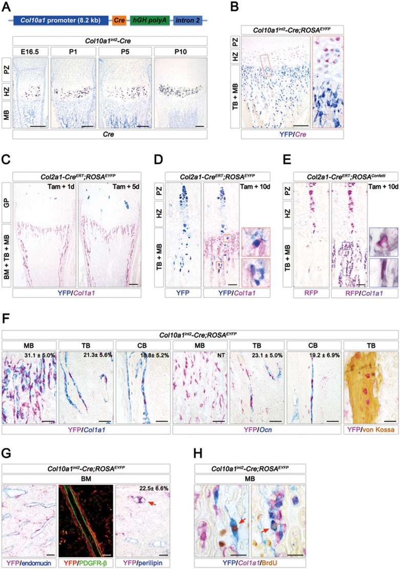
First, we generated a novel Col10a1int2-Cre transgenic mouse strain in which Cre expression was under the control of an 8.2 kb promoter and a 3.2 kb 2nd intron of the mouse type X collagen gene (Col10a1), a specific marker for hypertrophic chondrocytes (Figure 1A). In situ hybridization analysis of Col10a1int2-Cre transgenic mice showed that Cre+ cells were exclusively restricted to the hypertrophic zone, but not detectable in the metaphysis and bone marrow cavity from embryonic day 14.5 (E14.5) to postnatal day 10 (P10) (Figure 1A and Supplementary information, Figure S1A, S1B). Furthermore, double staining showed that all Cre+ cells co-expressed Col10a1, but not Col1a1 (a marker for osteoblasts), endomucin (a marker for endothelial cells) or perilipin (a marker for adipocytes) (Supplementary information, Figure S1A, S1B). In addition, RT-PCR detected Cre-hGH mRNA in growth plate cartilage, but not in bone marrow or cortical bone of Col10a1int2-Cre transgenic mice (Supplementary information, Figure S1C). These data demonstrate that this Col10a1int2-Cre strain is specific for hypertrophic chondrocytes.
Figure 1.
Multipotential fates of hypertrophic chondrocyte. (A) The transgenic vector contained an 8.2 kb promoter of mouse type X collagen (Col10a1), Cre cDNA, human growth hormone (hGH) polyadenylation signal and a 3.2 kb 2nd intron of Col10a1. Isotopic in situ hybridization assay was used to detect the transgenic Cre mRNA in tibia proximal growth plate from Col10a1int2-Cre transgenic mice at E16.5, P1, P5 and P10. (B) Double staining of YFP (blue) by immunohistochemistry (IHC) and Cre mRNA (fuchsia) by in situ hybridization in 10-day-old Col10a1int2-Cre;ROSAEYFP tibia. The right panel shows the high-magnification image of the area boxed in red on the left. (C) After 1 and 5 days following tamoxifen administration (50 mg/kg) at P5, Col2a1-CreERT;ROSAEYFP tibia was stained by YFP (blue) and Col1a1 (fuchsia) antibodies. The clonal columns of YFP+ cells were detectable in Col2a1-CreERT;ROSAEYFP chondrocyte zone. (D) After 10 days following tamoxifen administration at P5, Col2a1-CreERT;ROSAEYFP tibia was stained by YFP (blue) antibody, showing a column of YFP+ cells entering into the metaphysis. IHC double staining of YFP (blue) and type I collagen (Col1a1) (fuchsia) showed that some YFP+ cells underneath the growth plate were labeled with Col1a1 (red frames, high-magnification images of areas boxed on the left). (E) After 10 days following tamoxifen administration (50 mg/kg) at P5, Col2a1-CreERT;ROSAConfetti tibia was stained by RFP (fuchsia) antibody. Double staining of RFP (fuchsia) by IHC and Col1a1 (purple grey) mRNA by in situ hybridization showed that some RFP+ cells in metaphysis were Col1a1-positive (red frames, high-magnification images of areas boxed on the left). (F) Double staining of YFP (fuchsia) by IHC and the transcripts of Col1a1 or osteocalcin (Ocn) (blue) by in situ hybridization in metaphyseal, trabecular and cortical bones from 20-day-old Col10a1int2-Cre;ROSAEYFP tibia. The percentages of YFP+Col1a1+ and YFP+Ocn+osteogenic cells in total Col1a1+and Ocn+ cells from 3 images per mouse (n = 3) are shown in the top-right corner. Von Kossa staining showed that some YFP+cells (fuchsia) within the mineralized bone were osteocytes. (G) In 20-day-old Col10a1int2-Cre;ROSAEYFP bone marrow (BM), double immunostaining (left) of YFP (fuchsia) and endomucin (blue, marker for endothelial cells) as well as double immunofluorescence analysis (middle) using YFP (red) and PDGFR-β (green, marker for pericytes) antibodies indicated that some YFP+ cells were adjacent to the endothelial cells and vascular pericytes. Double immunostaining (right) of YFP (fuchsia) and perilipin (blue, marker for adipocytes) showed that some YFP+ cells were adipocytes (red arrow). (H) Triple staining of YFP (blue) and BrdU (brown) by IHC along with Col1a1 mRNA (fuchsia) by in situ hybridization in 20-day-old Col10a1int2-Cre;ROSAEYFP metaphyseal bones (MB). PZ, proliferation zone; HZ, hypertrophic zone; MB, metaphyseal bone; TB, trabecular bone; BM, bone marrow. Scale bars are 200 μm (A-C), 50 μm (D-F) and 25 μm (G, H).
To track the in vivo fate and spatial distribution of hypertrophic chondrocytes, we generated double transgenic mice harboring Col10a1int2-Cre and floxed ROSA26-loxP-stop-loxP-EYFP (herein ROSAEYFP) reporter alleles3. Cre-mediated recombination within the reporter locus activates YFP expression and thus indelibly marks the recombined cell and its progeny for their entire lifespan. YFP immunostaining showed that YFP+ chondrocytes occupied the lower hypertrophic zone in Col10a1int2-Cre;ROSAEYFP mice (Figure 1B). Unexpectedly, YFP+ cells were also localized throughout the metaphysis and bone marrow cavity beneath the growth plate where no Cre+ cells resided (Figure 1B and Supplementary information, Figure S1D). At the age of 8 months, YFP+ cells were still visible in metaphyseal, trabecular and cortical bones (Supplementary information, Figure S1D). This result indicates that beneath the growth plate cartilage, there are a considerable number of cells originated from hypertrophic chondrocytes.
To further strengthen this observation, we employed an inducible Col2a1-CreERT mouse line where the transgene expression is targeted to committed chondrocytes4. We first confirmed the restricted expression of Col2a1-CreERT in growth plate cartilage from P3 to P15 by in situ hybridization analysis (Supplementary information, Figure S1E). After a single dose of tamoxifen administration at P5, Col2a1-CreERT;ROSAEYFP mice gradually displayed unifying clonal columns of YFP+ cells (Figure 1C). On day 10 after tamoxifen induction, some YFP+ columns clearly extended from proliferating chondrocyte zone to the metaphysis (Figure 1D), implying that the progenies of chondrocytes translocated to metaphysis. Furthermore, we exploited the multicolor Confetti reporter system, a powerful tool for clonal analysis, in which Col2a1-CreERT-mediated recombination randomly activates one of its four reporters (Supplementary information, Figure S1F)5. After 5 days following tamoxifen treatment at P5, we observed the labeled clonal columns with representation of the different colors in the growth plate of Col2a1-CreERT;ROSAConfetti mice (Supplementary information, Figure S1G). On day 10 after tamoxifen exposure, some chondrocyte clones labeled by the same color elongated into metaphysis underneath the growth plate (Figure 1E and Supplementary information, Figure S1H), similar to the findings from the Col2a1-CreERT;ROSAEYFP mice. Taken together, these data strongly indicate that the progenies of chondrocytes entering into metaphysis and bone marrow cavity are still alive and active, raising a possibility that hypertrophic chondrocytes may give rise to other cell types.
To determine the YFP+ cell types within metaphysis and bone marrow cavity of Col10a1int2-Cre;ROSAEYFP mice, markers for osteogenic lineage cells and stromal cells were used. At the age of 10 days, 20 days and 8 months, some YFP+ cells presented as osteogenic lineage cells, as evidenced by co-expression of endogenous transcripts of Col1a1, osteocalcin (Ocn) and bone sialoprotein (Bsp), markers for osteoblastic differentiation (Figure 1F and Supplementary information, Figure S1D). Consistent with this, double labeled YFP+Col1a1+ cells were also observed in Col2a1-CreERT;ROSAEYFP mice and Col2a1-CreERT;ROSAConfetti mice (Figure 1D-1E and Supplementary information, Figure S1H). Quantification of the YFP+Col1a1+ proportion in total Col1a1+ osteoblasts in 20-day-old Col10a1int2-Cre;ROSAEYFP mice revealed that YFP+ cells accounted for approximately 31.1%, 21.3% and 18.8% of Col1a1+ cells in metaphyseal, trabecular and cortical bones, respectively (Figure 1F). Von Kossa staining suggested that at the age of 2 months, the YFP+ cells entrapped within the bone were osteocytes (Figure 1F). These data strongly demonstrate that hypertrophic chondrocytes directly give rise to osteoblasts and osteocytes during endochondral bone formation.
In addition to the osteogenic fate, hypertrophic chondrocytes exhibited multiple destinies. In the bone marrow of Col10a1int2-Cre;ROSAEYFP mice, some YFP+ cells displayed specific perivascular localization, as evidenced by their enfolding of the endomucin+ vascular endothelial cells and PDGFR-β+ pericytes (Figure 1G and Supplementary information, Figure S1D). Moreover, about 22.5% of the adipocytes labeled by perilipin co-localized with YFP staining (Figure 1G, red arrow), representing another fate of hypertrophic chondrocytes. Importantly, YFP+ cells in metaphysis showed proliferative ability, as evidenced by BrdU incorporation (Figure 1H, red arrow). As expected, some YFP+Col1a1+osteoblasts were BrdU-positive (Figure 1H, red arrow).
This study reveals the in vivo mutilpotential cell fates of hypertrophic chondrocytes. Supporting the transdifferentiation hypothesis based on an in vitro experiment 20 years ago6, our results demonstrate that the chondrocyte progeny have the capacity to turn into osteoblasts and osteocytes in the developing bone, providing new insight into the roles of hypertrophic chondrocytes in coupling chondrogenesis and osteogenesis. It is well accepted that by the production of regulatory factors, hypertrophic chondrocytes instruct adjacent perichondrial cells to differentiate into osteoblasts. Unexpectedly, the chondrocyte-to-osteoblast transition verified in our study also points to a direct role of hypertrophic chondrocytes in osteogenesis. Our observations differ from a previous lineage-tracing study that also used the Col2a1-CreERT transgenic line, which showed that chondrocytes do not contribute to the embryonic osteoblast pool7. Besides the different transgenic strains and induction time windows, the apparent discrepancy is probably due to the relatively short time frame of the previous study, which may have missed the ultimate fate of chondrocytes. Recent lineage tracing studies have confirmed that perichondrial osteoblast precursors and bone marrow mesenchymal stem cells contribute to trabecular osteoblasts in the developing and adult skeleton, respectively7,8. Here, our findings demonstrate that growth plate chondrocytes provide a new potent source of osteoblasts during bone growth. We also reveal that hypertrophic chondrocytes adopt perivascular cell and adipocyte fates in addition to the osteogenic fate. Given that endochondral ossification is required for hematopoietic stem cell niche formation9, the chondrocyte progeny might not only benefit bone formation but also support hematopoiesis.
Acknowledgments
This work was supported by the Chinese National Key Program on Basic Research (2012CB945103, 2012CB966904, 2011CB504202), and National Natural Science Foundation of China (31030040, 81241062, 31171249, 81272702). We thank Xizhi Guo for ROSA26EYFP reporter mice and Peng Li for perilipin antibody.
Footnotes
(Supplementary information is linked to the online version of the paper on the Cell Research website.)
Supplementary Information
Osteogenic fate of hypertrophic chondrocyte.
References
- Kronenberg HM. Nature. 2003. pp. 332–336. [DOI] [PubMed]
- Shapiro IM, Adams CS, Freeman T, et al. Birth Defects Res C Embryo Today. 2005. pp. 330–339. [DOI] [PubMed]
- Srinivas S, Watanabe T, Lin CS, et al. BMC Dev Biol. 2001. p. 4. [DOI] [PMC free article] [PubMed]
- Hilton MJ, Tu X, Long F. Dev Biol. 2007. pp. 93–105. [DOI] [PMC free article] [PubMed]
- Snippert HJ, van der Flier LG, Sato T, et al. Cell. 2010. pp. 134–144. [DOI] [PubMed]
- Descalzi Cancedda F, Gentili C, Manduca P, et al. J Cell Biol. 1992. pp. 427–435. [DOI] [PMC free article] [PubMed]
- Maes C, Kobayashi T, Selig MK, et al. Dev Cell. 2010. pp. 329–344. [DOI] [PMC free article] [PubMed]
- Park D, Spencer JA, Koh BI, et al. Cell Stem Cell. 2012. pp. 259–272. [DOI] [PMC free article] [PubMed]
- Chan CK, Chen CC, Luppen CA, et al. Nature. 2009. pp. 490–494. [DOI] [PMC free article] [PubMed]
Associated Data
This section collects any data citations, data availability statements, or supplementary materials included in this article.
Supplementary Materials
Osteogenic fate of hypertrophic chondrocyte.


