Figure 4.
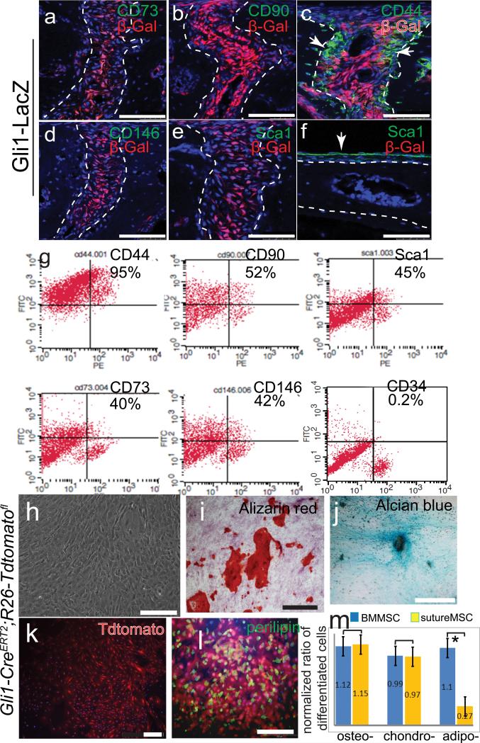
Gli1+ cells are MSCs in vitro. (a-f) Immunohistochemical staining of MSC markers CD73 (a), CD90 (b), CD44 (c), CD146 (d) and Sca1 (e) in the suture mesenchyme of Gli1-LacZ mice. Sca1 expression is also detectable in the periosteum (f). Arrows indicate expression. Dotted lines outline bone margins. (g) FACS analysis of suture mesenchymal cells harvested from one-month-old Gli1-CE;R26Tdtomato mice induced with tamoxifen. (h-l) Gli1+ cells form clones in culture. Positive clones were picked based on their fluorescence (k). Alizarin red (i), Alcian blue (j), and perilipin (l) staining indicates that cells from single clones can undergo tri-lineage osteogenic (osteo), chondrogenic (chondro), and adipogenic (adipo) differentiation. (m) Quantitation of the fraction of differentiated cells in the suture MSC culture normalized to that of BMMSCs under the same conditions. Values are plotted as mean ±SEM. *, student t-test, p=0.02, n=5 cultures derived from different mice. Scale bars, 100 μm.

