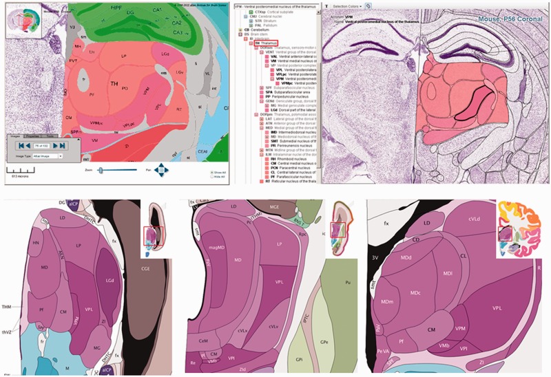
Figure 4.
Interactive Reference Atlas Viewer. The top left hand panel portrays an annotated coronal reference atlas plate from the P56 mouse (Allen Mouse Brain Atlas) in the High Resolution Image Viewer. At the top of the image is the hippocampus (green), and in the middle of the image is the thalamus (red) and its corresponding subdivisions (subnuclei). The top right hand panel presents the same annotated adult mouse reference atlas plate in the Interactive Reference Atlas Viewer. This viewer is divided into two windows, a collapsible hierarchical tree of brain structures on the left and annotated images of the Nissl-stained reference sections on the right that displays the images in a Zoom and Pan Image Viewer. Polygons scale and move with the image as one zooms in and out or moves the image in any direction. Moving the computer cursor over the image highlights individual structures; the structure name and acronym are displayed at the top of the window. Clicking on a structure also highlights the structure in the structure hierarchy. The lower panel shows coronal annotated reference atlas plates from the BrainSpan Atlas of the Developing Human Brain from a 15 pcw (post conception weeks), 21 pcw and 34 year reference atlas. The thalamus and its corresponding subregions (subnuclei) are purple in the human atlases. With the reference atlas viewers, one can examine neuroanatomy across time within an individual species (mouse or human), as well as across species (mouse versus human).

